Keywords
|
| Fundus Image,Glaucoma, Intra Ocular Pressure(IOP),Simple Linear Iterative Clustering (SLIC), KMeans clustering. |
INTRODUCTION
|
| Glaucoma is a chronic eye disease in which the optic nerve is progressively damaged. It is the second leading cause of blindness, and is predicted to affect around 80 million people by 2020.Glaucoma as a neuro degeneration of the optic nerve is one of the most common causes of blindness. Because revitalization of the degenerated nerve fibers of the optic nerve is impossible early detection of the disease is essential. This can be supported by a robust and automated mass-screening.A novel automated glaucoma detection system that operates on inexpensive to acquire and widely used digital color fundus images. After a glaucoma specific preprocessing, different generic feature types are compressed by an appearance-based dimension reduction technique. Subsequently, a probabilistic two-stage classification scheme combines these features types to extract the novel Glaucoma Risk Index (GRI) that shows a reasonable glaucoma detection performance. |
| Instead, it gives at the weakest point, which is the site in the sclera at which the optic nerve leaves the eye.The optic nerve is the part of eye that carries visual information to the brain. It is made up of over one million nerve cells, and while each cell is several inches long, it is extremely thin about one twenty-thousands of an inch in diameter. The death of these cells results in permanent vision loss. Early diagnosis and treatment of glaucoma can help to prevent this from happening. |
RELATED WORK
|
| Cheng proposed the optic disc and optic cup segmentation for glaucoma detection [1]. Optic nerve head assessment in retinal fundus images is both more promising and superior. Using this method the optic disc segmentation, histograms, and center surround statistics are used to classify each super pixel as disc or non-disc. And also segment the optic cup region. Calculating the cup to disc ratio value. However this method had poor visual quality. |
| Achanta proposed to improve the segmentation performance by using a super pixel algorithm,simple linear iterative clustering (SLIC) method[2]. It empirically compares five state-of-the-art super pixel algorithms for their ability to adhere to image boundaries, speed, memory efficiency, and their impact on segmentation performance. However this method increased the computations. |
| Joshi proposed an approach for an automatic OD parameterization technique based on segmented OD and cup regions obtained from monocular retinal images. In an active contour model is presented to get robust OD segmentation [3]. This has been achieved by enhancing Chan- vese (C-V) model by including image information at the support domain around each contour point. The drawback of this method was that it does not provide better quality.Babu proposed for the measurement of CDR[4].It is considered as a parameter for the diagnosis of glaucoma and 90% accuracy is obtained The CDR ratio is an important indicator of the risk of the presence of the glaucoma in an individual. |
| Arturo proposed to optic disc boundary using morphological, edge Detection, and feature Extraction techniques [5].It requires a pixel located within the OD as initial information. For this purpose, a location methodology based on a voting-type algorithm is also proposed. The algorithms were evaluated on many images and the results were fairly good. However this method had poor visual quality. |
OPTIC DISC AND CUP SEGMENTATION FOR CLUSTERING METHOD
|
| Using this technique, Cluster center can be applied. It aggregates the nearby pixel into super pixels. Select the pixel from an image and the centers move towards the lowest gradient position. In this technique searches for its best matching pixel from the neighborhood based on the color and spatial proximity. And then compute the new cluster center based on the found pixel. This iteration will be continue until the distance between the new center and previous one is small enough. |
| The output of the SLIC technique is given as input to the K-means Clustering algorithm. It classifies the input data into multiple clusters. And also apply the image in gabor filter technique. It is a linear filter used for edge detection. It is used to reduce noise. By applying thresholding technique to the segmented optic disc, optic cup will be segmented. The optic disc and optic cup diameter is measured to calculate the Cup to Disc Ratio (CDR). From the CDR value, the disease condition of the patient can be identified. |
Simple Linear Iterative Clustering Algorithm (SLIC)
|
| The super pixel algorithm, simple linear iterative clustering (SLIC), which adapts a k-means clustering approach to efficiently generate super pixels. Despite its simplicity, SLIC adheres to boundaries as well as or better than previous methods. At the same time, it is faster and more memory efficient, improves segmentation performance, and is straightforward to extend to superpixel generation. SLIC is simple to use and understand. |
| By default, the only parameter of the algorithm is k, the desired number of approximately equally-sized super pixels. For color images in the CIELAB color space, the clustering procedure begins with an initialization step where k initial cluster centers = li,ai,bi,xi,yi T are sampled on a regular grid spaced S pixels apart. To produce roughly equally sized super pixels, the grid interval is S= Nk . The centers are moved to seed. locations corresponding to the lowest gradient position in a 3 *3 neighborhood. This is done to avoid centering a super pixel on an edge, and to reduce the chance of seeding a superpixel with a noisy pixel. |
| Next, in the assignment step, each pixel i is associated with the nearest cluster center whose search region overlaps its location. This is the key to speeding up our algorithm because limiting the size of the search region significantly reduces the number of distance calculations, and results in a significant speed advantage over conventional k- means clustering where each pixel must be compared with all cluster centers. This is only possible through the introduction of a distance measure D, which determines the nearest cluster center for each pixel. |
K-Means Clustering
|
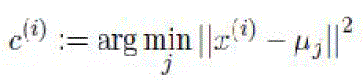
| K-Means algorithm is an unsupervised clustering algorithm that classifies the input data points into multiple classes based on their inherent distance from each other. The algorithm assumes that the data features form a vector space and tries to find natural clustering in them. The points are clustered around centroids μiAi = 1…k which are obtained by minimizing the objective. |
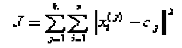 (1) (1) |
where  is a chosen distance measure between a data point (x)i(j) and the cluster centre cj, is an indicator of the distance of the n data points from their respective cluster centres. is a chosen distance measure between a data point (x)i(j) and the cluster centre cj, is an indicator of the distance of the n data points from their respective cluster centres. |
| • Compute the intensity distribution(also called the histogram) of the intensities. |
| • Initialize the centroids with k random intensities |
| • Repeat the following steps until the cluster labels of the image do not change anymore. |
| • Cluster the points based on distance of their intensities from centroid intensities replicated with the mean value within each of the array and then the distance matrix is calculated. |
 (2) (2) |
| • Compute the new centroid for each of the clusters. |
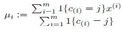 (3) (3) |
| where k is a parameter of the algorithm (the number of clusters to be found), i iterates over the all the intensities, j iterates over all the centroids and ���� are the centroid intensities.The algorithm assumes that the data features form a vector space and tries to find natural clustering in them. |
Gabor Filter
|
| Gabor filter is a linear filter used to detect the edge. It is used to reduce noise. Gabor filter can be tuned for specific frequencies and orientations which are useful edge detection. They act as low level oriented edge discriminators and also filter out the background noise of the image. Since that have directional pattern so 2-D Gabor filter is best option due to its directional selectiveness capability of detecting oriented features and fine tuning to specific frequencies. |
Cup Segmentation
|
| In cup segmentation,use thresholding or binarization for optic cup segmentation process. This process will convert the image into a B/W (Black & White) image where it can easily segment the optic cup from disc region. |
CDR Calculation And Diagnosis
|
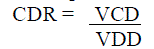
| After obtaining the disc and cup, various features can be computed. We follow the clinical convention to compute the CDR.It is ratio between the Vertical Cup Diameter (VCD) &Vertical Disc Diameter(VCD). |
 |
| The computed CDR is used for glaucoma screening. When CDR is greater than a threshold, it is glaucomatous otherwise healthy. |
SIMULATION RESULTS
|
DATA SETS
|
| Images are collected from Mahatma Eye care Hospital,Trichirapalli and the databases from the net. The images were acquired using a Canon CR5 non–mydriatic 3 CCD camera with a 45 degree field of view(FOV).Each image was captured using 8 bits per colour plane at 768 by 584 pixels.The FOV of image is circular with a diameter of approximately 540 pixels. For this database, the images have been cropped around FOV.For each image, a mask image is provided that delineates the FOV. |
| The VISUCAM NM/FA fundus camera is the perfect combination of a non-mydriatic fundus camera with superior image quality and an easy-to-use camera for fluorescein angiography. The VISUCAM NM/FA is enhanced with the hardware and software required to record,display and document fluorescein angiography. The VISUCAM NM/FA offers everything needed for the detailed diagnosis of typical diseases of the eye, e.g diabetic retinopathy, glaucoma and age-related macular degeneration (AMD).The camera documents the results and permits the subsequent management of the disease. |
GLAUCOMA SCREENING
|
| This paper evaluates the glaucoma detection for a patient |
CONCLUSION & FUTURE WORK
|
| In this paper presented super-pixel classification based optic disc and cup segmentations for glaucoma screening. This paper is presented and evaluated for Glaucoma detection in patients using multimodalities including simple linear iterative clustering (SLIC) algorithm, K-Means clustering and Gabor filter of the color fundus camera image to obtain accurate boundary delineation. Using structural features like CDR (Cut to Disc Ratio), the ratio value exceeds 0.6 shall be recommended for further analysis of a patient to the ophthalmologist. This shall help in patients worldwide by protecting further vision deterioration through timely medical intervention. We can increase the number of patients and analyze the performance. |
| |
Tables at a glance
|
 |
|
| Table 1 |
Table 2 |
|
| |
Figures at a glance
|
 |
 |
 |
| Figure 1 |
Figure 2 |
Figure 3 |
|
| |
References
|
- Jun Cheng, Jiang Liu, YanwuXu, Fengshou Yin &Ngan-Meng Tan, ‘Super pixel Classification Based Optic Disc and Optic CupSegmentation for Glaucoma Screening’, IEEE Transactions on Medical imaging, Vol. 32, NO. 6, pp.1019-1032, 2013.
- Achanta R , Shaji A, Smith K, Lucchi A &Susstrunk S,‘Slic super pixels compared to state-of-the-art super pixel methods’, IEEETransactions on Pattern Analysis and Machine Intelligence, Vol. 34, No. 11,pp.2274–2281, 2012.
- Joshi G D, Sivaswamy J &Krishnaas S.R, ‘Optic disk and cup segmentation from monocular color retinal images for glaucoma assessment’,IEEE Transactions on Medical Imaging, Vol. 30, No. 6, pp.1192–1205,2011.
- Ganesh Babu T.R & S.Shenbagadevi,‘Automatic detection glaucoma using fundus image’,European journal of scientistresearch,Vol.59,No.1,pp.22-32,2011.
- Aquino A, Gegundez M & Marin D, ‘Detecting the optic disc boundary in digital fundus images using morphological, edge detection, andfeature extraction techniques’, IEEE Transaction on Medical Imaging, Vol.29, No. 11,pp. 1860–1869, 2010.
- Meier J, Bock R, Michelson G, Nyl L G, &HorneggerJ,‘Effects of preprocessing eye fundus images on appearance based glaucomaclassification’, IEEE Transaction on Medical Imaging,Vol.69, pp. 165–172,2007.
|