ISSN ONLINE(2319-8753)PRINT(2347-6710)
ISSN ONLINE(2319-8753)PRINT(2347-6710)
VijayaRekha.R1, Sudha.S2, Sangeetha.J3, Shenbagarajan Anantharajan4
|
| Related article at Pubmed, Scholar Google |
Visit for more related articles at International Journal of Innovative Research in Science, Engineering and Technology
This project aims at detection of Tumor blocks and classifying the type of tumor using Decision Tree in MR images of the tumor affected patients with Astrocytoma type of Brain Tumor. The proposed technique consists of different stages namely, Preprocessing, Adaptive Thresholding Segmentation, GLCM Feature Extraction and Decision Tree Classification. The Image Processing techniques such as Median filtering, Sobel gradient edge detection, Adaptive Thresholding and feature extraction have been developed for detection of Brain Tumor in the MR images of cancer affected patients. The developed system classifies the images into a Grade of tumor belongs to Astrocytoma type of Brain Cancer. The system is found efficient in classification of these samples and responds on any abnormality noticed and added to it, the system eliminates the need for Biopsy for classifying the tumor grades efficiently
Keywords |
| Adaptive Thresholding, Decision Tree, Gray Level Co-occurrence Matrix, Magnetic Resonance Images, Median filter, Sobel gradient edge detection . |
INTRODUCTION |
| The Cancer is the next stage or effect of tumorous cells. It is dangerous and tedious to recover, sometimes it may lead to loss of life. Tumor is medically defined as an abnormal growth of cells. The tumor occurred within the brain or inside the skull then it is coined as Brain tumor. Brain tumors are great mimics of other neurological disorders. It may be of Least Aggressive or benign (non-cancerous) and Most Aggressive or Malignant (cancerous). They have been detected using either Computed Tomography(CT) or Magnetic Resonance Images(MRI)[4]. It is hard to conclude grade of tumor just by discussing the results of CT or MRI. A Biopsy is a surgical procedure in which a sample of tissue is taken from the tumor site and examined under a scientific microscope. The biopsy discovers the type and grade of a tumor. Hence C.A.D.S., eliminates the need for biopsy. |
II. LITERARATURE SURVEY |
| 1 .In [3] Hybrid Medical Image Classification Using Association Rule Mining With Decision Tree Algorithm. |
| ï½ The main focus is concerned with the classification of brain tumor in the CT scan brain images not in MRI. |
| ï½ In this paper the classification of brain images into normal, benign and malignant but not in grades. |
| 2. In [4] Classification Of Brain Tumors In MR Images |
| ï½ Feature extraction along with selection results in removal of reduntant features. |
| ï½ It classifies the results either Benign or Malignant. |
| But now-a-days there is a need for fine tuned classification. |
| In [1] Sonali Patil developed an diagnostic system for segmentation, feature extraction and classification. The technique used for segmentation is Square Based Segmentation and Component Labeling. Further Gray Level Co-Occurrence Matrix (GLCM). |
| As continued in [1] the Probablistic Neural Network (PNN) is implemented. But when considering PNN it is lagging in computation. |
| 1.PNN are slower when classifying new cases. |
| 2.PNN require more memory space to store the model. |
| 3.Backwarding is not possible. |
III. STRATEGICALMETHODS |
A.GENERALIZED VIEW |
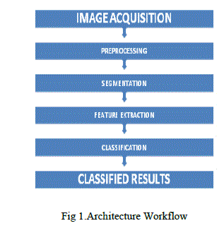
| The Architecture workflow depicts the steps involved and the corresponding input and output. |
 |
B.IMAGEPROCESSING |
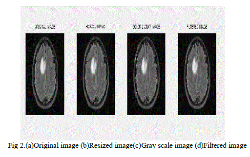
| The Image preprocessing[1],[7] is the technique of enhancing data images prior to computation processing. It is essential for reducing the complexity and computation time of the C.A.D.S.,. The input image may contain noise. It does Normalizing the intensity of the images, Resizing an image Color to Gray scale conversion and Filter the image from noise using Median filter. |
(i) Median Filter |
| As mentioned above the median filter is more effective than using the mean filter |
| If the values is odd then |
| Median = ((N+1)/2) |
| If the values is even then |
| Median = ((N/2)th value+((N+1)/2) th value). |
| In general, the median filter allows a great deal of high spatial frequency detail to pass while remaining very effective at removing noise on images where less than half of the pixels in a smoothing neighborhood have been effected. |
 |
C.IMAGE SEGMENTATION |
| The Image segmentation is the process of partitioning a digital image into multiple segments. The goal of segmentation is to simplify and/or change the representation of an image into something that is more meaningful and easier to analyze. |
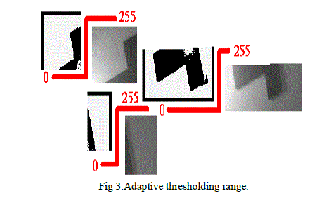
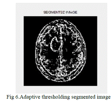
| The proposed work implements the Adaptive thresholding for segmentation[1],. Generally Thresholding is used to segment an image by setting all pixels whose intensity values are above a threshold to a foreground value and all the remaining pixels to a background value. The drawback of thresholding is dynamic change according to the pixel intensity cannot be achieved. |
| Adaptive thresholding typically takes a grayscale or color images as input and, in the simplest implementation, outputs a binary image representing the segmentation. For each pixel in the image, a threshold has to be calculated. If the pixel value is below the threshold it is set to the background value, otherwise it assumes the foreground value. |
 |
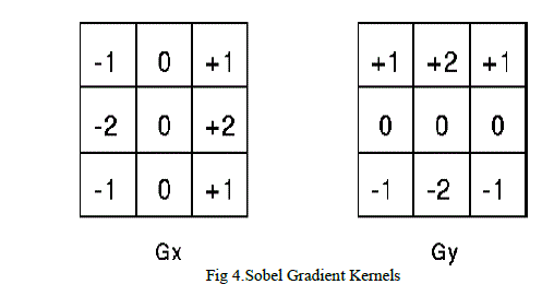
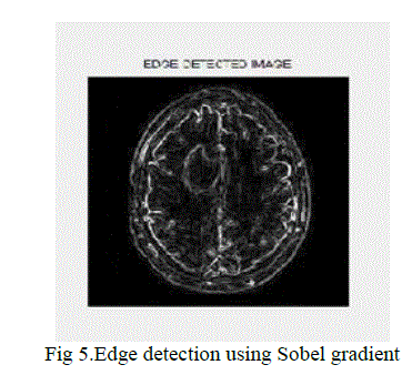
| For edge detection the Sobel gradient technique has been used. The Sobel gradient edge detection. The Sobel gradient performs 2-D spatial gradient measurement of an image and emphasizes the area of high spatially detectable that correspond to edges. Typically it is used to find the approximate absolute gradient magnitude at each point in an input grayscale image. |
 |
 |
D. Feature Extraction |
| GLCM - A statistical method of examining texture that considers the spatial relationship of pixels is the gray-level cooccurrence matrix (GLCM), also known as the gray-level spatial dependence matrix. |
| The third step of the proposed work is Feature extraction. The features may be texture based and intensity based. Here the GLCM[3],[5] technique is used for smart feature extraction. The main concept of feature extraction is based on the contrast value. |
| The values of the contrast and all other ranges from 0-1. |
| For each feature there is a mathematical calculation using formulae.Mathematically, a co-occurrence matrix C is defined over an n × m image I, parameterized by an offset (Δx,Δy), as: |
 |
| where i and j are the image intensity values of the image, p and q are the spatial positions in the image I and the offset (Δx,Δy) depends on the direction used and the distance at which the matrix is computed d. |
 |
E.CLASSIFICATION |
| The final step of our work is Classification. The classification[2],[6],[8] is done using Decision tree classifier. Based on the feature extracted values the brain tumor can be classified as Grade I-IV as per WHO. |
| ï· Grade I: The tissue is benign. The cells look nearly like normal brain cells, and they grow slowly. |
| ï· Grade II: The tissue is malignant. The cells look less like normal cells than do the cells in a Grade I tumor. |
| ï· Grade III: The malignant tissue has cells that look very different from normal cells. The abnormal cells are actively growing (anaplastic). |
| ï· Grade IV: The malignant tissue has cells that look most abnormal and tend to grow quickly. |
IV EXPERIMENTAL RESULTS |
| The figures depicts the preprocessing, segmentation, feature extraction and finally classification. |
 |
V CONCLUSIONS |
| The designed Brain Cancer Detection and Classification System use conceptually simple classification method using the Decision tree. The classification of tumors is in automated fashion. From the results of proposed work, it can be concluded that, The designed and implemented system provides precision detection and real time tracking by classifying the unknown sample Image into appropriate Astrocytoma type of Cancer, thus do not involve any pathological testing. This system provides precision Detection and Classification of Astrocytoma type of cancer. |
| The system has been tested with the Astrocytoma type of brain cancer Images only. The system can be designed to classify other types of cancers as well with few modifications. The proposed system delivers the prompt output efficiently. |
References |
|