ISSN ONLINE(2319-8753)PRINT(2347-6710)
ISSN ONLINE(2319-8753)PRINT(2347-6710)
G.P.GOKUL LAXMI 1, M.MEHARTAJ1, K.RAMANADEVI, M.SATHYA1, S.SOLASUBBU M.S 2
|
| Related article at Pubmed, Scholar Google |
Visit for more related articles at International Journal of Innovative Research in Science, Engineering and Technology
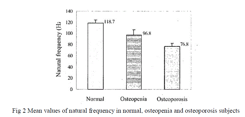
Osteoporosis and Osteopenia are increasingly being recognized as a health hazard now-a-days. The dynamic characteristics of bone which provides quantitative information concerning the mechanical properties of the bone along with measurement of bone mineral density (BMD) can help in the early detection and better diagnosis of osteoporosis. The stress waves are generated in the tibia bone by the impact of, impulse force hammer are monitored by accelerometers. The natural frequency of stress wave signal is significantly decreased in osteopenia and osteoporosis subjects indicating decrease in mechanical strength of the bone and bone mineral mass. The technique gives a better understanding of the dynamic behaviour of bone under impact force, and the natural frequency of stress wave Signal is a clear indication of mechanical stiffness of the bone under investigation.
Keywords |
| Impulse hammer, Accelerometers (ADXL 345), Coupler, PIC16F877A, MAX232. |
INTRODUCTION |
| Osteoporosis is a chronic, progressive condition associated with micro architectural deterioration of bone tissue .This would basically results in low bone mass leading to bone fragility that consequently increases the susceptibility to fractures [1, 2]. Osteoporosis usually remains asymptomatic until the weakened bone fractures. Osteopenia is an acute, treatable disease which would be treatable under certain medications and regular diet. This fractures lead to deformity, loss of mobility, frequent change in positions of bone and even death. With increasing population of elderly subjects, the assessment and treatment of osteoporosis and osteopenia have become a crucial problem in clinical gynaecology as well as osteology and other few related fields. Bone mineral loss occurs with various factors such as aging, menopause, and disuse. The decrease in bone mineral density with age is much more pronounced than the loss of bone mass due to perforations of other factors [3]. BMD measurements in conjunction with infuriation about the structure and elastic properties of bone will result in a good indication of its mechanical condition and susceptibility to fracture. Moreover, during accidental impact, our bones arc subjected to high strain rate loading. Since bone is a visco elastic material [4], its response to this type of loading cannot be assumed to be the saint as predicted by a static analysis. Therefore, it is important to study dynamic characteristics of bone under normal and diseased state in order to understand its response to more realistic loading condition [5]. Hence, the peed for impulse response tests, Stress waves generated by impulse force provide quantitative information concerning the mechanical properties of the bone in question. Development of a successful technique of this type would have many clinical applications such as in diagnosis of osteoporosis and osteopenia and the evaluation of fracture healing. |
| In the present work, dynamic behaviour of tibia bone is studied by stress wave signal generated by impulse force for assessment of osteoporosis and osteopenia. The natural frequency of stress wave signal is determined. The change in natural frequency with age is also evaluated. |
II.LITERATURE SURVEY |
| In the traditional method of measuring bone mineral density, bones are exposed to a dose of radiation i.e., using DXA. Equipment made by different companies will not give identical scores. Comparisons of measurements from different machines can lead to a false reading of an increase or decrease in BMD. This technique is time consuming, while the measurement is performed, a beam of low-dose x-rays passes through the area being measured. These xrays are detected by a device. The machine converts the information received by the detector into an image of the skeleton and analyzes the BMD, or “bone mineral density”. Takes longer time to produce the results ,the results are usually available in 2 to 3 days In traditional only present measured values can be obtained in the form of thin films. Accuracy will vary in some cases such as skeletal deformities like spinal curves or compression fractures, bowing in long bones and the presence of metal rods can significantly impair the results from a DXA. In this method there is no use of radiation; impulse response test is used to measure BMD. The stress waves arc generated in the tibial bone by the impact of impulse force hammer which are monitored by accelerometers are analyzed .Performance of this technique may not take much time. The results can be produced within few minutes. By means of storage past measured values can be obtained. Accuracy will be perfect. Children and adults with small bones, and/or very low bone density may get an accurate measurement because it can be able to identify the outline of the bones |
III.METHODOLOGY |
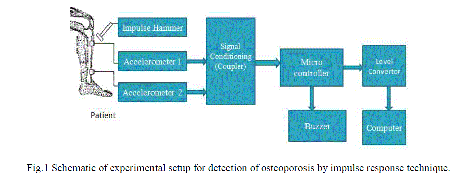
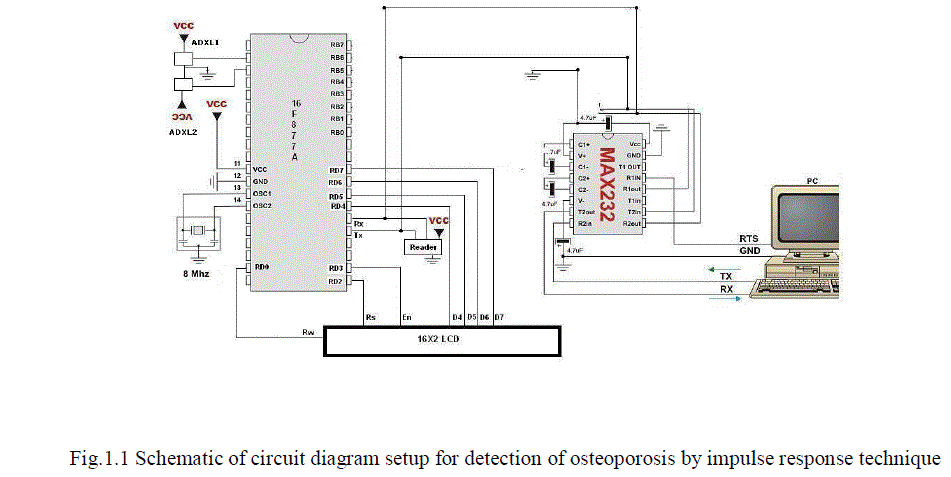
| The experimental setup comprises an impulse force hammer and two ceramic shear type accelerometers (ADXL 345) within built charge, amplifiers. The coupler provides constant current excitation required by accelerometers and decouples the DC bias voltage from the output signal. Storage oscilloscope is used for online observation of impulse and stress wave signal. High-speed PIC 16F877A is used to digitize and acquire the impulse response data into a personal computer for further processing and analysis. The experimental setup for in vivo assessment of osteoporosis and osteopenia by impulse response is shown in Figure 1. |
 |
| The in vivo impulse response measurements were performed non-invasively on diathesis of the left tibia. The choice of the tibia as a measurement site is validated by the fact that it contains approximately 25-30% trabecular bone and 70- 75% cortical bone by volume, and is subjected to extreme loading conditions during daily activities [6]. The trabecular bone is highly active in remodelling process and shows changes within bone tissue earlier than cortical bone. Observable changes are also seen in cortical bone during bone metabolism [7]. Men have been estimated to loose, on average, about 25% of their cortical bone, and women loose 35% of their cortical bone over their lifetime [SI]. So the net bone mineral loss occurring in the skeleton can be observed in tibia. Moreover, the medial side, of tibia is approximately flat and soft tissue thickness is minimum, making it an ideal choice for accelerometer mounting in impulse response study. The experimental arrangement was such that the subject was asked to sit on a chair with their leg well placed vertically on a vibration free wooden platform. The pre-selected points for the hammer strike and for the accelerometer mounting were determined on the skin surface and marked. |
 |
| The accelerometer-I is placed on the fascias medial is tibia at a distance of 8 cm from medial condoyle and accelerometer-2 is placed at a distance of 20 cm from the accelerometer- 1 using adhesive without imposing extra loading on bone. In this configuration of mounting, accelerometers are only sensitive to the axial acceleration, and no attempt was made to measure any other mode of acceleration. The stress waves are generated by the impact of hammer on the medial side of the proximal tibia at a distance of 4 cm from medial condoyle. The impact force applied through impulse hammer is standardized between 2 - 2.5N for each impact, which is obtained by dropping the hammer from a vertical distance of 5 cm. The above force is chosen to optimize the pulse width, maximize the signal to noise ratio and to ensure absolutely no pain to the subject. The application of the impact force is parallel to the axis of both accelerometers and therefore approximately perpendicular to the facieses medial is. The force and acceleration outputs were amplified by in-built amplifiers and observed simultaneously on oscilloscope. The data is acquired into a computer after digitization using ADC through software at a sampling frequency of 17 kHz per channel. This high sampling rate is chosen to acquire all information related to short duration impulses, high velocity stress waves and to reproduce the waveforms faithfully. The data is sampled for 5 seconds in which 4 impulses are applied (approximately at equal intervals) and corresponding response data are acquired. Repeatability of the test was ensured by conducting the experiment and recording three sets of data for each subject. Fresh data was taken if three consecutive readings were not almost identical. The data is analysed in Mat lab environment. The natural frequency of stress wave signal was obtained by taking, Fast Fourier transform (FFT) of the accelerometcr-2 output. The reproducibility of the natural frequency is tested in each subject by repeating the test three times. |
IV.RESULTS AND DISCUSSION |
| The values of natural frequency for normal, osteopenia and osteoporosis subjects were made analysed by this technique which was used to establish relationship between natural frequency, age, ultrasound stiffness index and Tscore. Frequency analysis of stress wave signal was carried out for all the subjects. It is found that the natural frequency is significantly decreased in osteopenia and osteoporosis subjects indicating low bone mineral mass, mechanical strength and stuffiness (Figure 2). The results show a significant difference in the item values between normal and osteopenia (P<0.0005), between normal and osteoporotic (P<O.OOOS) and also between osteopenia and osteoporotic (P<0.005) subjects. To evaluate the efficiency and reliability of the impulse response test in the diagnosis and assessment of osteoporosis, impulse response parameter (natural frequency) needs 'to be compared with output of standard and established osteoporosis test procedure. For this, all the subjects who had undergone impulse response test work also screened for osteoporosis using this technique. |
 |
| To evaluate the efficiency and reliability of the impulse response test in the diagnosis and assessment of osteoporosis, impulse response parameter (natural frequency) needs 'to be compared with output of standard and established osteoporosis test procedure. For this, all the subjects who had undergone impulse response test work also screened for osteoporosis using this technique. The result of linear regression analysis show a very good correlation (r = 0.92, P<0.0005) between natural frequency and SI .A significant correlation is also found between natural frequency and T-score (r = 0.9 1. P<0.0005) .This results strongly indicate that measurement of natural frequency from impulse response test can be used to find bone stiffness and to predict T-score based on which subjects can be diagnosed and classified into normal osteopenia and osteoporosis. |
| Regression analysis also shows a statistically significant negative correlation between natural frequency of impulse response and age (r = -0.79, P<O.OOOS). The natural frequency decreases with age at a rate of 9- IO‘%, per decade. The results show that the bone mineral loss and decrease in mechanical stuffiness of bone with increase in age is very well reflected by decrease in natural frequency. |
V.CONCLUSION |
| The impulse response technique for monitoring the stress wave propagation in tibia bone has been effectively used in the assessment of osteoporosis. The technique gives a better understanding of the dynamic behaviour of bone under impact force, and the natural frequency of stress wave Signal is a clear indication of mechanical stiffness of the bone under investigation. The study is non-invasive, reliable, easy to operate, inexpensive and has diagnostic potential in the early detection and assessment of osteoporosis and osteopenia. |
References |
|