International Journal of Innovative Research in Science, Engineering and Technology
ISSN ONLINE(2319-8753)PRINT(2347-6710)
ISSN ONLINE(2319-8753)PRINT(2347-6710)
Tijana LainoviÄ 1, Dr. Larisa BlažiÄ 1, Marko VilotiÄ 2, Dr. Damir Kakaš 2
|
| Related article at Pubmed, Scholar Google |
Visit for more related articles at International Journal of Innovative Research in Science, Engineering and Technology
Commercially available and the most widely used oral bionanomaterials in dental practice are resin-based composites containing oxide nanoparticles. The aim of this study was to determine the effect of diamond paste finishing on AFM surface texture parameters of dental nanocomposites polished by two different dental polishing protocols. Representative dental resin-based nanofilled and nanohybrid composite were tested in this study. The samples were polished by two dental polishing protocols using multi-step and one-step system. These protocols were than followed by diamond paste polishing. Samples were examined by Veeco di CP-II Atomic Force Microscope, in contact mode with CONT20A-CP tips. Average roughness (Ra) and maximum peak-to-valley distance (Rp-v) were analyzed and compared between samples. Multi-step polishing protocol produced significantly lower Ra and Rpv values, for both tested materials, than one-step polishing protocol, even when it was followed by diamond paste polishing.
Keywords |
| Nanocomposites, Composite Dental Resin, Microscopy, Atomic Force, Surface Roughness, Dental Polishing |
I. INTRODUCTION |
| Nanotechnology, as a revolutionary technology, has taken a wide range of applications in various fields [1]. Improvements in materials science and engineering have led to the development of modern and advanced nanomaterials for dental practice [2]. The most widely used dental nanostructured materials are resin-based composites containing oxide nanoparticles [3]. These materials are divided in two groups: nanofilled and nanohybrid composites [4]. Nanofilled materials are resin-based materials containing only nanometer-sized fillers, while nanohybrid composites include micrometer particles (0.4–5μm) mixed with nano-sized inorganic particles [5]. Evolution to nanostructured materials made dental composites more mechanically resistant materials, with reduced polymerization shrinkage, thermal expansion and water solubility, with improved optical and aesthetic characteristics, more wear resistant, with better polish ability and polish retention [2,6,7]. It is known that not only the materials properties are important, but also a high quality of working tools and processing mechanisms, in order to benefit from the characteristics of the new synthesized materials in the best possible way. |
II. RELATED WORK |
| The modern dental professional demands for quality of the restoration are increasing all the time, and for now, it is considered that the roughness below 0.2 μm is satisfactory feature for direct dental restoration [8,9]. This is the approximate limit under which the bacterial biofilm is not retained. The other demands include smooth and glossy surface, low retention possibility for stains and discolorations, lower wear during time of function [10,11]. On the other hand, there is a professional need for a more and more rapid clinical interventions with the introduction of a number of simplified procedures in every-day clinical work [12-14]. For this reasons, a method of materials polishing in a single step has been introduced in dentistry, so-called “single-step” or “one-step” polishing protocol [9,15]. This method supposed to push the multi-step polishing procedure in a second plan, by reducing of clinical chair-time. For this purpose, and in order to have a high quality of finishing procedure, a diamond paste polishers has appeared on the market [16-18]. |
| The aim of this study was to evaluate the effect of conventional multi-step polishing protocol on surface topography and roughness of resin-based nanocomposites comparing it with the effect of one-step polishing protocol. The diamond paste polishing effect on the surface features was also examined, following both of these previously conducted procedures. |
III. MATERIALS AND METHODS |
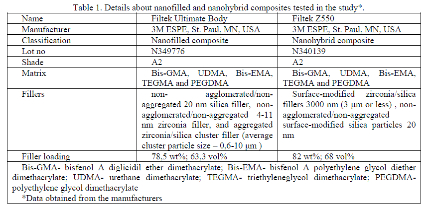
| Two contemporary dental resin-based nanocomposites were tested in this study: nanofilled (Filtek Ultimate Body, 3M ESPE), and nanohybrid composite (FZ550, 3M ESPE). Detailed information about the materials used in the study is shown in Table 1. |
 |
| Procedure for preparing the specimens |
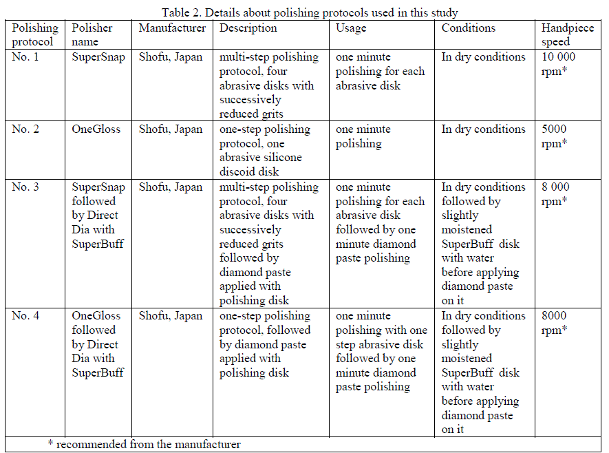
| Specimens were prepared in the cylindrical plastic molds (8 mm diameter x 2 mm depth) which were placed on the glass microscope slide, filled with material and covered with a polyester strip and a glass slide, taking care to obtain a flat surface without any defects and entrapped air. Material was then polymerized for 40 seconds with a SmartLite® IQTM 2 dental LED light curing unit (Dentsply Caulk). After removing glass plate and polyester strip from the top of the samples, After polymerization, the samples were polished by four dental polishing protocols: multi-step – MS (Super Snap, Shofu), multi-step followed by diamond paste polishing – MSD (DiamondDia, Shofu), one-step – OS (OneGloss, Shofu) and one-step followed by diamond paste polishing –OSD (See Fig 1. and Table 2. for details). |
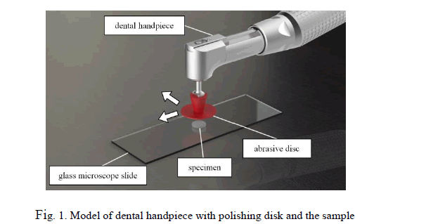 |
| During the polishing procedure, each abrasive disk was used only once for each material, in the dry condition, for 1 minute, using handpiece rotating 10 000, 8000 or 5000 revolutions per minute, depending on the polishing system and the manufacturer’s instructions. One single operator did all of the polishing treatments, simulating clinical finishing and polishing procedure. Two mutually perpendicular grinding directions were used during polishing procedure. |
 |
| AFM scanning |
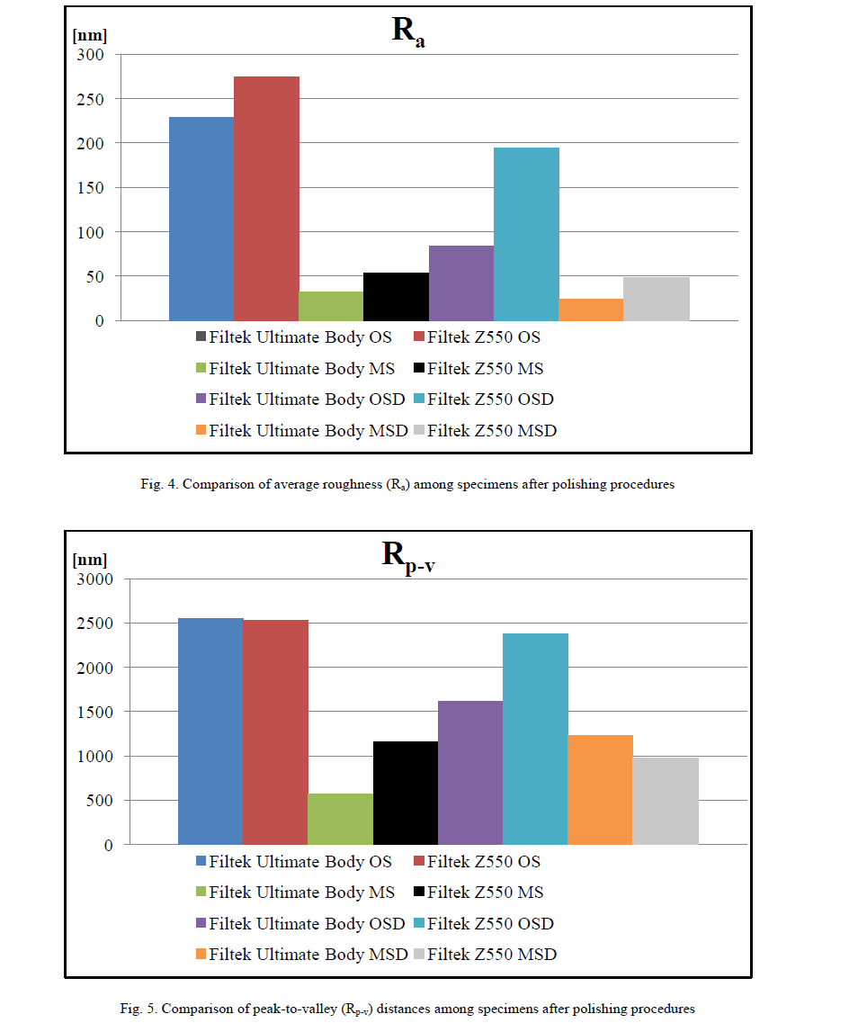
| After the polymerization and polishing procedures, samples were examined by Veeco di CP-II Atomic Force Microscope, in contact mode with CONT20A-CP tips. 1 Hz scan rate and 256 × 256 resolution were used to obtain topography on the 80 × 80 μm scanning area. Measured topography data were processed by Image Processing and Data Analysis v2.1.15 software. Following parameters were compared among specimens: average roughness (Ra) and maximum peak-to-valley distance (Rp-v). |
IV. RESULTS AND DISCUSSION |
| Topography of the tested materials |
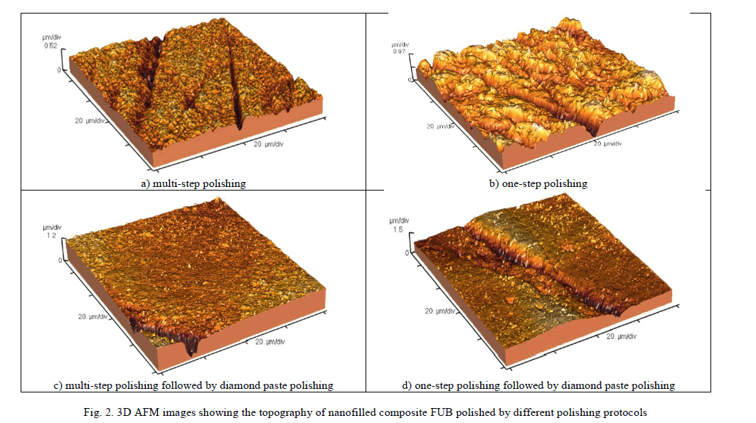
| Qualitative analysis of the 2D and 3D AFM images showed that multi-step polishing protocol produced lines and scratches on the tested materials' surfaces, which were results of the machining process with abrasive sandpapers (see Fig. 2 and Fig. 3). The abrasive wear mechanism of this polishing method created surface with furrowed and microploughed topography, with rare round local damages made as a consequence of the inorganic fillers dislodgement. One-step polishing procedure produced much higher elevations and depressions of the surface of both tested materials, but the damages were also linear and channel-type. Diamond paste polishing created smoother surfaces after both polishing procedures, by sweeping the lines and grooves, leaving only deep channels and a few new, hole-like defects of the surface. Nanofilled material had more regular surface with smaller holes, more shallow channels and less damages, than nanohybrid composite. Only one-step polishing system produced greater microplughing on nanofilled comparing to nanohybrid material. |
 |
| AFM surface texture parameters |
| Analysis of the quantitative results showed that multi-step polishing protocol produced significantly smoother surface and lower AFM surface texture parameters than one-step polishing protocol on the both tested nanomaterials. Calculated AFM roughness parameters (Ra and Rp-v) showed that both of the tested materials had significantly lower roughness values when samples were polished by diamond-paste polishing after the standard polishing protocols (see Fig. 4 and Fig. 5). This improvement was especially significant for the one-step polished samples, where this reduction was sometimes up to 200 nm in average roughness. Nanofilled material had lower Ra and Rp-v values nearly for all of the tested polishing protocols, than nanohybrid composite. |
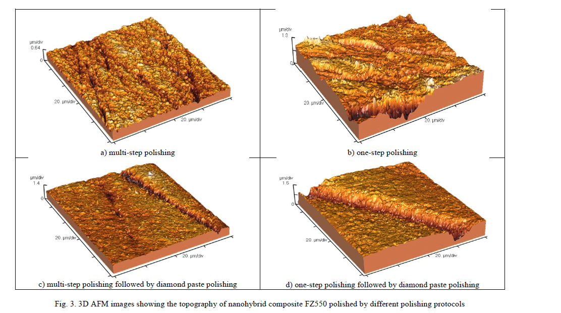 |
| The reasons for these results lay in the polishing mechanisms of differently designed polishers. The multi-step finishers and polishers are elastic silicone sandpapers, with four successively reduced grits, consisting of silicone carbide (Black and Violet sandpaper) and aluminium-oxide particles (Green and Red sandpaper). This polishing system turn out to be optimally designed for dental composite materials polishing, and shown great polishing results in other studies, also [19]. OneGloss is a one-step, aluminium-oxide impregnated silicone polisher, which is designed with the idea to save operative time, and to serve either for finishing, or polishing only by altering the contact pressure. In this study, it is shown that such a method, with rigid silicone polisher, can not provide the optimal polishing results, which is also the confirmation of some previous experimental results [20]. Polishing with this tool causes greater microploughing of the material and dislodging of filler or resin particles. This single-step protocol may be successfully used for dental ceramics of enamel polishing, but it is not suitable method for composite polishing. Only when it’s followed by diamond paste, it can reduce clinical chair time, and to create the acceptable surface topography and roughness parameters. The great wear capacity of the diamond paste reduced surface roughness of both tested materials, by erasing the scratched topography and leaving only sporadic deep grooves. Diamond paste removed both of the composite’s phases homogeneously, because the diamond components have been harder than the filler composite particles. This polishing protocol can save dental operative time and provide acceptable topography and roughness results. It depends on the size of the fillers, whether their dislodgement will leave the smaller or bigger holes and damages. Analyzing these experimental results, it can be concluded that the nano-sized particles in the material composition and the adequate polishing protocols have the major role in the final surface quality. |
 |
V. CONCLUSION |
| Multi-step polishing protocol produced significantly lower Ra and Rp-v values, for both tested materials, than one-step polishing protocol, even when it was followed by diamond paste polishing. The great wear capacity of the diamond paste enabled this polishing system to create smoother surface of the both tested nanocomposites and after both tested previously conducted polishing protocols. Nanofilled composite had lower roughness values than nanohybrid composites, due to its composition. There are two major factors which determine the smoothness of final composite restoration: nano-sized particles in the material composition and the adequate polishing protocols. Only the right choice of dental polishing mechanism, can lead to satisfying and good long-term clinical results. Although it is intended for reduction of clinical chair-time, one-step polishing protocol did not meet the high quality restoration demands of modern dental practice. In order to reduce chair time, dentists can use diamond paste polishing, after any multi-step or one-step polishing procedure, because it can reduce roughness in nearly every case creating smooth surface and satisfactory topography. Further studies need to investigate the effect of the other commercially-available polishing systems on the surface topography and roughness of different new dental composite materials containing nanoparticles. |
VI. ACKNOWLEDGEMENTS |
| This paper represents a part of the research realized in the frameworks of the projects: TR 035020 (Larisa Blaà ¾iÃÂ, Tijana LainoviÃÂ) and III-45006 (Damir Kakaš, Marko VilotiÃÂ) financed by the Ministry of Education, Republic of Serbia. The authors would like to thank 3M (East) AG company branch in Serbia, and Mikodental, Šabac – general dealers of Shofu, Japan for Serbia, for the material support. |
References |
|