International Journal of Innovative Research in Science, Engineering and Technology
ISSN ONLINE(2319-8753)PRINT(2347-6710)
ISSN ONLINE(2319-8753)PRINT(2347-6710)
Thilagavathi R1 and Gajalakshmi N2
|
| Related article at Pubmed, Scholar Google |
Visit for more related articles at International Journal of Innovative Research in Science, Engineering and Technology
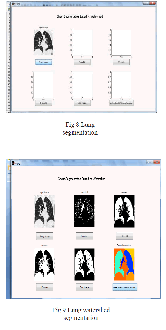
The objective of this work is to develop a segmentation model in order to remove the portion of lung for the treatment of certain illness such as lung cancer, and tumours. Image segmentation is the process of dividing an image into multiple parts. This method is vital to identify objects or other relevant information in digital images.During past few years we have gone across many region-based segmentation, data clustering, watershed segmentation, atlas based segmentation and edge-base segmentation algorithms, This work implements Marker based watershed transformation with the combination of both watershed transformation. In segmentation could be used for occlusion boundary,object recognition estimation within stereo or motion systems, image compression, image database look-up or image editing.Existing system segmentation is limited, But this System perform the segmentation for the lung image. Lung image contains the parts of Fissures, Vessels, and Bronchi.To segment this fissures, vessels and bronchi from the lung image, we perform the process of marker based watershed method.This segmentation technique is mainly used for find out the incomplete fissures. Thus this process provides the quality segmentation process.
Keywords |
| Fissure segmentation, lung lobe segmentation,Image segmentation |
INTRODUCTION |
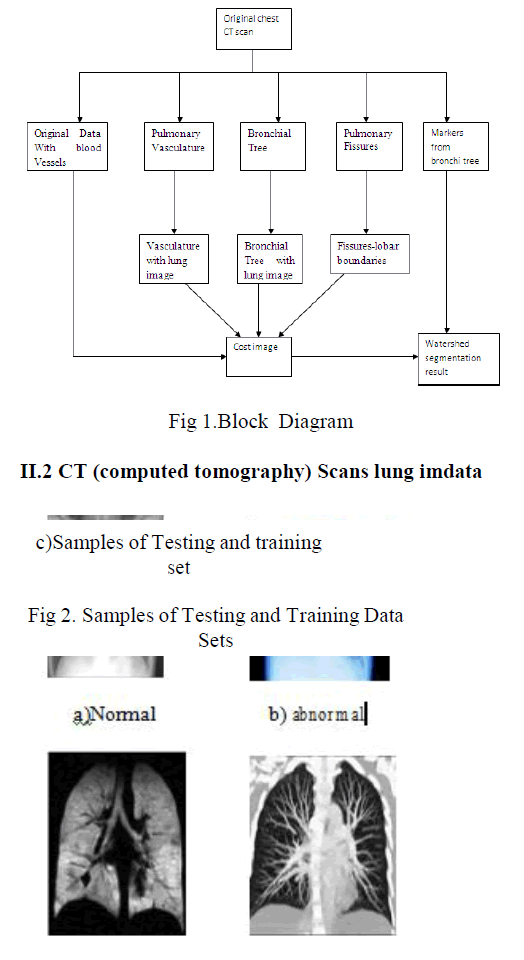
| A lobe segmentation method is developed which combines anatomical information from the lungs, vessels, airways, and lobar fissures to obtain the lobes using a Marker- based watershed segmentation method. Image segmentation is the first step . Segment the image based on Euclidean distance.This Euclidean distance is calculate the distance between the each parts. Then make the segmentations. System introduce the new system for segmentation and accuracy calculation. It performs the three types of operation: 1) The segmentations are combined into a cost image, 2) Markers for the watershed are computed, and 3) The lobes are segmented using a watershed transformation and post processing is applied.A lobe segmentation method is developed which combines anatomical information from the lungs, vessels, airways, and lobar fissures to obtain the lobes using a watershedbased segmentation method. Thus it provide the accurate results. |
PROPOSED METHODOLOGY |
| The proposed methodology consist of the stages, viz.CD SCAN image database,segmentation, Marker For Watershed, Lobe Segmentation, Graph Generation |
 |
Segmentation |
| Image segmentation is useful in many applications. It can identify the regions of interest in a scene or annotate the data. But all of these segmentation takes the more time to complete the process. Here we introduce the new technique of marker based watershed transformation technique. This marker based watershed transformation method |
| i Collect the lung images. |
| ii Load the lung image in the process. |
| iii Segment the image based on Euclidean distance. |
| iv This Euclidean distance is calculate the distance between the each parts. Then make the segmentations. |
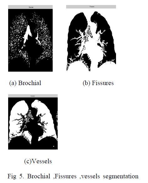
| v Segmented images are original image with blood vessels, fissure, vessel and bronchi. |
 |
Marker For Watershed |
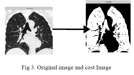
| Lung image contains the parts of Fissures, Vessels, and Bronchi.To segment this fissures, vessels and bronchi from the lung image, we perform the process of marker based watershed method.This segmentation technique is mainly used for find out the incomplete fissures.To generate the markers, the different sub trees belonging to the lobes and lobar segments are identified in the airway tree. This marker for watershed method is also be the cost image. Here it get the input from the segmented lung image. That is it get the output from the segmentation.Input for this process is segmented fissures, vessels, bronchi's and original lung image with blood vessels.Here it fix the segmented parts in the original image. Experiment results with comparison of existing method shows better accurate result less convergence rate. |
 |
Lobe Segmentation |
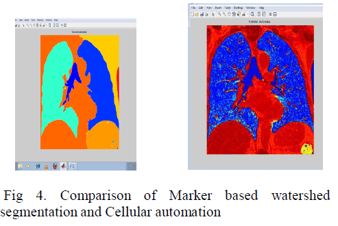
| A lobe segmentation method is develobed which combines anatomical information from the lungs,vessels,airways and lobar fissures to obtain the lobes using watershed transformation method.Here system apply the marker based watershed transformation. After finding the cost image, this lobe segmentation takes the input of cost image and marker from bronchi tree.The applied watershed algorithm separates regions with local maxima in between and can be used with an arbitrary number of markers.The borders between the obtained lobes after the watershed segmentation are not always smooth due to local variations in the cost image. The label value that occurs most often under the kernel is set to the voxel. To obtain the lobe segmentation on the original resolution, the segmentation results are up sampled using nearest neighbor interpolation. |
 |
Graph Generation |
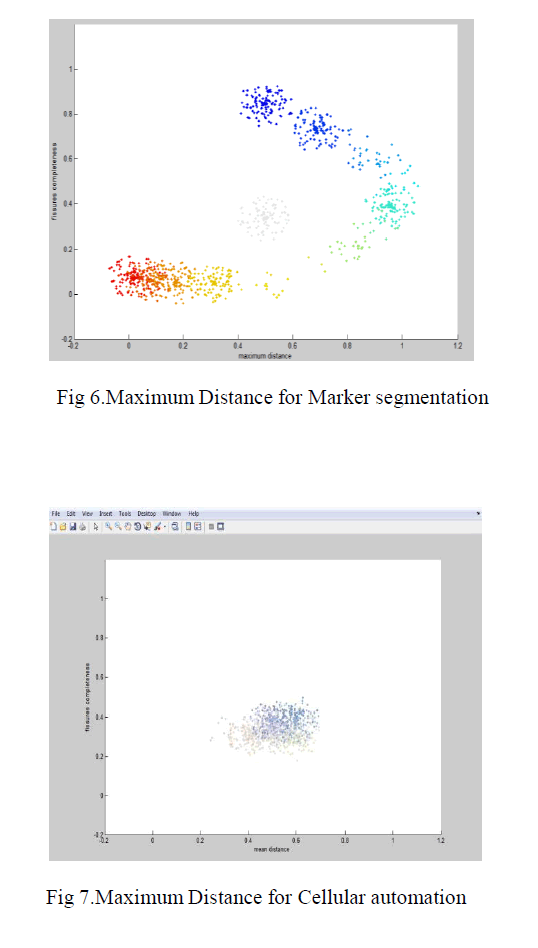
| System generate the three tables and two graphs for the solution.Two graphs are the mean distance graph and the maximum distance graph.Mean distance graph is generate between the mean distance and fissures completeness.Maximum distance graph is generate between the maximum distance and fissure completeness.For generating the table, system use the values of mean values, median values, distance between the each lobes and etc. |
 |
RESULTS AND DISCUSSIONS |
| The algorithm is developed using Matlab 2010a, and it is tested with the database of approximately 30 testing images. The sample input image taken for analysis is segmentation using Marker based watershed segmentationTo get the segmented image (lung image)calculate Mean, distance, SD and medians valuesfinally construct the Graph for mean distance and maximum distance. |
 |
CONCLUSION |
| Most of the segmentation system is used in the medical field. But the problem is takes more time to complete the process or makes the inaccurate segmentation results.To avoid this types of problem, system introduces the new method of marker based watershed transformation and cellular automation method. This method takes the less time compared with the existing system.And makes the accurate results.Finally this provides the graphs and accuracy table and the segmented image. |
References |
|