|
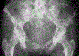
| Figure 4: Anteroposterior view of the pelvis showing patchy, poorly defined areas of sclerosis over the entire pelvis and proximal femar. Subtle, more focal areas and diffuse areas of sclerosis are visible. This pattern is most typical of sclerotic bone metastasis (e.g. prostate cancer). A subtle underlying pattern of small, rounded lesions can also be seen. |