Keywords
|
| Nanostructure, Cellular, Polymer, Carbon nanotubes, Apoptosis, Proliferation. |
INTRODUCTION
|
| Carbon nanotubes (CNT) present remarkable optical, electrical, and mechanical properties [1,2] which have been explored for their applications in biological areas [3] such as trackers for detection in cellular micro environments [3], as vehicles for delivery of agents [4], or in cell differentiation by electrical stimuli [5]. Nowadays, nanoparticles are becoming an important component as biomaterial for tissue regeneration [6-8]. In fact, CNTs have already been successfully used as scaffolding for cellular or tissue growth [6-11]. In order to obtain biocompatible features from CNTs, and because of their hydrophobic behavior, CNTs are commonly subject to different functionalization processes. The most common of these involves an acid treatment, generating carboxylic groups at the walls of the CNTs, producing functionalized CNTs (fCNT) [12,13]. However, has been found in some cases that this treatment causes cytotoxic effects in specific cell lines [10,13,14]. |
| Another way to generate functional groups on CNTs, is by modification with dendritic and hyper branched biocompatible polymers [4,15,16] consists of generating chains similar to molecules of the extracellular matrix [16], allowing biocompatibility and an easier interaction between the polymerized CNTs and the cells. In addition, some polymers also improve the solubility in water and the dispersion in biological fluids [14]. For example, polyethylene terephthalate (PET) and polyurethane (PU), have shown a favorable cellular response in MSCs [17]. Synthetic polyethylene glycol (PEG) together with chondroitin sulfate (CS), has been used as scaffolding on chondrogenesis induced by MCSs [18]. Using PEG, Nayak,[11] has demonstrated that a thin film of nonaligned PEGylated multiwalled CNTs (MWNT) influences the proliferation and morphology of human MSCs (hMSC), leading, in the absence of biochemical inducing agents, to their final differentiation into osteoblasts [11]. However, some results of the toxicity of polymerized CNTs with PET, PU and PEG [11,17,18] suggest that these polymerizations cause toxic effects on MSCs, leading to the ongoing search for different, non-cytotoxic polymers. Recently, poly-citric acid (PCA) [4,15,19-24], a hyper-branched polymer with a highly biocompatible surface [4], has been proposed to build scaffolding and to increase the hydrophilicity of CNTs [15,21], reducing their aggregation and size polydispersity, and consequently diminishing their cytotoxicity [4,22,23] Additionally, it has been found that PCA polymerization process does not generate pollutant particles [20]. The polymerization of MWNTs with PCA, starts with the acid functionalized MWNTs (fMWNT) and is followed by the addition of PCA, which covalently joins carboxylic functional groups of PCA and fMWNTs via a cleavable ester, obtaining poly citric acid-MWNTs (PWNTs) an interesting material for nano-medicine applications [3,15,20,21,24]. |
| In this work, the cellular model chosen for testing biocompatibility with PCA were MSCs due to the following two factors: |
| First, the proliferative capacity of MSCs is a fundamental aspect for the design and production of MSCs scaffolding [25]. However, recent experiments have shown that, due to lack of anchorage between tissue and supplied cells, the proliferative capacity of MSCs applied by suspension directly in damaged areas, produces little or null improvements on the surrounding damaged tissue [26], thus the production of adequate scaffolds for proliferation and anchorage of MSCs is a fundamental aspect for future study [25]. MSCs cultured on scaffolding are a real option in the field of tissue engineering [27] for the formation of new tissue or for replacing or regenerating the damaged one [6,28]. The interaction of polymerized CNTs with the MSCs can influence cell behavior and promote adhesion [16,29]. Interestingly, this material can also lead to changes on a stem cell´s shape [30,31], providing signals that may promote proliferation or differentiation [16]. Second, the work with MSCs derived from primary cultures (bone marrow), ensures a clean and clear genetic background without the presence of genetic disorders such as those accumulated in cell lines previously pre-established and characterized, like those of traditional cell lines [32]. Therefore MSCs are an excellent model to evaluate several biological parameters such as proliferation, apoptosis and cytotoxicity [25] due to their high sensitivity inducing changes in their multipotent differentiation patterns [29,30,31] and proliferation [16]. Based on the reports of cytotoxic effects on fCNTs [10,13,14] and polymerized CNTs (PCNT), with PEG, PU and PET [11,17,18], prior to considering PCNTs as biological scaffolding for MSCs, it is mandatory and fundamental to perform cytotoxic assays of cells in contact with fCNTs and PEGylated CNTs. The aim of this work is thus to generate and characterize a set of functionalized and PCA polymerized CNTs to evaluate their cytotoxicity capacity on a MSC cellular in-vitro model [33,34], because it is mandatory prior to considering PCNTs as biological scaffolding for MSCs. The CNTs are fully characterized after functionalization and PCA polymerization. Then, cell viability and apoptosis assays are performed, and by confocal microscopy the cell morphology and presence of CNTs are evaluated. |
EXPERIMENTAL
|
Synthesis of CNTs
|
| MWNTs and nitrogen doped nanotubes (CNx) were synthesized by chemical vapor deposition (CVD) following procedures similar to those reported by Botello [35]. Mainly a tubular furnace with a quartz tube reactor is heated under a 0.2 l/min Ar flow. After reaching 850°C, micro-droplets of ferrocene/toluene (3.5/96.5 wt%) and ferrocene/benzylamine (3.5/96.5 wt%) solutions were supplied as Fe, N and C feedstock by a spray pulverization chamber (Pyrosol 7901, France) at a flow rate of 2.5 l/min for MWNTs and CNx production respectively. After 15 minutes of CVD reaction, the system is allowed to cool down to room temperature and the CNTs are scratched from the reactor. |
Functionalization
|
| After synthesis both types of CNTs, were independently acid treated for functionalization [36]. 0.6 g of the CNTs were sonicated (at 750 W and 20 kHz /kwatts) for 4 hours [37] in 100 ml of an acid solution of H2SO4/HNO3 3:1 by 3M [10]. The solution was filtered and washed with NaOH 3M and bi-distilled water. Finally solutions of fMWNTs and functionalized CNx (fCNx) at 1000 ng/ml, 100 ng/ml and 10 ng/ml were prepared using bi-distilled water in an ultrasonic processor for 4 hours [37]. |
Citric Acid Polymerization onto fMWNTs and fCNxs
|
| A total of 0.1 g of the respective functionalized CNTs, either fMWNTs or fCNxs, were added to a round bottom flask, simulating the polymerization ampule equipment was set on a water bath with magnetic stirrer and vacuum inlet. Five grams of monohydrate citric acid was added to the flask which was sealed under vacuum. The mixture was heated up to 120°C and stirred at this temperature for 30 min. After removing the water with a vacuum line, the reaction temperature was raised to 140°C and stirred at this temperature for 1 h. Again, the produced water was removed by the vacuum line, and the reaction temperature was raised to 160°C [21]. Hence citric acid could be polymerized onto fMWNTs and fCNxs through poly condensation reaction. Polymerization was continued in this temperature under dynamic vacuum for 1.5 h. following a procedure similar to the reported Sarlak[19] Due to the poor solubility of fCNTs and citric acid in the organic solvents, melting esterification is the best method for a poly condensation reaction between fCNTs and citric acid [20,21]. We used monohydrate citric acid to overcome the problem of the decomposition of citric acid problem because its melting point is 100°C. At this temperature, poly condensation reaction can occur partially between PCA and fMWNTs and fCNxs. Then water can be removed and increasing the reaction temperature leads to hyper branched poly citric acid grafted onto fCNTs, obtaining PCAylated MWNTs (PMWNT) and PCAylated CNx (PCNx) respectively. The mixture was cooled and dissolved in acetone and the product was precipitated in bi-distilled water. Finally for the biological tests, solutions of fMWNTs, fCNxs, PMWNTs and PCNxs at 1000 ng/ml, 100 ng/ml and 10 ng/ml were prepared using bi-distilled water in an ultrasonic processor for 4 hours. |
| CNT Characterization |
| The characterization of the four types of CNTs was done using several instruments. For morphological characterization, A Dual-Beam scanning electron microscope (SEM, Nova 200 Nanolab, USA) was used, coupled with an X-ray Si (Li) ultra-thin window energy dispersive spectrometer (EDS) for low atomic number detection. The quantitative estimation of the elemental atomic percent was done with the ZAF method implemented on the EDAX EDS Genesis software. A transmission electron microscope (TEM, Tecnai G2 F30 S-TWIN, UK) was used to analyze the CNT structure. Statistical evaluation of diameters was carried out on samples containing fMWNTs, fCNx, PMWNTs and PCNxs. |
| The Raman analysis was performed with a (Horiba Jobin Yvon, Labram HR800, France) long 633 instrument. The IR spectra were examined with a FT-IR spectrometer (Thermo Scientific, Nicolet iS5 ID5 ATR, USA). Thermo-gravimetric analysis (TGA) were performed with a TGA (Q500, USA) instrument, under a N2 flow of 50 ml min-1 following this method: equilibration from room temperature to 15°C in 6 min, isothermal heating at 100°C for 30 min, then thermal increase from 15°C to 200°C in 90 min with a heating rate of 10°C min-1. |
| Cell Culture |
| MSCs derived from rat bone marrow were isolated and cloned from a heterogeneous culture[38], seeded at 104 cell/ml suspensions and grown in 3.0 cm2 plate dishes with α-MEM medium (Sigma-Aldrich, USA) with supplemented fetal bovine serum (10%), penicillin (10,000 U), streptomycin (10 mg/ml) and amphotericin B (25 mg/ml) (Sigma-Aldrich, USA). Then, the culture was incubated at 37°C and 5% CO2 with three different concentrations of fMWNTs, PMWNTs, fCNxs and PCNxs, i. e., 10, 100 and 1000 ng/ml. |
| Cellular Proliferation |
| Following the cell culture preparation, a trypan blue exclusion test (TBET) [13] was performed at 0, 24, 48, 72 and 144 hours. The cells were trypsinized and re-suspended in fresh culture medium. Finally cells were counted on a hemocytometer. The cellular proliferation test was done in triplicate. |
| Apoptosis Assay |
| The cells treated at different concentrations (10, 100 and 1000 ng/ml) of fMWNTs, PMWNTs, fCNxs and PCNxs, MSCs during 48,72 and 144 hours were trypsinized and re-suspended using a Annexin V/propidium iodide assay kit (ellEvent®, USA). The cells were washed with Phosphate Buffered Saline/Bovine Serum Albumin (PBS/BSA) and incubated at room temperature with a solution of annexin-V-FLUOS and propidium iodide for 30 min as suggested by Jiang [38]. The cells were then analyzed by flow cytometry, monitoring the fluorescence emission at 395/525 nm for annexin V-FITC and 483/659 nm for PI in a flow cytometer (FACSCalibur; Becton Dickinson, USA). The cells were seeded by triplicate; the assay was carried on in untreated MSCs as pure control and MSC positive control containing, 25, 12.5 and 2.5 μl of hydrogen peroxide respectively. |
| Confocal |
| Cell growth on cover slips treated at 100 ng/mL of each type of nanotubes after 72 hours of incubation time, which was washed with PBS and fixed with 4% w/v paraformaldehyde (PFA) (Sigma-Aldrich, USA), a nuclear immunofluorescence staining was performed with Draq7 (bio status). The presence of nanotubes was revealed by auto-fluorescence using a lesser beam at 488 and an emission filter 505-30 LB, while nuclei were seen also at 488 and 633 LB. Images were captured using a confocal microscopy (Zeiss, Germany) and analyzed by the Zen 2009 software and a confocal microscope Leica Mod. TCS sp 8x with a LAS AF 3.3.0 software. |
| Statistics |
| Three independent repetitions of each biological experimental treatment were done. Data are expressed as a mean standard deviation of the experiments. Statistics analysis was performed by analysis of variance (ANOVA). Statistical significance was inferred at p < 0.05. |
RESULTS
|
Nanotubes Characterization
|
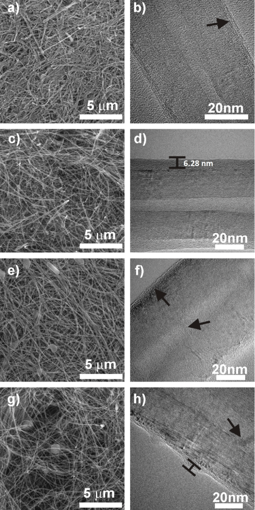
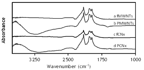
| The morphology and structure of fMWNTs, PMWNTs, fCNxs and pPCNxs are shown in the micrographs of Figure 1. The average width of the nanotubes after the functionalization is shown in Table 1, suggesting that PCA increases the diameter by 30% in fMWNTs and almost 50% in fCNxs, wider than those synthesized by Terrones [2]. The TEM micrographs in Figure 1b and 1f show some degree of disorder on the external walls of the acid treated CNTs, which complementing the infrared (IR) spectroscopy information (Figure 2), indicates the presence of carboxyl functional groups, generated during the acid functionalization [36,37]. A different kind of disorder is also noticeable as bamboo-like structures in the inner part of the CNTs as shown in Figure 1f and 1h, indicating the presence of nitrogen as a doping agent [2]. During synthesis of CNxs, the nitrogen tends to incorporate pentagons on the hexagonal structure of the nanotube, affecting its crystallinity and curvature of the inner surface, generating the bamboo shaped structures in the interior [39,40]. Currently, CNxs are being investigated, since the effects of these irregularities may favor the interaction with polymers [2]. The polymerization of the CNTs by PCA is noticeable in Figure 1d and 1h, where a thin layer of polymer covers the corresponding nanotubes, similar to those reported by Jiang [38]. |
| The elemental compositions for each of the four types of CNTs are shown in the EDS spectra of Table 1, confirming that the nanotubes do not contain enough weight percentage of Fe pollutants that may cause cytotoxicity [2,14]. The Fe nanoparticles are found inside the CNTs, and thus not in contact with the environment and finally not with the MSCs, avoiding chance of cytotoxicity by Fe. The functionalization has been found to remove most of the catalyst nanoparticles or other compounds that are in contact with the nanotube during growth [4]. |
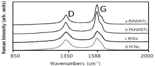
| Raman spectra in Figure 3 yield data about the types of nanotubes and their defects[1]. Impurities and defects of carbon nanotubes are usually understood on the basis of the D-band at about 1350 cm-1, as reported by Datsyuk [41] where the intensity (I) of the D-band is related to the concentration amount of defects. In contrast, the G-band is associated with the graphitic or crystalline structure of the tubes and is located at 1588 cm-1. The ID/IG ratio has been obtained for the four types of nanotubes and it relates the defects and the crystallinity of the tubes. Functionalization and polymerization increases the amount of disordered carbon caused by the partial destruction of fMWNTs and fCNxs during the exfoliation of bundles and the polymerization treatment[40,42].The intensity of the D and G bands are almost constant for the MWNTs and the CNx respectively (Figure 2); i.e. only a small change in the ID/IG ratio appears when functionalized nanotubes are polymerized. As expected, fMWNTs and PMWNts show a higher intensity in G-band in comparis on with fCNxs and PCNxs. The ID/IG ratios of fMWCNTs and fCNxs are 0.41 and 0.89 respectively, while the ratio of PMWNTs and PCNxs are ≈0.43 and ≈0.82 respectively (Table 1) results derived from the mechanism of oxidation during functionalization, more effective for the external walls of the CNxs.The chemical compositions of the 4 types of nanotubes were further confirmed by IR spectroscopy (Figure 3). |
| All the samples show the main absorbance bands between 1750 and 2200 cm−1 related to C-C bonds of the CNT structure. While an increase in the intensity of this band is found in Figure 3b-3d due to the bonds of the polymer. The carbonyl groups of the OH bands, obtained from the functionalization, are slightly marked at 2500 cm−1 in the 4 samples. The broad absorbance bands of PMWNTs and PCNxs (Figure 3b and 3c), around 3200 and 3500 cm−1, are related to the OH associated with acidic and alcoholic hydroxyl functional groups of the polymer [21,23]. |
| After SEM, TEM and Raman characterization, confirming the structure of the functionalized and polymerized nanotubes, the IR results shown in Figure 3, consistent with other studies [21-23], further confirm the adequate functionalization and polymerization performed on the CNTs used in this experiment. Furthermore, data from thermal gravimetric analysis (TGA) (data not shown) indicate that the amount of carboxylic acid groups and PCA bound to the CNTs do not change with the temperature rise; all the samples keeping a constant weight in the range from 15°C to 200°C with only 1% of variation, consistent with Vicente[36]. Taking into account that the temperature required for the biomedical experiments is between 25 and 45°C, the TGA results ensures that, if for some reason there are temperature fluctuations during the process, the chemical conformation of CNTs will not be affected. |
| Biological Assays |
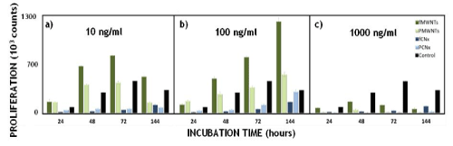
| Once the different CNT samples were characterized, we proceeded to carry out the biological assays with derived rat bone marrow-MSCs as primary cultures. The effect on cell proliferation of MSCs for different concentrations of CNTs is shown in Figure 4. At 10 ng/ml (Figure 4a) the most significant proliferative result comes from fMWNTs, which in comparison with the control sample, almost doubles the amount of cells at 48 and 72 hours. PMWNTs show less proliferation with similar behavior with respect to the control sample at 10 ng/ml for both: 48 and 72 hrs. In contrast fCNxs and PCNxs do not show proliferative capability at 10 ng/ml. |
| At 100 ng/ml (Figure 4b), fMWNTs and PMWNTs present a high proliferative growth behavior, especially at 144 hours. In contrast, fCNxs and PCNxs remain with a lower proliferative capacity than the control sample at all times. Using the highest concentration, 1000 ng/ml, Figure 4c it was remarkable that cell growth was not present in any sample, with the exception of the control one. The results of proliferative analysis for fMWNTs and PMWNTs, differs with the behavior reported by Y. Jiang, for PEGylated MWNTs in macrophage model cells [38], with our MWNTs showing a higher proliferative rate than the macrophage cells. The flow cytometry study performed in this work provides data on cellular death by apoptosis quantifying annexin V. |
| From the apoptosis results of the 10 ng/ml concentration (Figure 5), it is evident from the beginning of the experiment that fCNxs causes cell death. The effect of the polymer is clear, with PCNxs presenting a lower level of apoptosis than fCNxs. The apoptotic tendency of fCNxs is confirmed by the proliferative assay shown in Figure 4a), where at the same concentration and time, both fCNxs and PCNxs do not proliferate in comparison with the control sample. The cytotoxicity of CNxs could be related to the high degree of defects confirmed by the ID/IG ratio reported in Table 1. Correspondingly, at 10 ng/ml, fMWNTs show apoptotic behavior in a similar relation to the control, while PWNTs present a lower apoptotic behavior, i.e. fMWNTs and PMWNTs present higher proliferative capacity in contrast to fCNxs and PCNxs (Figure 4a). |
| The apoptotic behavior of fCNxs at 100 ng/ml is the highest among the four types of CNTs solutions (Figure 5b), similar to the behavior at 10 ng/ml. PCNxs at 100 ng/ml also presents a low apoptotic behavior with respect to fCNxs, as with the 10 ng/ ml concentration, still presenting a higher apoptotic percentage in contrast to the positive control. Accordingly, the proliferative behavior of both fCNxs and PCNxs at 100 ng/ml always presents lower levels than the control. Thus, it is very significant that for all CNxs, when inducing apoptosis, its proliferative capability decreases. In contrast, fMWNTs and PMWNTs, at 100 ng/ml, present a higher apoptotic level than the positive control (still lower than fCNxs and PCNxs). In spite of this, their proliferative tendency does not to diminish, but increases at all times; i.e. they activate the proliferation in a significant way, particularly the fMWNTs. It seems that MSCs, at 100 ng/ml of MWNTs, show resistance to cell death and even enhance their proliferation. As time passes for the 100 ng/ml concentration, proliferation increases in all cases and the apoptosis decreases (Figures 4b and 5b). Remarkably, the PMWNTs solution presents the lowest apoptotic induction and a high proliferative capacity at all times, including at 144 hours. Kroustalli [9] reported similar results, showing from the assay that PMWNTs support adhesion and proliferation of hMSCs. Other studies indicate that polymerized CNTs present less impact in the activation of the oxidative responsible for the apoptotic pathway [43-45]. |
| Finally at the highest concentration of 1000 ng/ml shown in Figures 4c and 5c, all the samples had the lowest proliferative capacity sharing the same apoptotic tendency. Interestingly, the most significant results for both proliferation and apoptosis assays, are at concentrations of 100 ng/ml, shown in Figures 4b and 5b respectively, and proved statistically by three independent experiments (p<0.05), showing high proliferation and apoptosis for MWNTs and CNxs respectively). The next step was to analyze by confocal microscopy the behavior of MSCs in cultures performed with CNTs from each group, focusing mainly in the 100 ng/ ml concentration. Figure 6 shows the evaluation of cellular morphology and intracellular incorporation of external agents for the cultured MSCs with CNT solutions at 100 ng/ml. Starting with the behavior of the control sample for reference in Figure 6 (i-iv), a standard morphology of the nucleus is observed in red fluorescence (Figure 6a (i)). Since, no CNTs are present in the control, no fluorescence from CNTs is observed in Figure 6a (ii), and a normal morphology of the whole cell is observed in the bright field in panel (iv) of Figure 6a. The effect of 100 ng/ml solutions for all CNTs: fMWNTs, PMWNTs, fCNxs and PCNxs is shown in Figure 6b and 6e respectively. Panels (i) of Figure 6b and 6d correspond to the red fluorescence from fMWNTs and fCNxs respectively; both showing an apoptotic behavior represented by the nucleus destruction indicated by white circles. In comparison, panels (i) of Figure 6c and 6e, show a well-ordered nucleus, morphologically identical to the control (Figure 6a (i)). These panels 6c (i) and 6e (i) correspond to PMWNTs and PCNxs respectively, clear evidence that the polymer of the PCNTs reduces the apoptotic behavior of fCNx shown in Figure 5b. |
| Panels (ii) in Figure 6b-6e show the green fluorescence of the corresponding nanotubes inside the MSCs. In particular, the case of fMWNTs and fCNxs in Figure 6b and 6d, show an evident presence of nanotubes in green, indicated by white arrows. Panels (iii) in Figure 6 result from merging corresponding images of panels (i) and (ii), illustrating the exact position of the nanotubes with respect to the nucleus. Finally bright field panels (iv) are included, where the fibrilar structure of fMWNTs and fCNxs are clearly shown inside the cells. In addition to apoptosis decrease in treated cells with PMWNTs, interesting was the fact that these nanotubes in a major quantity remain out of cell (Figure 7). |
DISCUSSION
|
| Comparing with other nanotubes, the facility to be internalized into cells [4], it has been proved that MWCNTs differ in their intracellular incorporation mechanism [46]. MWCNT capability to incorporate into cells is highly dependent on their diameter and length; the nanotubes with the smallest dimensions present the higher capacity to enter the cell. Accordingly, as shown in Figure 6b and 6d, fMWNTs and fCNxs localize inside the cell, a similar situation to that reported by Adeli[4]. However, PMWNTs and PCNxs are encountered in fewer occasions inside the cells, although they present the required dimensions to be incorporated. The fact that PMWNTs and PCNxs do not enter the cell can be attributed to the high presence of oxygenated groups, like those encountered in the hyperbranched PCA surface, which interact with the serum proteins of the extracellular matrix, reducing the cellular endocytosis mechanism [43]. These could be the reasons why CNTs do not enter the cell, even though non PCAylated CNTs present similar morphology and dimensions form Table 1 and Figure 7a reinforces the fact that the polymerized nanotubes enter in less quantity into the cell, remaining in the periphery. Also from Figure 6, it is shown that the effect on the morphology of the MSCs, in contact with PMWNTs and PCNxs, does not present major changes, contrasting with the results of hyper branched polymers like PEG, reported by Murphy [16]. |
| Given these results, fCNTs entering the cell [4,47-49] opens a prospect of study opportunities for the creation of drug carrier vehicles [49-51], cancer therapy [4], as well as for studies of cytotoxic effects[50]. The green fluorescence of the confocal images inside the cells confirms the fCNx uptake, a phenomenon that can be used for optical stimulation of CNTs inside living cells to afford multifunctional biological transporters of CNTs. Thus, the transporting capacity of CNTs combined with a suitable chemistry functionalization and their intrinsic optical properties can lead to novel nanomaterial’s for drug delivery and cancer therapy.On the other hand, a subject of great relevance in this field is to continue the study of the interaction between PCNTs and MSCs, with a view to the scaffolding production and design [51-53], but there are some interesting questions to answer beforehand e. g. Can the interaction between PMWNTs and MSCs could induce growth factors and secretion of substances from the cells [16] Can the polymer interact with molecules found in the extracellular matrix [54,55], such as glycoproteins? Will this allow binding to a range of growth receptors with high affinity [16], or simply increase the adhesive capacity of the material [9,56]. |
| Due to the physic-chemical characteristics of CNTs coated with PCA, these provide a clear advantage for the design of scaffolds in comparison to uncovered CNTs: coated CNTs do not cause intracellular damage, because the PMWNTs cannot be internalized inside cells. So the scaffolds could be able to promote novel tissue formation, favoring cell adhesion and proliferation. Related to our results is the suggestion to use nanotube-based films linked by a bi-or tri-dimensional network of PCA to prevent individual nanotubes from being dislodged and entering the cell, expecting to resemble the tissue extracellular matrix [51-53], providing adequate structural support and favoring a good biomedical development. Finally, in order to continue the analysis of carbon materials with the presence of MSCs, the use of PCA polymerized graphene structures could be proposed as a biomaterial [57], hoping that grapheme properties could provide different and interesting cellular responses. |
CONCLUSION
|
| In summary, to the best of our knowledge, this is the first time that PCA polymerized CNTs have been used with a MSCs cellular model. Our results show that apoptotic and proliferative behavior is similar between cells treated with fCNxs and PCNxs (Figure 4), presenting the lowest proliferative rates and the highest apoptotic levels (Figure 5), thus proving less viable for applications such as cell scaffolding, but suitable for the transport of substances inside the cell. In contrast MWNTs, both functionalized and polymerized, present better results for possible biomedical applications in scaffolding, since at 100 ng/ml, both present high proliferative rates and relatively low apoptotic behavior. Building a structure based on PCA and CNTs, where nanotubes are well fixed to the matrix, could result in a stable scaffold for MCS cells. Furthermore, the morphology [58,59] of the MSCs in contact with PMWNTs and PCNxs does not present major changes, contrasts with hyperbranched polymerized CNTs[9] effects on cells. This work contributes to the better understanding of the behavior of MSCs on various CNT cultures, suggesting alternatives for the design of novel scaffold systems. |
ACKNOWLEDGEMENTS
|
| G.R.L. thanks Felix Martínez and the Engineering Faculty of Panamericana´s University for support during the project. S.H.G thanks funding from the CONACyT-Project 101596. C.S.F. thanks funding from the Physics and Mathematics Department, the Resaearch Direction and FICSAC, UIA. Authors are thankful to S. Rosas-Meléndez, M. Ballesteros-Villareal, F.A. Iñiguez-Rábago. S. Macias, R. Estrada and V. Garibay-Febles for fruitful discussions and support with experiments and characterization. |
Tables at a glance
|
 |
| Table 1 |
|
Figures at a glance
|
 |
 |
 |
 |
| Figure 1 |
Figure 2 |
Figure 3 |
Figure 4 |
|
|
|
| Figure 5 |
Figure 6 |
Figure 7 |
|
References
|
- Dresselhaus M, et al.Carbon nanotubes, synthesis, structure, and applications, Nanotechnology.2001;80:391.
- Terrones M, et al.Doped Carbon Nanotubes: Synthesis, Characterization and Applications, Topics in Applied Physics. 2008;111:531
- Heister E, et al. Are Carbon Nanotubes a Natural Solution? Applications in Biology and Medicine, Appl Mater Interf.2013;5:1870.
- Adeli M, et al.Carbon nanotubes in cancer therapy: a more precise look at the role of carbon nanotube–polymer interactions,ChemSoc Rev. 2013;42:5231.
- MooneyE,et al.The electrical stimulation of carbon nanotubes to provide a cardiomimetic cue to MSCs Biomaterials. 2012;33:6132.
- Harrison BandAtala A.Carbon nanotube applications for tissue engineering, Biomaterials 2007; 28:2344.
- Mooney E, et al.Carbon Nanotubes and Mesenchymal Stem Cells: Biocompatibility, Proliferation and Differentiation, Nano Lett.2008;8:2137.
- Yi C, et al. Gold Nanoparticles Promote Osteogenic Differentiation of Mesenchymal Stem Cells through p38 MAPK Pathway, ACS Nano. 2010;4:6439.
- Kroustalli A, et al. Cellular Function and Adhesion Mechanisms of Human Bone Marrow Mesenchymal Stem Cells on Multi-walled Carbon Nanotubes, Ann Biomed Eng.2013;41:2655.
- Correa-Duarte M, et al. Fabrication and Biocompatibility of Carbon Nanotube-Based 3D Networks as Scaffolds for Cell Seeding and Growth, Nano Lett. 2014;4:2233.
- Nayak TR, et al.Thin films of functionalized multiwalled carbon nanotubes as suitable scaffold materials for stem cells proliferation and bone formation, ACS Nano.2010;4: 7717.
- Boehm HP. Surface oxides on carbon and their analysis: a critical assessment, Carbon. 2002;40:145.
- Gutiérrez-Praena D, et al. InïìÃâuence of carboxylic acid functionalization on the cytotoxic effects induced by single wall carbon nanotubes on human endothelial cells,Toxicol In Vitro. 2011;25:1883
- Isobe H,et al. Preparation, Purification, Characterization, and Cytotoxicity Assessment of Water-Soluble, Transition-Metal-Free Carbon Nanotube Aggregates,AngewChem Int.2006;45:6676.
- Adeli M, et al. Carbon nanotubes-graft-polyglycerol: Biocompatible hybrid materials for nanomedicine, Polymer. 2009;50:3528
- MurphyWm, et al. Materials as stem cell regulators,Nature Materials.2014;13:547.
- Gustafsson Y, et al.Viability and proliferation of rat MSCs on adhesion protein-modified PET and PU scaffolds, Biomaterials 2012;33:8094.
- Varghese S, et al. Chondroitin sulfate based niches for chondrogenic differentiation of mesenchymal stem cells, Matrix Biol. 2012;27:12.
- Sarlak N, et al. Quantitative study on the interaction of Ag+ and Pd2+ with CNT-graft-PCA (polycitric acid) in aqueous solution, J of Molecular Liquids. 2013;180:39.
- Bahari A, et al. Carbon nanotube-graft-poly (Citric acid) containing silver and palladium nanoparticles, Nano. 2009;4:217.
- Adeli M, et al.Carbon nanotube Graft-poly (citric acid) Nanocomposites, Nano.2008;3:1
- Zhao B, et al.Synthesis and Characterization of Water Soluble Single-Walled Carbon Nanotube Graft Copolymers, J Am Chem Soc. 2005;127:8197.
- Kainthan R, et al. In vitro biological evaluation of high molecular weight hyperbranchedpolyglycerols, Biomaterials.2007;28:4581
- Sohhani Z,et al.Increased paclitaxel cytotoxicity against cancer cell lines using a novel functionalized carbon nanotube,Nanomed. 2011;6:705.
- Lee J, et al. Directing stem cell fate on hydrogel substrates by controlling cell geometry, matrix mechanics and adhesion ligand composition, Biomaterials. 2013;34:8140.
- Hodgkinson C, et al.Genetic Engineering of Mesenchymal Stem Cells and Its Application in Human Disease Therapy, Human Gene Therapy.2010;21:1513.
- Dalby MJ, et al.The Control of Human Mesenchymal Cell Differentiation Using Nanoscale Symmetry and Disorder, Nature Materials.2007;6:997.
- Engler AJ, et al. Matrix Elasticity Directs Stem Cell Lineage Specification, Cell. 2006; 126:677.
- Bot PT,et al. Hyaluronic acid:targeting immune modulatory components of the extracellular matrix in atherosclerosis,Curr Med Chem. 2008;15:786.
- Tsimbouri P, et al.Nanotopographical effects on mesenchymal stem cell morphology and phenotype, J CellBiochem.2014;115:380.
- Khetan S, et al. Degradation-mediated cellular traction directs stem cell fate in covalently crosslinked three-dimensional hydrogels, Nature Materials.2013;12:458.
- MiyagawaS, et al. Tissue Cardiomyoplasty Using Bioengineered Contractile Cardiomyocyte Sheets to Repair Damaged Myocardium: Their Integration with Recipient Myocardium, Transplantation.2005;11:1580.
- Xin X, et al. Continuing Differentiation of Human Mesenchymal Stem Cells and Induced Chondrogenic and Osteogenic Lineages in Electrospun PLGA Nanofiber Scaffold, Biomaterials.2007;28:316.
- L. Zhang, and C. Chan, Isolation and enrichment of rat mesenchymal stem cells (MSCs) and separation of single-colony derived MSCs, J Vis Exp.2010;37:1852.
- Botello-Méndez A, et al. Controlling the dimensions, reactivity and crystallinity of multiwalled carbon nanotubes using low ethanol concentrations,ChemPhysLett.2008;453:55.
- Vicente J, et al. Effect of acid oxidation treatment on adsorption properties of arc-disharge synthesized multiwall carbon nanotubes, The Journal of the Argentine Chemical Society.2011;98:29.
- Liu J, et al. Fullerene Pipes, Science. 1998;20:1253.
- Jiang Y, et al. Modulation of Apoptotic Pathways of Macrophages by Surface-Functionalized Multi-Walled Carbon Nanotubes,PLoS One 2013;10:1371.
- Gong K, et al. Nitrogen-Doped Carbon Nanotube. Arrays with High Electrocatalytic Activity for Oxygen Reduction, Science. 2009;323:760.
- Bower C, et al. Intercalation and partial exfoliation of single-walled carbon nanotubes by nitric acid,ChemPhysLett. 1998;288:48.
- Datsyuk V, et al. Chemical oxidation of multiwalled carbon nanotubes, Carbon 2008;46: 833.
- Kuhlmann U, et al.Thomsen, Infrared active phonons in single-walled carbon nanotubes,ChemPhysLett.1998;294:237.
- Zhang Y, et al. Mechanistic toxicity evaluation of uncoated and PEGylated single-walled carbon nanotubes in neuronal PC12 cells, ACS Nano. 2011;5:7020.
- Wang X, et al. Dispersal state of multiwalled carbon nanotubes elicits profibrogenic cellular responses that correlate with fibrogenesis biomarkers and fibrosis in the murine lung, ACS Nano.2011;5:9772.
- Tabet L,et al.Coating carbon nanotubes with a polystyrene-based polymer protects against pulmonary toxicity, PartFibreToxicol. 2011;8:13.
- Kang B, et al. Cell Response to Carbon Nanotubes: Size-Dependent Intracellular Uptake Mechanism and Subcellular Fate Chen, Small. 2010:6:2362.
- ChenX, andSchluesener HJ.Multi-walled carbon nanotubes affect drug transport across cell membrane in rat astrocytes, Nanotech. 2010;21:100-104.
- Liu Z, et al. Carbon Nanotubes in Biology and Medicine: In vitro and in vivo Detection, Imaging and Drug Delivery, Nano Research 2009;2:85.
- Johnston H, et al. A critical review of the biological mechanisms underlying the in vivo and in vitro Functionalized MWCNTs on Apoptosis of Macrophages,Nanotoxicology. 2013;4:207.
- Cheng C, et al.Toxicity and imaging of multi-walled carbon nanotubes in human macrophage cells, Biomaterials.2009;30:4152.
- Huebsch N, et al.Harnessing traction-mediated manipulation of the cell/matrix interface to control stem-cell fate, Nature Materials.2010;9:518.
- Trappmann B, etal.Extracellular-matrix tethering regulates stem-cell fate, Nature Materials. 2012;11:642.
- Li X,et al. The use of carbon nanotubes to induce osteogenic differentiation of human adipose- derived MSCs in vitro and ectopic bone formation in vivo, Biomaterials. 2012;33:4818.
- Del Gaudio C, et al.Are synthetic scaffolds suitable for the development of clinical tissue-engineered tubular organs, J Biomed Mater Res. 2013;102:24277.
- Liu L, et al. Novel strategy to engineer trachea cartilage graft with marrow mesenchymal stem cell macroaggregate and hydrolysable scaffolds,Artif Organs. 2010;34:426.
- Kloxin AM, et al. Photodegradable hydrogels for dynamic tuning of physical and chemical properties, Science 2009;324:59.
- Li N, et al. Three-dimensional graphene foam as a biocompatible and conductive scaffold for neural stem cells,Sci Rep. 2013;3:1604.
- McBeath R,et al. Cell shape, cytoskeletal tension, and RhoA regulate stem cell lineage commitment,Dev cell. 2004;6:483.
- Kilian K, et al. Geometric cues for directing the differentiation of mesenchymal stem cells,ProcNatlAcad Sci. 2010;107:4872.
|

 Menu
Menu
 555-666-0606