Research & Reviews: Journal of Pharmacology and Toxicological Studies
e-ISSN: 2322-0139 p-ISSN: 2322-0120
e-ISSN: 2322-0139 p-ISSN: 2322-0120
1Amala Cancer Research Centre, Amala Nagar, Thrissur, Kerala, 680555, India
2Assistant Professor, Department of Chemistry, Baselius College, Kottayam, Kerala, India
3Principal, Mookambika College of Pharmaceutical Sciences and Research, Muvattupuzha, Kerala, India
Received Date: 30/10/2016 Revised Date: 24/11/2016 Accepted Date: 30/11/2016
Visit for more related articles at Research & Reviews: Journal of Pharmacology and Toxicological Studies
The chemical compositions of essential oils from Lagerstroemia speciosa L flowers obtained by hydrodistillation yielded 0.085% and was analysed by GC and GC-MS. Out of forty five compounds identified the major volatiles in flowers were α-pinene, myrcene, α-terpineol, a β-bisabolene etc. Our findings demonstrate for the first time the potential benefits of this medicinal plant flower as a rich source of high therapeutic value compounds for medicines, cosmetics, supplements and as a health food. As concerning volatile fraction, that can be often a helpful tool to discriminate between different taxa also. Cytotoxicity assay was carried out using Dalton’s Lymphoma Ascites cells (DLA) and Ehrlich Ascites Carcinoma cells (EAC). L. speciosa essential oil at a concentration of 50 μL/mL produced 13.33% and 31% cytotoxicity to DLA and EAC cells respectively.
Lagerstroemia speciosa, DLA cells, GC-MS, Cytotoxic activity
Lagerstroemia speciosa L. (L. speciosa) belonging to the family Lythraceae, commonly called as banaba (queen’s flower or Crapemyrtle), the Pride of India is a medicinal and ornamental handsome decidous small tree native of East Asia, India, China and Australia commonly cultivated in gardens throughout for beautiful flowers. Crape myrtle blooms from May through August in India.
Banaba tea is a traditional health drink in Philippines since ancient times. It was found to have therapeutic effects on various ailments such as diabetes, kidney and other urinary problems. Besides being an exotic health drink, banaba tea has a soothing and pleasant taste and Japanese nowadays consume banaba in various forms such as tea, juice, health drinks, and as food supplement. The potential antidiabetic activity of ethyl acetate and ethanolic extracts were studied in the leaves of L. speciosa [1,2] Nephroprotective [3], free radical scavenging, anti-inflammatory [4] and hepatoprotective activity [5] of Lagerstroemia speciosa (L) extracts were studied recently.
L. speciosa seeds contain caprylic, lauric, myristic, palmitic, steric, arachidic, behenic, lignoceric, oleic, and linoleic acids in the oil [6]. The active principles 9-keotoctadec-cis-11-enoic acid has been isolated from seed oil as well [7]. Chemical investigation of amino acid components in banaba seed oil has been performed in earlier studies [8]. Components nonanedioic acid, 12-acetyloxy-9-octadecenoic acid, and 16-methyl-heptadecandic acids present in seed extracts have been identified as having antibacterial activity [9].
An essential oil is a concentrated, hydrophobic liquid containing volatile aroma compounds from plants. An oil is "essential" in the sense that it carries a distinctive scent, or essence, of the plant. The natural products industries are currently looking for natural therapeutics and preservatives that can replace synthetic preparations. The scientific literature has identified new applications and uses of both traditional and exotic essential oils. Interest in essential oils has revived in recent decades with the popularity of aromatherapy, a branch of alternative medicine which claims that the specific aromas carried by essential oils have curative effects.
There is a tremendous historical legacy in folklore use of plant preparations in medicine. Novel, safer and effective compounds with cytotoxic and anti tumour activities will be of immense clinical benefit. A wide variety of biologically active compounds from plants have been used as effective chemotherapeutic agents [10]. Around 60% of the current anticancer drugs, in use today are of natural origin. Vinblastine, vincristine, etoposide, teniposide, taxol, topotecan and irinotecan are some of the approved anticancer drugs.
The diverse therapeutic potential of essential oils has drawn the attention of researchers to test them for anticancer activity. From leaves of L. speciosa, a new triterpenoid was isolated along with four known compounds of virgatic acid, corosolic acid, ursolic acid and β-sitosterol glucoside [11] . Using the brine shrimp (Artemia salina) lethality bioassay, the ethanol fruit extract of L. speciosa showed prominent cytotoxic activity [12].
The present study was aimed to identify the components from the essential oils of the L. speciosa flowers and to screen the cytotoxicity for which has not explored yet to our knowledge.
Plant Materials
The flowers of Lagerstroemia speciosa L. were collected from the premises of Amala Ayurvedic Hospital, Thrissur, Kerala, India. The plant material was identified by Dr. C.N. Sunil, Department of Botany, of S.N.M. College, Maliankara and was authenticated at Botanical Survey of India, Coimbatore. The voucher specimen (L. speciosa BSI No. 62373) has been kept in Fr. Gabriel Herbarium, Amala Ayurvedic Hospital and Research Centre, Thrissur, Kerala, India.
Cell Lines
Dalton’s Lymphoma Ascites (DLA) and Ehrlich Ascites Carcinoma (EAC) cells were inoculated in peritoneal cavity of Swiss albino mice at Amala Cancer Research Centre. Weekly intraperitoneal inoculation of 106 cells in Swiss albino mice was done to maintain the cell-line. Animal experiment was carried out after approval by Institutional Animal Ethics Committee (Reg. No. 149/1999/CPCSEA) and followed the guidelines of CPSCEA, Govt. of India.
Isolation of Essential Oil
Essential oils were obtained by hydrodistillation of fresh flowers of L. speciosa using a Clevenger-type apparatus for 6 h and n-Hexane (10 mL) was used as the collector solvent. After evaporation of the solvent, the oil was dried over anhydrous sodium sulphate and stored in sealed vials protected from the light at −20°C before analyses.
Identification and Quantification of Volatile Components
The identification of volatile components was based on computer matching with the WILEY275 and NIST05 libraries, as well as by comparison of the mass spectra and retention indices (RI) with those reported in the literature. In addition, a home-made library, constructed based on the analyses of reference oils and commercial available standards, was used as well.
Gas Chromatography (GC) Analysis
Essential oil (10 μl) was dissolved in hexane (100 μl) and 2 μl of the solution was injected into a GC-17A with Flame Ionization Detector (FID) and C-R6A-chromatopac integrator (Shimadzu Co, Japan). The column used was SUPELCOWAX fused silica (film thickness: 0.2 μm, Supelco USA, 60 m x 0.25 mm). Column temperature was programmed to range from 40 to 250 °C with a rate of 6°C/min. Injector and detector temperatures were 220 and 280 °C, respectively. The carrier gas was hydrogen. Quantification was carried out by % peak area calculations (GC/FID using a non-polar Column).
Gas Chromatography – Mass Spectrometry
For GC/MS analysis, a GC–17A with a QP5050 mass spectrometer (Shimadzu Co, Japan) was used. Helium was used as carrier gas at a flow rate of 0.6 ml/min; the mass spectrometer was operated at 70 eV. Column temperature was programmed from 70 to 200°C at 10°C/min; injector temperature was 250°C. Mass spectra correlations were carried out with Wiley, NBS, NIST and private aroma library spectra. Identifications were determined by comparing the retention indices Kóvats Index (KI), of each compound with that of known compounds [13].
Cytotoxicity assay- Trypan blue exclusion method
The dye exclusion test is used to determine the number of viable cells present in a cell suspension. It is based on the principle that live cells possess intact cell membranes that exclude certain dyes, such as trypan blue whereas dead cells do not. Tumor cells were aspirated from peritoneal cavity and pelleted by centrifugation (1000 × g, for 5 min). The cells were washed with sterile phosphate-buffered saline (PBS) and counted and made up at a concentration of 10 million cells per millilitre. DLA cells (1 million cells/0.1 mL) were incubated with essential oils (dissolved in DMSO) at different concentrations in a total volume of 1 mL made up with PBS. Cells were incubated at 37°C for 3 h. After incubation, 0.1 mL of trypan blue (1%) was added and cytotoxicity was determined by counting live and dead cells using a haemocytometer [14].
%Cell Death = No. of dead cells/ No. of viable cells + No. of dead cells x 100
The chemical composition of the oil of L. speciosa flowers was investigated using both GC and GC-MS techniques. Hydrodistillation of the sample yielded pale yellow oil (0.085%).
The components identified from the essential oil and their retention indices (RI), and the percentages are shown in Table 1. The fragmentation patterns of mass spectra were compared with those stored in the spectrometer database using the NBS54K.L and WILEY.L built in libraries and with those reported in the literature. Comparison of the fragmentation patterns in the resulting mass spectra with those published in literature and using mass spectral database of the gas chromatograph computer is represented as (a,b) in Table 1.
| Identified Compounds | GC components % area L. speciosa | Kic | |
|---|---|---|---|
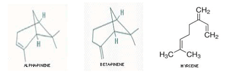
| α-pinene | 10.38 | 932 | a,b |
| β-pinene | 8.45 | 976 | a,b |
| myrcene | 6.76 | 990 | a,b |
| limonene | 2.60 | 1027 | a,b |
| Cis- β-ocimene | 1.33 | 1039 | a |
| trans- β ocimene | 2.12 | 1048 | a |
| linalool | 1.22 | 1095 | a,b |
| terpinolene | 0.16 | 1099 | a |
| 8-hydroxy linalool | 1.12 | 1186 | a,b |
| α-terpineol | 12.76 | 1187 | a |
| Benzene acetaldehyde | tr | 1041 | a |
| unknown | 0.47 | 1086 | a |
| borneol | 2.18 | 1166 | a,b |
| p-cymen-8-ol | 0.70 | 1180 | a,b |
| cis-dihydrocarvone | 1.23 | 1192 | a |
| trans-dihydrocarvone | 1.7 | 1199 | a |
| α -copaene | 1.14 | 1370 | a |
| γ-Elemene | 0.38 | 1430 | a,b |
| unknown | 1.96 | 1456 | a |
| humulene | 0.89 | 1472 | a,b |
| a β-bisabolene | 5.97 | 1508 | a |
| γ-cadinene | tr | 1513 | a |
| β-selinene | 3.54 | 1519 | a,b |
| δ-cadinene | 2.47 | 1524 | a |
| caryophyllene oxide | 1.69 | 1581 | a,b |
| Humulene oxide | 0.79 | 1595 | a,b |
| α -bisabolol | 3.14 | 1681 | a,b |
| Unknown | 0.97 | 1695 | a |
| Nootkatone | 1.90 | 1742 | a,b |
Table 1. Chemical constituents of the Essential oil from the flowers of L. speciosa.
The results showed that L. speciosa flower oil contains the monoterpenes like α-pinene, β-pinene, myrcene etc. α-pinene (10.38%), β-pinene, (8.45%) myrcene (6.76%), limonene (2.6%), α –bisabolol (3.14%) etc as major components. L. speciosa oil contains (a β) bisabolene of 5.97 % and 1.9% percentage of nootkatone. Also peaks of some unknown compounds were identified in this oil.
The cytotoxicity assay was carried out using trypan blue exclusion method and the percentage of dead cells encountered with essential oil treatment at different volumes of oil is shown in Figure 1 L. speciosa essential oil at a concentration of 50 μL/ mL produced 13.33% and 31% cytotoxicity to DLA and EAC cells respectively. This result emphasized that the oil possesses cytotoxicity at higher concentration. The results obtained from the present study show that the essential oil from L. speciosa is cytotoxic to DLA and EAC cell lines. Our results revealed 77.02% of the constituents and more detailed analysis will help to find out the unknown compounds which may be more active. Of the 45 compounds identified, those present in the largest quantities were α-pinene, myrcene, α-terpineol, a β-bisabolene etc.

Figure 1: Cytotoxic effects of the oils of L. speciosa flower against DLA and EAC cells.
This study describes the chemical composition of essential oil obtained from the flowers of Lagerstroemia speciosa. Studies have shown differential sensitivities to several natural compounds between tumor and normal cells in vitro or in vivo, and the results obtained from the present study shown that the essential oil from L. speciosa is cytotoxic to DLA and EAC cell lines.
Lymphoma is a disease of the lymphocytes in the lymphatic system, which includes the spleen, thymus, as well as other lymphatic tissues. Both EAC and DLA are rapidly growing transplantable tumor cells with aggressive behavior [15]. Dalton’s ascites lymphoma is transplantable, poorly differentiated malignant tumor which appeared originally as Lymphoma in a mouse. It grows as both solid and ascitic forms [16]. The tumor implantation initiates a local inflammatory reaction, with increasing vascular permeability, which results in an intense ascetic fluid accumulation [17]. Ascitic fluid constitutes the direct nutritional source for tumor cells [18]. In untreated DLA tumor bearing mice, an increase in ascitic tumor volume was observed.
The toxic effects verified in this study against DLA and EAC cell lines might not be attributed to any of its specific constituents, but to the synergism between the cytotoxic effects of some components of the essential oil from the plant. Thus, it is possible to observe that the essential oil from flowers of L. speciosa presents an interesting biological activity concerning to its selective cytotoxic activity against DLA and EAC cell lines.
Monoterpene hydrocarbons like α-pinene and myrcene, an oxygenated monoterpene linalool; etc was earlier reported to possess cytotoxic effects against A-549 and HeLa cell lines [19] Tatman and Mo [20], verified that both linalool and α -pinene present cytotoxic activity against murine B16 melanoma and human HL-60 leukemia cells. Some other tested compounds, such as ß-caryophyllene and α -humulene, showed high cytotoxic activity against this same cells [21,22] In fact, Sylvestre et al., [23] showed that myrcene is a low cytotoxic compound against DLD-1 cells. Sibanda et al [24] demonstrated that linalool is a compound with some antimicrobial activity, but not cytotoxic against SK-MEL-28, MDA-MB-231, MCF7, PC-3 and Hs 578T cells.
Isolation of active compounds and studies involving different cell lines and animal models might characterize the presence of other substances potentially active against several types of tumors and other therapeutic effects. The present study concludes that the active principles of the flower oil proved to have cytotoxic effect with the cancer cells. It is suggested that in future, the studies to be continued to find out the anticancer property of the oil using animal models.
The authors are grateful to Sophisticated Test and Instrumentation Centre, CUSAT, Kerala, India and National Textiles Committee, Kannur, Kerala, India for the help rendered during the GC-MS analysis.