ISSN ONLINE(2319-8753)PRINT(2347-6710)
ISSN ONLINE(2319-8753)PRINT(2347-6710)
D Gowrishankar1, G Ramprabu2, E Bharathi3, R Divya4, S Thulasi5
|
| Related article at Pubmed, Scholar Google |
Visit for more related articles at International Journal of Innovative Research in Science, Engineering and Technology
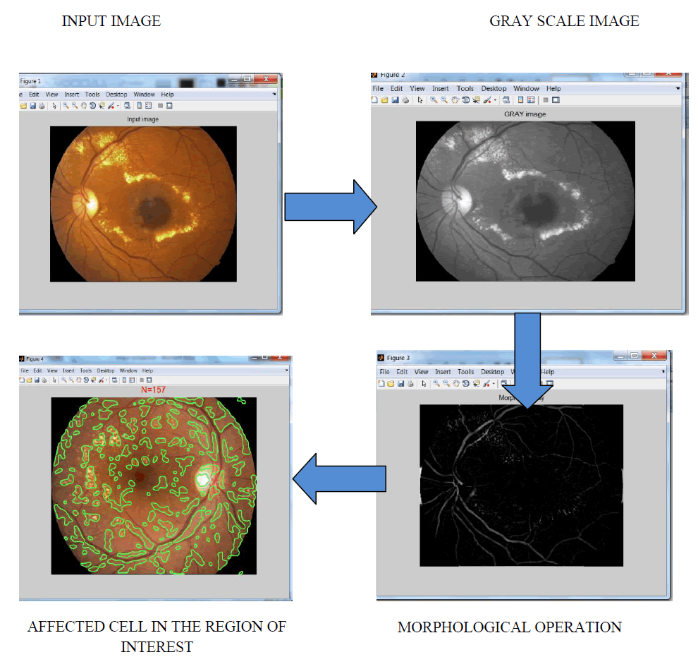
Diabetic retinopathy is one of the pre-eminent causes of blindness among the diabetic affected patient. It is caused by the light sensitive tissue in the retina because of the damaged blood vessels.The substances of lipoprotein gets leaked out of the blood vessels which are damaged and are deposited in the intra retinal part. The nerves of optics beneath the substances are interrupt from being excited by rays of light and are failed to produce any nerve impulse to brain and leads to loss of vision partially. Exudates are the one which is a lipoprotein substancein the retinal region which are yellow in colour.It is the important symptom of the retinopathy disease. Final stage detection of the retinopathy leads to loss of vision completely .By visual inspection, it is very difficult to detect exudates. In this paper, different image processing methods , include image thresholding,median filtering,morphological process are involved.By counting the number of affected cell in the retina by selecting the region of interest ,it shows the occurrence of retinopathy diseases.
Keywords |
| Retina,thresholding,cell count,image processing techniques. |
INTRODUCTION |
| Image processing is a process of an image using many image processing methods. Digital image can be represented as a composition of acquisition, coding, and editing. Images are produced by a various physical devices like stills cameras and video, radar , electron microscopes, X-ray and ultrasound. They are widelyuse for many application, including medical business and entertainment, military,industry,security,and scientific. Manual detection of affected region in the retina by ophthalmologists is time consumption. In developing countries like India, well trained ophthalmologists are less compared to other field. Diabetic retinopathy is one of the most serious complications of diabetes mellitus.It pre-dominantly leads to blindness. |
II. PROPOSED METHODOLOGY |
| They had been tested on a set of retinal images. For the analysis,database were collected from the images of retina.The algorithm based on the structure of blood vessels and little bit information on the optic disk. It help ophthalmologists to find the region of affected cells for detection of diabetic retinopathy disease. |
III. SCOPE OF THE PROJECT |
| Analysis of retinal image is one of the difficult task in medical image prosessing. The substances of lipoprotein get leaked out of the damaged blood vessel and are deposited in the intra retinal part. The nerves beneath the substances are interrupt from being excited by light rays and fails to produce any nerve impulse to brain which automatically leads to loss of vision partially. Difficult to detect by visual inspection because of small inner diameter of retina. It help ophthalmologists to find the number of affected cell in the region of interest. |
IV. BASE PAPER EXPLANATION |
| The main objective is to present a new computationally simple and automatic method to extract the region of interest from which the number of affected cell is observed from retinal blood vessels.It shows the severity stages in the retinopathy patient. They constitute several basic image processing techniques namely edge enhancement by morphological operation, image thresholding, standard template,noise removal,image and classification of object. |
V. FUNCTION OF THE EYE |
| In the science of optic, both camera and human eye are compared. The light reflected from an object is targeted on the retina after through the cornea, pupil and lens, which is similar to light passing through the camera optics to the film . The received information is received by the photoreceptor cells .The details are transmitted to the brain via the optic nerve from the region of retina, where the sensation is produced. During the transmission, the information is processed in the retinal layers. A cross-section of the eye and the structures present in the image formation. There are three features in the camera which are analogous to the function of the eye:camera lens, camera sensor aperture. There is a region of transparent cornea,in the human eye. The coloured iris intiaties the light entering the eye by varying the size of the pupil. The presence of pupil which is allowing the maximum amount light which enters in the dark region, and in the bright region the pupil is protecting the eye to receive an excess amount of light. |
 |
VII. DIABETIC RETINOPATHY |
| Diabetic retinopathy is a microvascular complication of diabetes, leads to abnormalities in the part of retina.Basically, there are no earlier symptoms in the early stages, but the severity and number increases predominantly in time. |
| The diabetic retinopathy typically starts as small changes in the capillaries of retina. The smallest detectable abnormalities, micro aneurysms (MA), appear as red dots in the retina. Because of the capillary vessels damage, the blood vessels in the retina may damage and leads to intraregional haemorrhages (HA). In the retina, the haemorrhages may appear either as small red dots different from micro aneurysms or larger round-shaped blots showing irregular outline. |
| Further it increases the permeability of the capillary walls which tends to retinal oedema and hard exudates (HE). Hard exudates are the formations of lipids leaking from the unstrengthen blood vessels and appear yellowish in colour with well-defined borders. when local oxygen and local capillary support fail because of obstructed blood vessels, the area shows pale indistinct margins appear in the part of the retina. They are small micro infarcts called as soft exudates (SE).Intraregional microvascular abnormalities (IRMA) and venopathy are symptoms of severe stage of diabetic retinopathy, where intraregional microvascular abnormalities visualize as dilation in the capillary system and venopathy as shape changes in veins and artery. The growth of these new vessels is known as neovascularisation. Maculopathy may occur at any stage of the diabetic retinopathy, it is more likely in the more advanced stages of the disease. In the final stage, it can result of fovea .A retina with no proliferative retinopathy is illustrated in fig. |
 |
VII. GRAY SCALE PROCESS |
| Grayscale images are different from one-bit bi-tonal white and black images, which in the reference of computer imaging .The images with only the two colours, black, and white ( bi-level). They have variety of shades of gray in between. |
VIII. MORPHOLOGICAL PROCESS |
| Morphology is a process of image processing based on shape and object’s form.Morphological methods apply a element of structure to an image input, creating an image of the output at the same size. The pixel value in the input image is based on a comparison of the corresponding pixel of the image input.By selecting the shape and size of the neighbour, it will create a morphological operation that is sensitive to specific shapes in the input image. Morphological operations can be defined on images of gray scale where the source image is one-channel. The concept can then be expanded to full-colour images. They undergo m operations which includes dilation,closing,opening and erosion. Combinations of these operations are used to performance analysis of the image. There are many useful operators defined in morphological mathematical operation.They are dilation, erosion, opening and closing. It applies structuring elements to an input image, creating an image of the output which is of the same size. |
IX. EXPERIMENTAL RESULTS |
 |
X. CONCLUSION |
| It gives a common segmentation algorithms for retinal image analysis.. The segmentation in the retinal images from the blood vessels using Morphological filters. It reveals the appearance of vessels is highly sensitive in the image of gray scale containing only the wavelength of green color. Because of this, segmentation of vessels was done using only RGB colour image of green channel. Morphological filter, whose application in the sector of and automatic object detection and machine were lead to detect and enhance image enhancement of the retina.It gives a better result. The morphological operations include dilation, erosion, opening, and closing. Dilation is a process involved in morphological operation that thickens objects in a binary image. The result shows that the proposed algorithm can be used for detection of affected cell in the retina from which tha retinopathy diseases occurrence can be detected. |
References |
|