International Journal of Innovative Research in Science, Engineering and Technology
ISSN ONLINE(2319-8753)PRINT(2347-6710)
ISSN ONLINE(2319-8753)PRINT(2347-6710)
Pazhanirajan.S1., Dhanalakshmi.P2
|
| Related article at Pubmed, Scholar Google |
Visit for more related articles at International Journal of Innovative Research in Science, Engineering and Technology
In recent years humans suffer from various neurological disorders such as headache, dementia, traumatic brain injuries, strokes and epilepsy. Nearly 50 million people of the world population in all ages suffer from epilepsy. To diagnose epilepsy an automatic seizure detection system is an important tool. In this paper we present a new approach for classification of Electroencephalogram (EEG) signals into two categories namely epilepsy and non epilepsy. The features of the EEG images are extracted using Discrete Cosine Transform (DCT) and Discrete Wavelet Transform (DWT). The extracted features are used in the model generation. The pattern classification model of SVM observes the distribution of the EEG features of classes. The experiment on various EEG image illustrate that the results of SVM are significant and effective.
Keywords |
| Electroencephalogram (EEG), Electroencephalogram Classification, Discrete Cosine Transform (DCT), Discrete Wavelet Transform (DWT), Support Vector Machine (SVM). |
I. INTRODUCTION |
| Electroencephalogram (EEG) is a complex human brain signal consisting of high information about brain function and neurological disorders. |
| Epileptic seizures affect the daily life of the patient due to its unpredictable and abrupt nature. Particularly, for drugresistant epileptic patients, the possibility to predict forthcoming seizures could be very useful, not only for the patient’s safety, but also to have the possibility to stop the unwanted event. Pharmacological or electrical treatment on demand could, in fact, be applied to stop an oncoming seizure (Guttinger et al., 2005), (Esteller et al., 2004). |
| Epilepsy is a seizure disorder that affects the nervous system. Seizure is caused if there is any disturbance in the normal pattern of neuron activity. Detection of epilepsy by visual scanning of EEG signal is very time consuming and may be inaccurate, particularly for long recordings. The detection of epileptic seizures in EEG signals is an important part in the diagnosis of epilepsy. |
| The raw EEG signal is highly difficult to classify for the prediction process. The EEG signal is captured as EEG images and DCT and DWT is applied to these images for extracting features. The extracted features are applied to SVM for classification. |
II. RELATED WORK |
| In [1] the authors have proposed a epileptic seizure detection system in EEG signals based on Discrete Wavelet Transform (DWT) and Swarm Negative Selection (SNS) algorithm. |
| An immune feature weighted SVM is proposed in [2] to overcome the difficulties in the traditional SVM for EEG classification. In this study, the authors have used IA in searching for the optimal feature weights and the optimal parameters of SVM and its kernel function simultaneously. Combining SVM with IA and feature weights, immune feature weighted SVM (IFWSVM) is used to multiclassify five different mental tasks. In [3] the authors have developed an automatic seizure detection system that diagnoses epilepsy. The proposed detection system is based on generalized autoregressive conditional heteroscedasticity (GARCH) model. |
| A novel wavelet-chaos-neural network methodology is presented [4] for classification of electroencephalogram (EEGs) signals. The EEG signal is decomposed into sub-bands using Wavelet analysis. The classification accuracies of the following techniques are compared: 1) unsupervised–means clustering; 2) linear and quadratic discriminant analysis; 3) radial basis function neural network; 4) Levenberg–Marquardt backpropagation neural network (LMBPNN). |
| In [5] the authors have proposed a multiclass support vector machine (SVM) with the error-correcting output codes for the multiclass electroencephalogram (EEG) signals classification problem. Classification was performed in two stages; in the first stage features are extracted by computing the wavelet coefficients and the Lyapunov exponents, in stage two classification is done using the classifiers on the extracted features. |
| In [6], features which are usually employed in automatic speech recognition (ASR) are used for the detection of seizures in newborn EEG. In particular, spectral envelope-based features, composed of spectral powers and their spectral derivatives are compared to the established feature set which has been previously developed for EEG analysis. In [7] the authors have addressed the prediction of epileptic seizures from the online analysis of EEG data. The proposed solution relies in a novel way on autoregressive modeling of the EEG time series and combines a least-squares parameter estimator for EEG feature extraction along with a support vector machine (SVM) for binary classification between preictal/ictal and interictal states. |
III. FEATURE EXTRACTION |
| A. Discrete Cosine Transform |
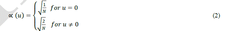
| DCT exploits the fact that pixels in an image exhibit certain level of correlation with their neighbouring pixels. The 2D DCT is denoted in the equation given below. |
 |
| The 2D DCT is applied on the image which transforms the image into an image with de correlated image data. The two main properties that describe DCT are De Correlation and Energy Compaction. De Correlation is the property of removal of redundancy between neighbouring pixels. DCT exhibits high level of de correlation property. Energy compaction is the ability to pack input data into as few coefficients as possible. DCT exhibits excellent energy compaction for highly correlated images. These properties of the DCT provide a means for using DCT in the feature extraction from the image. This will extract the important features of the image leaving the unwanted redundant data from the image. |
| B. Discrete Wavelet Transform |
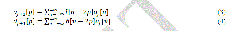
| The discrete wavelet transform (DWT) [13] is a linear transformation which operates on a data vector which has an integer length that is power of two, transforming it into a numerically different vector of the same length. DWT’s feature |
| is that it segments the data into different frequency components, and then studies each component with resolution matched to its scale. DWT is computed with a cascade of filtering followed by a factor 2 sub sampling Fig.1. |
 |
| Elements aj are used for next step (scale) of the transform and elements dj, called wavelet coefficients, determine output of the transform. l[n] and h[n] are coefficients of low and high-pas filters respectively One can assume that on scale j+1 there is only half from number of a and d elements on scale j. This causes that DWT can be done until only two aj elements remain in the analyzed signal. These elements are called scaling function coefficients. |
IV. MODELLING TECHNIQUES FOR EEG CLASSIFICATION |
| Support vector machine (SVM) is based on the principle of structural risk minimization. SVM learns an optimal separating hyper plane from a given set of positive and negative examples. This is in contrast to traditional pattern recognition techniques (Mourad Adnane et al., 2012; Marcus Musselman et al., 2010) of minimizing the empirical risk, which optimizes the performance on the training data. SVM can be used for pattern classification. For linearly separable data SVM finds a separating hyper plane which separates the data with the largest margin. For linearly inseparable data, it maps the data in the input space into a high dimension space. |
 |
| with kernel function Φ(x), to find the separating hyper plane. SVMs are evaluated as popular tools for learning from the given data. The reason is that SVMs are more effective than the traditional pattern recognition approaches based on the combination of a feature selection procedure and a conventional classifier. |
| An example for SVM kernel function Φ(x) maps 2-Dimensional input space to higher 3-Dimensional feature space as shown in Fig.2. SVM was originally developed for two class classification problems. The N class classification problem can be solved using N SVMs. Each SVM separates a single class from all the remaining classes[14]-[16] |
| SVM generally applies to linear boundaries. In the case where a linear boundary is inappropriate SVM can map the input vector into a high dimensional feature space. By choosing a non-linear mapping, the SVM constructs an optimal separating hyper plane in this higher dimensional space, as shown in Fig.2. The function K is defined as the kernel function for generating the inner products to construct machines with different types of non-linear decision surfaces in the input space. |
 |
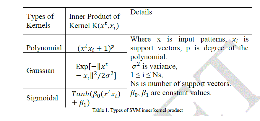
| The kernel function may be any of the symmetric functions that satisfy the Mercer’s conditions (Courant and Hilbert, 1953). There are several SVM kernel functions as given in Table 1. |
 |
V. EXPERIMENTAL RESULT |
| A. Dataset |
| An EEG data as shown in Figure.3, collected from Raja Muthiah Medical College and Hospital is used in this work. The EEG data consists of five subsets (denoted as C, T, O, P and F), each containing 100 single-channel EEG signals, each one having 5 minutes duration. From this data the dataset is formed by collecting 100 images of 10 seconds duration which identifies the seizure and another 100 images of 10 seconds duration which is randomly selected. The EEG signal is analysed manually and if there is any abnormality then the signal is captured as 10 second image. |
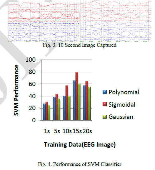 |
| The DCT and DWT each extracts 40 features from the EEG image. The extracted features are applied to the SVM classifier, which classifies the image into two models 0 and 1. The EEG images are separated into training and testing groups each consisting of 100 images. The SVM learning is used to calculate the classification parameter. The images are classified into their respective models by the training process. Model 1 represents epilepsy and model 0 represents non epilepsy. Using the test images the accuracy of the SVM classifier is found. The results show that the SVM classification of EEG images using the features extracted from DWT has higher classification accuracy than the SVM classification that uses the features extracted by DCT. |
References |
|