International Journal of Innovative Research in Science, Engineering and Technology
ISSN ONLINE(2319-8753)PRINT(2347-6710)
ISSN ONLINE(2319-8753)PRINT(2347-6710)
SivaSankari.S 1, Sindhu.M 2 , Sangeetha.R 3 , ShenbagaRajan.A4
|
| Related article at Pubmed, Scholar Google |
Visit for more related articles at International Journal of Innovative Research in Science, Engineering and Technology
Magnetic resonance imaging (MRI) is a medical imaging technique used in radiology to visualize internal structures of the body in detail. MRI provides good contrast between the different soft tissues of the body, which makes it especially useful in imaging the brain, muscles, the heart, and cancers compared with other medical imaging techniques such as (CT) or X-rays. By using this MRI we are going to extract the optimal features of brain tumor by utilizing GLCM, Gabor feature extraction algorithm with help of k-means Clustering Segmentation. The brain tumor characterize by uncontrolled growth of tissue. It can be easily cured if it is found at early stage.
Keywords |
|||||||||||||||||||||||||||||||||||||||||||||||||||||||
| MRI, Brain Tumour, segmentation, k- means Clustering, Feature extraction, GLCM, Gabor. | |||||||||||||||||||||||||||||||||||||||||||||||||||||||
INTRODUCTION |
|||||||||||||||||||||||||||||||||||||||||||||||||||||||
| This paper deals with the concept for brain tumour segmentation and feature extraction . Normally the anatomy of the Brain can be viewed by the MRI scan or CT scan. In this paper, | |||||||||||||||||||||||||||||||||||||||||||||||||||||||
| The MRI scanned image is taken for the entire process. The MRI scan is more comfortable than CT scan for diagnosis. It is not affect the human body. Because it doesn't use any radiation. But they may have some drawback in segmentation. In this paper, k-means algorithm is used for segmentation. So it gives the accurate result for tumor segmentation. Tumour is due to the uncontrolled growth of the tissues in any part of the body [1][8]. | |||||||||||||||||||||||||||||||||||||||||||||||||||||||
II. RELATED WORKS |
|||||||||||||||||||||||||||||||||||||||||||||||||||||||
| The existing method is based on the thresholding and region growing. The thresholding method was ignored the spatial characteristics [4][5][8]. Normally spatial characteristics are important for the malignant tumour detection. In the thresholding based segmentation the image is considered as having only two values either black or white. But the bit map image contains 0 to 255 gray scale values. So sometimes it ignores the tumour cells also. In case of the region growing based segmentation it needs more user interaction for the selection of the seed [7][6].Seed is nothing but the centre of the tumour cells; it may cause intensity in homogeneity problem. And also it will not provide the acceptable result in our feature extraction for all the images. So we are avoiding thresholding and region growing method it is not suitable for feature extraction technique.[1] | |||||||||||||||||||||||||||||||||||||||||||||||||||||||
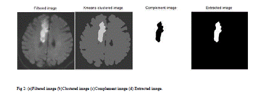
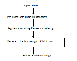
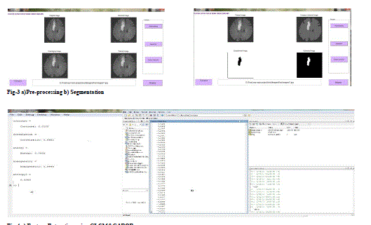
| The proposed system has mainly three modules: pre-processing, segmentation and Feature extraction. Pre processing is done by median filtering. Segmentation is carried out by K-means clustering algorithms. Feature extraction is an, approximate reasoning method to recognize the tumour shape and position in MRI image using edge detection method. In the existing method many algorithms were developed for segmentation. But they are not good for all types of the MRI images.[1][3][7][8]. | |||||||||||||||||||||||||||||||||||||||||||||||||||||||
III. METHODS USED IN PROPOSED SYSTEM |
|||||||||||||||||||||||||||||||||||||||||||||||||||||||
A.IMAGE PREPROCESSING |
|||||||||||||||||||||||||||||||||||||||||||||||||||||||
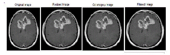
| According to the need of the first level the pre processing step convert the image. It performs filtering of noise in the image. RGB to grey conversion and Reshaping also takes place here. It includes median filter for noise removal. The possibilities of arrival of noise in modern MRI scan are very less. It may arrive due to the thermal effect. The main aim of this paper is to extract optimal features provide efficient result in feature extraction | |||||||||||||||||||||||||||||||||||||||||||||||||||||||
(i). MEDIAN FILTER |
|||||||||||||||||||||||||||||||||||||||||||||||||||||||
| In this paper we are using median filter for removing noise from an image. The median filter is a non linear digital filtering technique, is often used to remove noise. Median filtering is very widely used in digital image processing because, under certain conditions, it preserves edges while removing noise. The median filter is normally used to reduce noise in an image, somewhat like the, mean filter. However, it often does a better job than the mean filter. | |||||||||||||||||||||||||||||||||||||||||||||||||||||||
| The median is a more robust average than the mean and so a single very unrepresentative pixel in a neighbourhood will not affect the median value significantly. Since the median value must actually be the value of one of the pixels in the neighbourhood, the median filter does not create new unrealistic pixel values when the filter straddles an edge. For this reason the median filter is much better at preserving sharp edges than the mean filter.[5] | |||||||||||||||||||||||||||||||||||||||||||||||||||||||
 |
|||||||||||||||||||||||||||||||||||||||||||||||||||||||
B.SEGMENTATION USING K-MEANS CLUSTERING: |
|||||||||||||||||||||||||||||||||||||||||||||||||||||||
| Image segmentation methods can be classified into three categories: Edge-based methods, region-based methods, and pixel-based methods [3].The K-means clustering technique is a pixel-based method, it is one of the most simple techniques, it's complexity is relatively lower than other region-based or edge-based methods. Furthermore, Kmeans clustering is suitable for biomedical image segmentation as the number of clusters is usually known for images of particular regions of the human anatomy[3]. Combined with the existing methods and aiming to get better results, it is useful to take segmentation method into account.There is a two-phase iterative algorithm to minimize the sum of point-to-centroid distances. | |||||||||||||||||||||||||||||||||||||||||||||||||||||||
| ïÃâ÷ . Batch updates: Each iteration consists of reassigning points to their nearest cluster centroid, all at once, followed by recalculation of cluster centroids. | |||||||||||||||||||||||||||||||||||||||||||||||||||||||
| ïÃâ÷ . Online updates: Points are individually reassigned; in doing so the sum of distances is reduced, and cluster centroids are recomputed after each reassignment. | |||||||||||||||||||||||||||||||||||||||||||||||||||||||
|