International Journal of Innovative Research in Science, Engineering and Technology
ISSN ONLINE(2319-8753)PRINT(2347-6710)
ISSN ONLINE(2319-8753)PRINT(2347-6710)
J. Kavitha 1, Dr. D. Saravanan 2
|
| Related article at Pubmed, Scholar Google |
Visit for more related articles at International Journal of Innovative Research in Science, Engineering and Technology
The skin disease namely psoriasis affects a large number of people in the country, there is a need for improvement in the field of therapeutics which would actually be able to cure the disease. Hence my work has been designed in order to use the segmented scaling images for enhancing the treatment for psoriasis. The approach is to reduce the problem of segmenting scaling to a binary classification problem by removing erythema from consideration and then classifying the remaining pixels as either skin pixels or scaling pixels. Thus there are two stages as said which are namely feature extraction stage and scaling segmentation stage. Feature extraction is done using gabor filter. Segmentation of scaling is done using training set from the filtered images. A Markov random field (MRF) is used to smooth a pixel-wise classification from a support vector machine (SVM) that utilizes a feature space derived from image colour and scaling texture.
Keywords |
| Feature extraction, image segmentation, Markov Random Field (MRF), psoriasis, Support Vector Machine (SVM). |
INTRODUCTION |
| Psoriasis is a chronic skin disease that affects an estimated 125 million people worldwide. In this project, I have proposed a method of segmentation of scaling in 2-D psoriasis skin images. Here Markov Random field (MRF) is used to smooth a pixel-wise classification from a support vector machine (SVM) that utilizes a feature space derived from image colour and scaling texture. This method is expected to give reliable segmentation results when evaluated with images in different lighting conditions, skin types, psoriasis types. This system is believed to be the first to localize scaling directly in 2-D digital images. |
| The paper presents what we believe to be the first algorithm to automatically segment scaling directly from skin and erythema in 2-D digital images. The approach is to reduce the problem of segmenting scaling to a binary classification problem by removing erythema from consideration and then classifying the remaining pixels as either skin pixels or scaling pixels. The feature space used in the classification is derived from the colour contrast between scaling and erythema, and the image texture describing the roughness of scaling which is determined by the aggregated result from a bank of Gabor filters. Our evaluation indicates that our combination of Markov random fields (MRFs) with support vector machines using an appropriate feature space can solve a wide range of scaling segmentation problems that include variations in lighting conditions, variations in skin type and variations in the types of psoriatic lesions. |
| Scaling typically appears as white or creamy coloured scales on regions of red and inflamed skin (erythema) but can also appear in isolation without the accompanying erythema. When psoriasis appears without discernibly white or creamy flakes on normal skin. Scaling can present as small spots or as patches scattered within erythema. Fig. 1 shows some examples of the variation in the appearance of scaling. The variation makes it difficult to identify scaling boundaries through more conventional boundary detection algorithms and as a consequence we use a pixel based classification and labelling approach. |
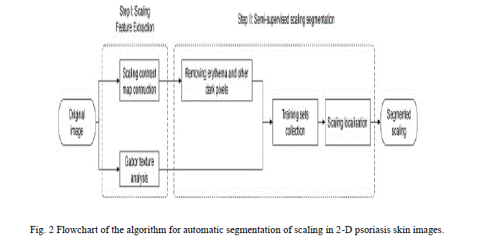
| Moreover, the colour of scaling may be very similar to that of normal skin, especially if the skin is fair, making it difficult to differentiate between scaling and normal skin using on colour alone. However, the rough textured surface of scaling is markedly different from normal skin. The algorithm uses a feature space derived from both colour and texture to classify pixels. The result is a pipeline that is essentially a pixel labelling algorithm that identifies scaling in 2-D digital skin images without the need for locating psoriasis first. It is composed of two main stages: 1) a feature extraction stage and 2) a scaling segmentation stage. The two stages are the accompanying erythema it appears as described as follows (see Fig. 2). |
| Step 1) The algorithm first analyzes skin colour and skin texture using an appropriately chosen color space and bank of Gabor filters to create a feature space for the image. |
| Step 2) The algorithm next removes erythema pixels from consideration and resamples the image to collect training samples for the classification process. The segmentation is achieved by using a MRF and the hyperplane derived from a support vector machine (SVM). |
II.RELATED WORK |
TITLE: AUTOMATED FEATURE EXTRACTION FOR EARLY DETECTION OF DIABETIC RETINOPATHY IN FUNDUS IMAGES, X.ZHANG AND O. CHUTATAPE, SEPT. 1996. |
| Automated detection of lesions in retinal images can assist in early diagnosis and screening of a common disease: Diabetic Retinopathy. A robust and computationally efficient approach for the localization of the different features and lesions in a fundus retinal image is presented in this project. Since many features have common intensity properties, geometric features and correlations are used to distinguish between them. We propose a new constraint for optic disk detection where we first detect the major blood vessels first and use the intersection of these to find the approximate location of the optic disk. This is further localized using color properties. We also show that many of the features such as the blood vessels, exudates and microaneurysms and hemorrhages can be detected quite accurately using different morphological operations applied appropriately. These compare very favorably with existing systems and promise real deployment of these systems. |
TITLE: THE DIARETDB1 DIABETIC RETINOPATHY DATABASE AND EVALUATION PROTOCOL, HEIKKI KALVIAINEN AND JUHANI PIETILA, 2002. |
| Automatic diagnosis of diabetic retinopathy from digital fundus images has been an active research topic in the medical image processing community. The research interest is justified by the excellent potential for new products in the medical industry and significant reductions in health care costs. However, the maturity of proposed algorithms cannot be judged due to the lack of commonly accepted and representative image database with a verified ground truth and strict evaluation protocol. In this study, an evaluation methodology is proposed and an image database with ground truth is described. The database is publicly available for benchmarking diagnosis algorithms. With the proposed database and protocol, it is possible to compare different algorithms, and correspondingly, analyze their maturity for technology transfer from the research laboratories to the medical practice. |
TITLE: IMPROVING MICROANEURYSM DETECTION USING AN OPTIMALLY SELECTED SUBSET OF CANDIDATE EXTRACTORS AND PREPROCESSING METHODS,BALINT ANTAL, ANDRAS HAJDU, JANUARY 2012 |
| In this project, I present an approach to improve microaneurysm detection in digital color fundus images. Instead of following the standard process which considers preprocessing, candidate extraction and classification, we propose a novel approach that combines several preprocessing methods and candidate extractors before the classification step. We ensure high flexibility by using a modular model and a simulated annealing-based search algorithm to find the optimal combination. Our experimental results show that the proposed method outperforms the current state-of-the-art individual microaneurysm candidate extractors. |
III.MATERIALS AND METHODS |
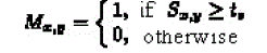
| The work presented here consists of five modules: 1) Preprocessing 2) Feature Extraction 3) Removing erythema 4) Training sets collection and localization 5) Segmentation |
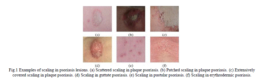 |
| Fig.1 Examples of scaling in psoriasis lesions. (a) Scattered scaling in plaque psoriasis. (b) Patched scaling in plaque psoriasis. (c) Extensively covered scaling in plaque psoriasis. (d) Scaling in guttate psoriasis. (e) Scaling in pustular psoriasis. (f) Scaling in erythrodermic psoriasis. |
 |
1. Preprocessing: |
| Preprocessing is removal of noise contents present in the image. It is done using the preprocessing algorithm „Scaling Contrast MapâÃâ¬ÃŸ. A scaling contrast map is developed to enhance the contrast of scaling from erythema. The map aims to enhance the contrast of scaling especially in situations where scaling is scattered in erythema. |
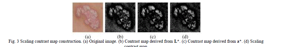 |
| Fig. 3 Scaling contrast map construction. (a) Original image. (b) Contrast map derived from L*. (c) Contrast map derived from a*. (d) Scaling contrast map. |
| L*a*b* color space is used to develop a pair of multi-scale center-surround filters that increase the contrast between scaling and erythema. The L* dimension specifies lightness where an L* value of 0 is black and an L* value of 100 is a diffuse white. The a* dimension is the red–green dimension, where a positive value of a* is red and a negative value of a* green, and the b* dimension is the blue–yellow dimension, where a positive value of b* is blue and a negative value of b* is yellow. |
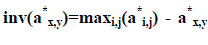
| The color of scaling correlates well with higher values of L* and erythema with positive values of a*. Shadows result in smaller values but do not necessarily affect the other dimensions. Furthermore, by inverting the dimension the color difference between scaling and the surrounding erythema or skin can be increased. With this in mind a scaling contrast map can be defined as follows: |
 |
2. Feature Extraction: |
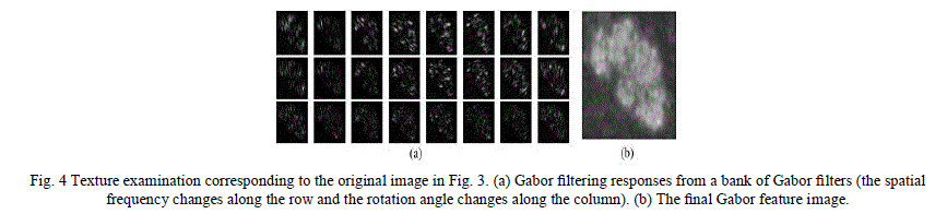
| Feature extraction is extracting the number of important objects present in the image. Algorithm of extraction is Gabor Texture analysis. The algorithm uses a bank of 24 Gabor filters designed to respond well in a variety of skin and scaling texture conditions. Finally, the Gabor texture image is obtained by summing the smoothed output. |
 |
3. Removing erythema: |
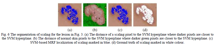
| Take out the dark pixels representing erythema, hair, moles and other blemishes are removed using the scaling contrast map. The first step is to threshold out the dark pixels representing erythema, hair, moles and other blemishes using the scaling contrast map S. Scaling and normal skin pixels remain in consideration after the application of the contrast map because they result in a significantly high value of S. We define a binary image M by |
 |
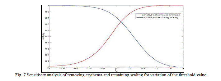
| where tx is the threshold for dark pixels. Pixels labelled with 1 are retained for further analysis while pixels labelled with 0 denote darker pigments and are removed from further consideration. |
| The scaling contrast map is applied to the image and the resulting image is processed to thresh-hold out all dark pixels representing darker pigments in the skin and including erythema, hair, moles, and other blemishes. |
4. Training sets collection and localization: |
| The training set data is collected by following steps |
a) Approximate Localization of Erythema |
| The location of erythema is identified by gray-scale intensity using the scaling contrast map where low values are indicating red pixels; normal skin would show negative values in the scaling contrast map. |
| The location of erythema is identified by gray-scale intensity using the scaling contrast map S |
| where low values S of indicate red pixels. A rough segmentation of erythema, but one that serves our purposes, can be obtained by empirically labelling a pixel to be erythema if |
b) Obtaining a Sample of Scaling and Skin Pixels |
| Localization of erythema is to collect a sample of skin pixels and scaling pixels. Using the fact that scaling is often surrounded, or partially surrounded, by erythema, we use dilation and erosion operations to create regions of scaling enclosed by boundaries of erythema. Dilating by a disk U of radius p pixels widens each of the erythema regions. The (Ãâò) algorithm erodes the erythema regions back to their original width while keeping the closed “doughnut” shape. The variable η is used for counting how many dilation phases have taken place so that we know how many erosion phases need to take place to regain the original erythema width (line 9, where ηU is a disk-shaped structure with radius ηp). |
c) Soft-Constrained K -Means Clustering |
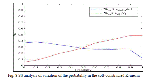
| The algorithm uses a soft-constrained K-means clustering to select training data from the candidate sets. The constraints are defined as the probability of data being in each cluster in the initial stage. A cluster of scaling pixels C1 and a cluster of skin pixels C2 are formed from Lscaling U Lskin, and then within each of these clusters the pixels with the great likelihood of being scaling and normal skin respectively are chosen. The clustering algorithm partitions the feature set into the set of features into those that are closer to ∂1 and those that are closer to ∂2 with respect to the Euclidean norm ||.||. |
 |
 |
| MAD values closer to 0 indicate a better training set, and SS values closer to 1 indicate a better training set. Table II hows the analysis results. For skin, the soft-constrained K-means has a better MAD value than the traditional K-means and the Fuzzy C-means. For scaling, the soft-constrained K-means shows an obvious advantage to the Fuzzy C-means in their MAD, but a slight inferiority to the K-means. Moreover, the soft constraints K-means has a much better SS over both the skin and scaling clusters. |
 |
II. CONCLUSION |
| In this paper, we present a general framework for automatic localizing scaling in psoriasis images. The result indicates that our algorithm makes progress towards the aim of automatic scaling segmentation. Scaling localization is implemented by a semi-supervised classification in this study. Two features are used: one is the scaling contrast map, which enhances the conspicuousness of scaling against erythema, and the other is a Gabor feature, which differentiates between scaling and normal skin based on image texture. Training sets for the classification are collected by a softconstrained K-means to avoid the human interference. At the end, we combine the advantages of the SVM and the MRF to separate scaling from skin images. The SVM shows good performance in classification, but does not involve spatial information. Normal skin pixels around psoriatic lesion boundaries exhibit similar misclassified by the SVM. By integrating the SVM into our adaptation of the MRF, the internal structure of images are considered and that increases the classification accuracy. When we compare the algorithm to manually collected training sets, the proposed method presents a slightly weaker sensitivity to the SVM and the MRF. In the future, we will further investigate the algorithms for training set collection to improve the classification results. Moreover, scaling features need to be researched further, especially for the very vague scaling, which remains difficult to be detected using the algorithm in this paper. |
References |
|