ISSN ONLINE(2319-8753)PRINT(2347-6710)
ISSN ONLINE(2319-8753)PRINT(2347-6710)
Dr. Vipin Arora 1, Dr. Vineeta Nikhil 2, Dr. Shefali Sawani 3, Dr. Pooja Arora4
|
| Related article at Pubmed, Scholar Google |
Visit for more related articles at International Journal of Innovative Research in Science, Engineering and Technology
One of the critical goals of adhesive dentistry is to restore the peripheral seal of dentine that is interrupted when enamel is lost as a result of developmental sequelae, trauma, caries or operative intervention such as preparatory excision. For coronal lesions the exposed strata may be bounded by dentine, enamel or both. Manufacturers continue to work vigorously on resin formulations that will restore this peripheral seal with operative ease and absolute durability. Difficulties with Class II restorations led to the development of open-sandwich restorations: a glass ionomer cement (GIC) or a resin-modified glass ionomer cement (RMGIC) placed between the dentin gingival margins and occlusal composite restorations. GIC presents two interesting features in restorations by bonding spontaneously to dentin and releasing fluoride. These sandwich restorations are less sensitive to technique than composite restorations and show a high percentage of gap-free interfacial adaptation to dentin.
Keywords |
| Class II restorations, Open sandwich technique, sandwich restorations |
INTRODUCTION |
| With the increasing demand for esthetic treatment options in restorative dentistry, an interest in longevity and reliability of resin composite restorations has grown. Resin composites represent the material most commonly used as an alternative to amalgam for class II restorations. [1] |
| Microleakage is one of the most frequently encountered problems for posterior composite restorations, in particular, at the gingival margins of class II cavities extending onto the root. [2] Direct class II restorations are known to show more leakage around enamel and dentin margins than indirect restorations. Unfortunately, several factors account for marginal microleakage when using composite. The enamel around the proximal box is often of poor quality or totally absent. [3] Furthermore, some voids within the materials and at the gingival margin have been reported. Adequate polymerization of the material and, therefore, clinical success, depends on the factors related to the material itself, such as the type of monomer or its shade, and on clinical factors, such as the incremental technique, distance from the light source,[4] the type of curing unit and blood and salivary contaminations. Together, this renders the class II restorations technique sensitive to operator skill. [5] Difficulties with class II restorations led to the development of open-sandwich restorations: a glass ionomer cement (GIC) or a resinmodified glass ionomer cement (RMGIC) placed between the dentin gingival margins and occlusal composite restorations. GIC presents two interesting features in restorations by bonding spontaneously to dentin and releasing fluoride. These sandwich restorations are less sensitive to technique than composite restorations and show a high percentage of gap-free interfacial adaptation to dentin. [6] Since, there are conflicting views regarding the clinical performance of open sandwich restorations, this review attempts to highlight the intricate details about this technique and critically evaluates the literature regarding clinical performance of the restorations. |
THE OPEN SANDWICH TECHNIQUE FOR RESTORATIONS |
| McLean and Wilson first described the open sandwich technique in 1977, proposing it as a method to improve adhesion of resin composite restorations. The technique was developed to limit the shortcomings of posterior composite restorations, particularly their lack of permanent adhesion to dentine, which could result in microleakage and postoperative sensitivity. Mount [7] advocated that the glass-ionomer (GI) at the cervical margin be left exposed to allow release fluoride to protect the surrounding tooth structure. This became to be known as the Open-Sandwich Technique. This so-called “Sandwich” of glass ionomer, dental adhesive and composite resin was proposed as an effective technique for both anterior and posterior resin based restorations by several clinicians as a means for pulpal protection from the acid-etch technique as well as a mechanism for sealing the cavity in the absence of good dentin adhesion available with the materials of the time. [8] The Open-Sandwich Technique for placement of a Class II posterior composite restoration has all layers of restorative material exposed to the oral cavity at the proximal margins, which are areas of primary concern for long-term clinical success. A self- or dual-cured composite resin material, glass ionomer, or resin-modified glass ionomer is placed as a base that covers the entire proximal box including all the dentin and cervical margin up to about one-third to one-half the height of the matrix band. After an initial polymerization period of this base, a top layer of a light-cured composite resin is placed to complete the restoration to full anatomic form and function. [9] |
| A. Clinical Technique for Open-Sandwich Restoration with Glass Ionomer Cement (GIC) |
| The Open Sandwich Technique involves layering of GIC and composite to obtain better results. After the removal of caries, isolate the tooth and place sectional matrix. After a 2 second, etch of the dentin with 37% phosphoric acid and then rinse. Apply glass ionomer cement in the proximal box areas to a point just apical to the contact area. Condense the first increment of composite resin to place using a non-serrated amalgam plugger. This increment extends from the flowable layer to the occlusal side of the proximal contact. Further, apply composite resin to facial and palatal enamel in increments and sculpt the desired occlusal anatomy. Then, smooth the resin layer prior to curing. |
| B. Modifications in the technique for Open-Sandwich Restoration |
| 1. Composite resin co cure technique |
| Bond a thin layer of a resin modified glass ionomer cement (RMGIC) bonding agent directly onto etched enamel and dentine. Next place a second layer of resin modified glass ionomer cement bonding agent followed immediately by the application of a composite resin prior to light curing. The first layer of resin modified GIC bond cures all the HEMA and seals the cavity while the second layer acts as a polymerization stress release during photo initiation of the composite resin. For cavities over 2mm deep a further layer of resin modified glass ionomer cement bonding agent can be used as a stress breaker between layers of composite resin. [10] |
| 2. Glass ionomer cement co cure technique |
| Following cavity preparation and etching of dentine and enamel surfaces, an increment of auto cure glass ionomer cement is placed into the proximal box and over the floor of the cavity extending up to the dento-enamel junction around the perimeter of the preparation or just short of the cavo-margin at the base of the proximal box. |
| Either prior to or at the immediate set of the auto cure glass ionomer cement, a layer of resin modified glass ionomer cement bonding agent is brushed over the auto cure glass ionomer cement and up to the outer perimeter of the preparation. [10] An increment of composite resin is next placed over the auto cure glass ionomer cement to fill the cavity followed immediately by photo curing the preparation. Upon photo initiation the composite resin cures and undergoes polymerization shrinkage before the resin modified glass ionomer bond has cured resulting in a stress free bond to tooth structure at the cavity perimeter. Resin modified glass ionomer cement chemically bonds composite resin to glass ionomer cement. The exothermic setting reaction of the composite resin heats the auto cure glass ionomer cement initiating a cascade setting reaction of the auto cure glass ionomer cement to occur between 20 to 40 seconds depending on the ambient temperature.[10] |
| C. Rationale for Use of GIC |
| Given the advances in dentine-bonding agents and resin composites, one would think that the technique would by now have become obsolete. However, the clinical success of posterior composite restorations is still limited with respect to leakage and longevity [11],[12] and this has meant that the sandwich technique is still in use today. The restoration of deep approximal cavities also requires that several problems must be overcome, the difficulty of placement of a rubber dam, the time-consuming incremental packing technique, and the intricate handling required by some dentine bonding systems.[13] It is therefore relevant to look at these relatively old materials with the aim of solving current problems in adhesive bonding because the GI family is naturally self-adhesive to tooth structure. [14] |
| The open-sandwich technique failed clinically when conventional GI’s were used to restore the cervical margins of Class II restorations, mainly because of a continuous loss of material. Consequently, the then newly developed resin-modified glass ionomers (RMGI) were used in place of conventional GI. The inclusion of resin in the GI formulation allowed these newer materials to polymerise upon light activation. The resin also supplemented the chemical bond that GI achieves with tooth structure by bonding micromechanically. This double adhesion mechanism is the main determinant of the retention and marginal sealing capacity of the material. It has been reported that higher bond strengths were achieved with RMGI than with conventional GI. [15],[16],[17] It is assumed that better sealing produced by RMGIC is a result of the formation of resin tags into dentinal tubules allied to the ion exchange process present in the interface between dentin and RMGIC, as previously reported. [18] Although some studies do not testify the presence of these resin tags or even the formation of a hybrid layer, [19] this assumption stands to be the reason for the superior performance of the RMGIC. In addition, the presence of HEMA in the RMGIC is responsible for the increased bond strengths to resin composite. [20] |
| The use of RMGIC as base material in Open Sandwich restoration reduces considerably the bulk resin composite used, so, the amount of shrinkage polymerization of resin composite is decreased and the marginal adaptation may be improved. A further advantage of the sandwich technique is the fluoride releasing property of GIC, which is considered to have some inhibitory effect on caries formation and progression around the restoration. [21] GIC is still considered the only material that self-adhere to tooth tissue and it has been previously shown that GIC and resin composite can adhere effectively to each other, regardless the limitations concerning this system. [22] Other authors suggest that the use of resin-modified glass ionomer could change the configuration factor to a more favourable internal shape, minimizing the polymerization contraction effects. [23],[24] The intrinsic porosity of this material, introduced by hand mixing, can increase the “within-material” free surface area, which also contributes to stress relief. Furthermore, the higher water sorption and hygroscopic expansion that occur with this kind of material may decrease the gaps developed in the bonding interface. [25] The low elasticity modulus of this material is also considered by some authors as another reason for the good seal it provides. Its relative “flexibility” can compensate the internal stress and the high stiffness of the composite resins after cure, preventing the adhesive interface from debonding. [26] This fact has been correlated with a better marginal adaptation. |
| Recently, Kleverlaan, Duinen V and Feilzer investigated the mechanical properties and compressive strength of GI’s that were either chemically cured, ultrasonically activated or heat cured, and concluded that the mechanical properties of GI’s significantly improved after ultrasound or heat curing. An ultrasonically cured GI demonstrated increased hardness, a decrease in the soft surface layer and negligible creep at a significantly shorter time after placement, suggesting that the curing process may be accelerated immediately after ultrasonic activation. [27] |
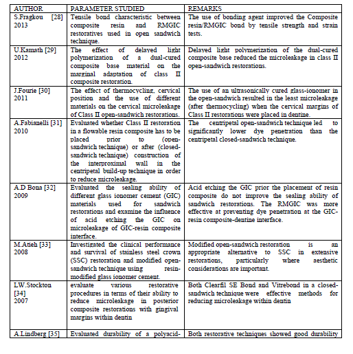
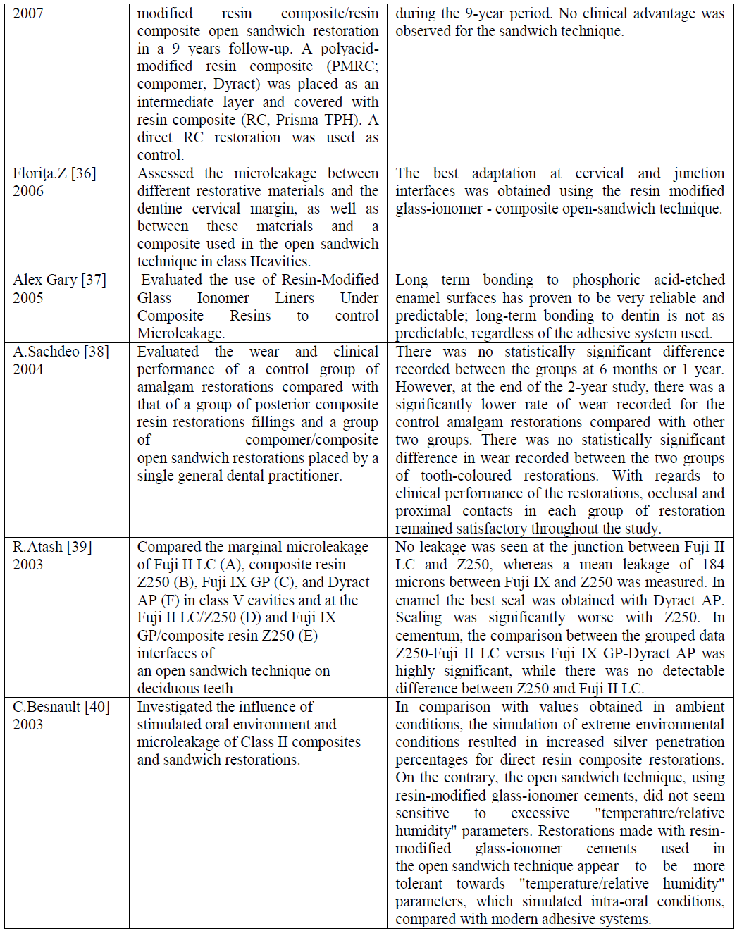
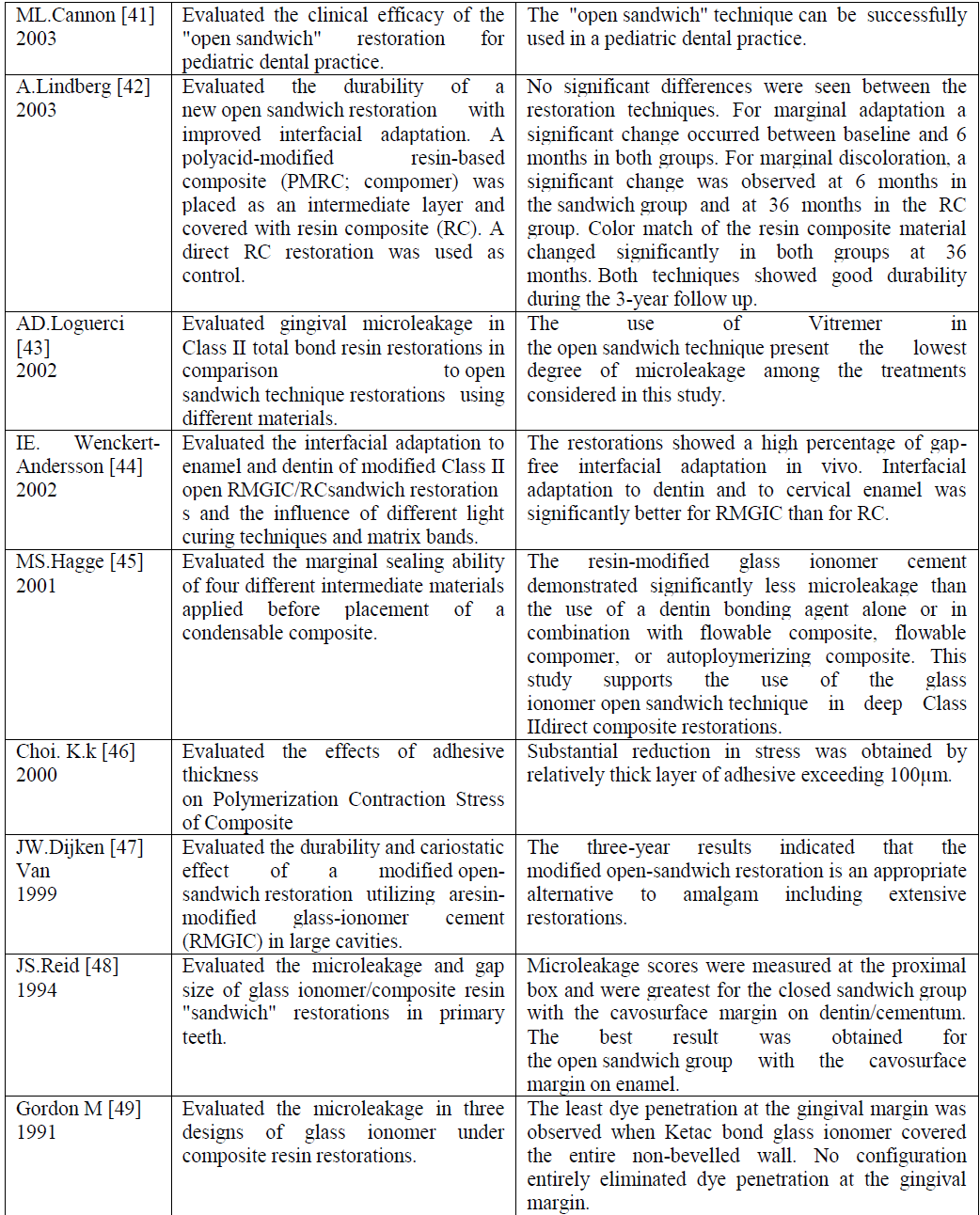
| D. Critical Analysis |
 |
 |
 |
CONCLUSION |
| One of the many questions that is still debated in dentistry relates to the optimal current methods of restoring Class I and Class II restorations directly. There are many practitioners who adopt a resin only approach and there are others that follow a combination of glass ionomer/bonding regime. The latter approach employs the philosophy that in restoring teeth, one treats dentin and enamel as separate entities and with such an approach, maximizes different materials to achieve optimum long term success. In using the sandwich technique the operator selects a dentin substitute (glass ionomer) and an enamel analog (resin composite). Glass ionomers in this technique are utilized for dentin replacement and offer the following characteristics: |
| Long term fluoride release that can create fluoro-appetite in replacement of damaged dentin and have long term caries inhibition effects. |
| • Similar thermal expansion properties as dentin. |
| • Insulation from the affects of higher temperature from curing lights. |
| • Insulation from the potential of uncured monomer from bonding agents that could seep into dentin tubules and create |
| negative outcomes. |
| • Less shrinkage and stress than composites. |
| • A family of materials that have demonstrated less microleakage than adhesion products and thus ultimately creating better internal seals with dentin. |
| • Overall a far less technique sensitive procedure that eliminates the issues of hydrophilic and hydrophobic properties of adhesion materials. [50] |
| Previous studies have shown that the inability of conventional GICs to produce an effective seal depends on two factors: 1) the material’s sensitivity to moisture during placement and early set; and 2) the dehydration after setting, resulting in crazing and cracking. Yet, it is assumed that the better sealing produced by RMGIC is a result of the formation of resin tags into the dentinal tubules allied to the ion exchange process present in the interface between dentin and RMGIC, as previously reported. [51] Although some studies do not testify the presence of these resin tags or even the formation of an hybrid layer [52],[53]. This assumption stands to be the reason for the superior performance of the RMGIC. In addition, the presence of HEMA in the RMGIC is responsible for the increased bond strengths to resin composite [54] and should contribute to prevent dye penetration through the interface of these materials. |
References |
|