Research & Reviews: Journal of Medical and Health Sciences
ISSN: 2319-9865
ISSN: 2319-9865
Sebin Tom Scaria, Satheesh Kumar Bhandary, Rajeshwary A and Vadisha Srinivas Bhat*
Department of ENT, KS Hegde medical Academy, Deralakatte, Mangalore-575018, Karnataka, India
Received: 19 February 2014 Accepted: 25 March 2014
Visit for more related articles at Research & Reviews: Journal of Medical and Health Sciences
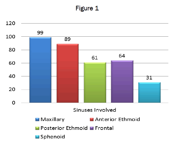
This study is aimed to find out the frequency of involvement of various paranasal sinuses in Chronic Rhinosinusitis. Hundred adult patients with Chronic Rhinosinusitis were selected. . All selected patients underwent non-contrast coronal and axial CT scanning of paranasal sinuses. Involvement of various paranasal sinuses in CT images of all patients recorded. Out of total 100 patients, 52 were males and 48 females. Patients were in the age range of 18 and 65 years. Maxillary sinus was involved in 99% cases, Anterior Ethmoid in 89% cases, Frontal sinus in 64% cases, Posterior Ethmoid Sinus in 61% cases and Sphenoid sinus in 31% cases. The most involved sinus was Maxillary sinus and the least involved was Sphenoid sinus. When multiple sinuses were involved, the most common combination was Maxillary, Anterior Ethmoidal, Posterior Ethmoid and Frontal sinuses in 50 patients, followed by Maxillary, Anterior Ethmoid, Posterior Ethmoid, Frontal and Sphenoid sinusitis in 26 cases and Maxillary and Anterior Ethmoid sinusitis in 19 cases. Isolated maxillary sinus is seen in 17 cases, isolated Frontal sinusitis in 3 cases and isolated Sphenoid sinus in 2 cases. Maxillary sinus is the most commonly involved paranasal sinus in Chronic Rhinosinusitis and sphenoid sinus least involved.
Chronic, Rhinisinusitis, CT scan, Paranasal sinus.
Chronic Rhinosinusitis (CRS) is a widely prevalent condition globally. According to National Institute of Allergy and Infectious Diseases (NIAID), 134 million Indians are suffering from CRS [1]. CRS is the 5th common medical condition where antibiotics are prescribed [2].
Rhinosinusitis refers to a group of diseases characterized by inflammation of mucosa of Nose and Paranasal sinuses [3,4]. The term CRS is used when duration of symptoms occur more than 12 weeks [3-5].
Computed Tomography (CT) of the Paranasal Sinuses the radiological investigation of choice for diagnosis of Sinonasal diseases [6,7]. Unlike plain radiography, CT shows an excellent anatomical soft tissue and bony details, helps in the diagnosis and gives detail of sinonasal anatomy for safe surgery. CT plays an important role in the diagnosis and management for CRS due to its ability to delineate mucosal disease, to demonstrate primary obstructive pathology and to image distal structures such as posterior ethmoid and sphenoid sinus that cannot be viewed with direct endoscopy.
100 cases of CT of Paranasal sinuses were studied and with the aim of determining the frequency of involvement of various sinuses in chronic Rhinosinusitis.
This study is a prospective study conducted on 100 patients with Chronic Rhinosinusitis attending Department of E.N.T, from June 2011 to December 2013. All patients with established clinical criteria for CRS were treated with antibiotic course for three weeks prior to CT scan. All selected patients underwent non-contrast coronal and axial CT Scan of Paranasal sinuses with 16 slice MDCT Bright Speed GE machine. Involvement of various paranasal sinuses was recorded.
Out of 100 patients studied, 58 were males and 42 were females. The minimum age was 18 and the maximum age was 65. Maximum number of patients were in the age range of 31 – 40 years.
Among 100 cases of CRS, CT scan of paranasal sinuses showed Unilateral sinusitis in 25% cases and Bilateral sinusitis 75% cases. Maxillary sinus was involved in 99 (99%) cases, Anterior Ethmoid in 89 (89%) cases, Frontal sinus in 64 (64%) cases, Posterior Ethmoid Sinus in 61 (61%) cases and Sphenoid sinus in 31 (31%) cases. The most involved sinus was Maxillary sinus and least involved was Sphenoid sinus. (Figure 1)
In unilateral disease, Maxillary sinus was the most common involved and Posterior Ethmoid sinus was least involved. Anterior Ethmoid sinus is the most common sinus involved bilaterally and Sphenoid sinus was the least common. (Table 1) .

Table 1: Incidence of Unilateral and Bilateral sinusitis in each paranasal sinus
Among 200 Maxillary sinuses in 100 patients, haziness was present in 155(77.5%) and absent in 45(22.5%) sinuses. Among 155 maxillary sinuses which showed haziness, partial haziness was present in 89(44.5%) and complete haziness was present in 66(33%).
Among 200 anterior ethmoid sinuses in 100 patients, haziness was present in 148(74%) sinuses and absent in 52(26%) sinuses. Among 148 anterior ethmoid sinuses which have haziness, partial haziness was present in 100(50%) and full haziness was present in 48(24%).
Among 200 posterior ethmoid sinuses in 100 patients, haziness was present in 106(53%) sinuses and absent in 94(47%) sinuses. Among 106 posterior ethmoid sinuses having haziness, partial haziness was present in 81(40.5%) and full haziness was present in 25(12.5%).
Among 200 frontal sinuses in 100 patients, haziness was present in 105(52.5%) sinuses and absent in 95(47.5%) sinuses. Among 105 frontal sinuses which have haziness, partial haziness was present in 75(37.5%) and full haziness was present in 30(15%).
Among 200 sphenoid sinuses in 100 patients, haziness was present in 39(19.5%) sinuses and absent in 161(80.5%) sinuses. Among 39 sphenoid sinuses with haziness, partial haziness was present in 31(15.5%) and full haziness was present in 8(4%).
Among 100 cases of CRS, most common combination of sinuses involved in sinusitis was Maxillary, Anterior Ethmoidal, Posterior Ethmoidal and Frontal sinusitis in 50 cases, followed by Maxillary, Anterior Ethmoid, Posterior Ethmoid, Frontal and Sphenoid sinusitis in 26 cases and Maxillary and Anterior Ethmoid sinusitis in 19 cases. Isolated maxillary sinus is seen in 17 cases, isolated Frontal sinusitis in 3 cases and isolated Sphenoid sinus in 2 cases. (Table 2).

Table 2: Combination of paranasal sinuses involved in Chronic Rhinosinusitis
Chronic Rhinosinusitis is a widely prevalent condition globally. The chronicity of the conditions and its multifactorial origin makes the detection of the underlying mechanism of disease difficult, and renders the management challenging. The management of CRS depends on an accurate and timely diagnosis.
The introduction of head and neck CT imaging and current wider use of this modality have undoubtedly helped the clinician. CT has become a useful diagnostic modality in the evaluation of the paranasal sinuses and an integral part of surgical planning. It is also used to create intra operative road maps. Today, CT is the radiological examination of choice in evaluating the paranasal sinuses of a patient with CRS.
In our study, the disease was Unilateral in 25% cases and Bilateral in 75% cases. In a study by Sathish Nair, Unilateral CRS was seen in 42.9% cases and Bilateral CRS was seen in 57.1% cases [8]. GL Fadda et al found Unilateral CRS in 25% cases and Bilateral CRS in 75% cases [9]. In most of these studies Bilateral CRS is more common than Unilateral.
In our study, Maxillary sinus (99%) was the most commonly involved sinus in our study, followed by Anterior Ethmoids (89%), Frontal (64%), Posterior Ethmoids (61%) and Sphenoid sinus (31%). In Sathish Nair’s study, Maxillary sinus (72.9%) was the most commonly involved, followed by Ethmoids (65.8%), Frontal sinus (55%) and Sphenoid sinus (35%) [8]. In the study of GL Fadda et al, Maxillary sinus (67.1%) was the most commonly affected sinus followed by, Anterior Ethmoids (22.1%), Frontal (22.1%), Posterior Ethmoids (10%) and Sphenoid sinus (10%) [9]. The Maxillary sinus was the most commonly affected sinus in the study of T Bhattacharyya et al [10] and Shika et al [11]. Sphenoid sinus was the least involved sinus in our study, as well as study of Sathish Nair [8] and GL Fadda et al [9].
We have observed that involvement of the sphenoid sinus is quite rare compared to the other sinuses. In multi sinus involvement sphenoid sinus is usually spared. Anterior ethmoid sinuses are more commonly involved than posterior ethmoids. Our studies show that, in multi sinus involvement, anterior group of sinuses are more commonly involved than posterior group of sinuses. Even in isolated sinusitis, maxillary sinus is the most commonly involved sinus.
Most of the studies show that Maxillary sinus is the most commonly involved sinus in CRS, followed by Anterior Ethmoidal sinus, Frontal sinus, Posterior Ethmoidal sinus and Sphenoid Sinus. Sphenoid sinus will be the least involved sinus by CRS. So findings of our study are similar to most of the studies conducted in this regard.
Computed Tomography of paranasal sinuses is the gold standard investigation in case of Chronic Rhinosinusitis. In Chronic Rhinosinusitis, among the paranasal sinuses, maxillary sinus is more commonly involved and sphenoid is the least involved sinus. Multi sinus involvement is more compared to isolated sinus involvement in Chronic Rhinosinusitis.