International Journal of Innovative Research in Science, Engineering and Technology
ISSN ONLINE(2319-8753)PRINT(2347-6710)
ISSN ONLINE(2319-8753)PRINT(2347-6710)
Dr.K.Meenakshi Sundaram 1, D.Sasikala2 , P.Aarthi Rani3
|
| Related article at Pubmed, Scholar Google |
Visit for more related articles at International Journal of Innovative Research in Science, Engineering and Technology
Digital mammogram becomes the most effective technique for early breast cancer detection modality and processing these images requires high computational capabilities. Computer image processing techniques will be applied to enhance images. This paper attempts to study about pre-processing is the most important step in the mammogram analysis due to poor captured mammogram image quality. Pre-processing is very important to correct and adjust the mammogram image for further study and processing. Different types of filtering techniques are available for pre-processing. Filters are used to improve image quality, remove the noise, preserves the edges within an image, enhance and smoothen the image. The experimental results concludes that the adaptive median filter is best for mammogram image noise removal and gives better performance by estimating the PSNR values
Keywords |
| Median filter, Adaptive median filter, Peak Signal to Noise Ratio, Mean Squared Error. |
INTRODUCTION |
| A Mammogram is an x-ray of the breast that can reveal abnormalities like benign or malignant. The procedure involves compressing the breast between two plates and then applying a small dose of radiation to produce an x-ray image. Mammograms can be used for screening and for diagnosis. Screening Mammogram is performed to attempt to detect breast cancer before symptoms occur. The goal of screening mammography programs is to decrease mortality from breast cancer. Diagnostic Mammogram is performed to help detect breast cancer if a woman has symptoms, such as a lump that can be felt in her breast. Breast Cancer is one of the most common cancers, leading to cause of death among women, especially in developed countries. Mammography is currently the most effective imaging modality for breast cancer screening. Mammography is a radiographic imaging technique which is used to obtain breast images for diagnostic and screening purpose for early stage detection of cancer and the images are called mammograms which are obtained by using low dose radiation levels between specific intervals. Mammography plays an important role to detect abnormalities in the breast. It gives detailed information about anatomy, morphology and pathologies of breast for screening and diagnosis of breast cancer. There is a difficulty to detect masses in mammograms because sometimes masses seemed to be similar to normal breast tissues on mammograms. It is difficult to distinguish between malignant and benign masses. Irregular shapes have a higher probability of being malignant and regular shapes have a probability of being benign. Difference in regions of the right and left breast is known as bilateral asymmetry of the breast. |
II. RELATED WORKS |
| This research work have discusses about the methodology for pre-processing. The related works are retrieved, analyzed are presented. Cervinka T [1] the proposed method of the histogram of the intensity in CT images down sampled. Therefore, the low contrast and blurring regions in CT images enhanced. A Markov Random Field model, which is consider the geometrical constraints of the processed image used to develop the accuracy resulting from the downsampling procedure. Median filtering open morphological operation and contrast enhancement [2] used to reduce noise and also image enhancement. Lai [3] used four selective averaging schemes and a modification of median filtering called selective median filtering. The Pre-processing technique used in medical images to remove special markings and unwanted noises.Morrow [4] the contrast of each region calculated with respect to its individual background. Background noise removing while preserving the edge information of suspicious areas can enhance a digital mammogram. Muller [5] the noise, poor image contrast, in homogeneity, weak boundaries and special mark existing in the medical image segmentation process extremely difficult to remove the noise and special markings that exist in medical images. The pre-processing method [6] including cutting out background area and normalization for CT brain images. In the proposed approach, an elliptical structure constructed based on skull contour and then the inclineimaging angles corrected |
III. EXISTING METHODOLOGY |
| Image pre-processing techniques are necessary, in order to find the orientation of the mammogram, to remove the noise and to enhance the quality of the image. Before any image-processing algorithm can be applied on mammogram, preprocessing steps are very important in order to limit the search for abnormalities without undue influence from background of the mammogram. Digital mammograms are medical images that are difficult to be interpreted, thus a preparation phase is needed in order to improve the image quality and make the segmentation results more accurate. The main objective of this process is to improve the quality of the image to make it ready to further processing by removing the unrelated and surplus parts in the back ground of the mammogram. Breast border extraction and pectoral muscle suppression is also a part of pre-processing. The types of noise observed in mammogram are high intensity rectangular label, low intensity label, tape artefacts etc. Generally filters are used to filter unwanted things or object in a spatial domain or surface. In digital image processing mostly the images are affected by various noises. The main objectives of the filters are to improve the quality of image by enhancing is to improve interoperability of the information present in the images for human visual. Image filtering is useful for many applications, including smoothing, sharpening, removing noise and edge detection. A filter is defined by a kernel, which is a small array applied to each pixel and its neighbours within an image. |
Median Filter |
| A median filter is a nonlinear filter is efficient in removing salt and pepper noise median tends to keep the sharpness of image edges while removing noise. The several of median filter is Centre-weighted median filter Weighted median filter, Max-median filter, the effect of the size of the window increases in median filtering noise removed effectively. |
Mean Filter |
| The mean filter replaces each pixel by the average value of the intensities in its neighbourhood. It can locally reduce the variance and is easy to implement. It has the effect of smoothing and blurring the image and is optimal for additive Gaussian noise in the sense of mean square error. Speckled image is a multiplicative model with non-Gaussian noise and therefore, the simple mean filter is not effective in this case. |
Adaptive Mean Filter |
| In order to alleviate the blurring effect, the adaptive mean filters have been proposed to achieve a balance between straightforward averaging and all-pass filtering. They adapt to the properties of the image locally and selectively remove speckles from different parts of the image. The uses of local image statistics such as mean, variance and spatial correlation to effectively detect and preserve edges and features. The speckle noise is removed by replacing it with a local mean value. The adaptive mean filters outperform mean filters, and generally reduce speckles while preserving the edges. |
Histogram Equalization |
| This technique corresponds to redistribution of gray levels in order to obtain uniform histogram. In this case every pixel is replaced by integral of the histogram of the image in that pixel. Histogram equalization is a method in image processing of contrast adjustment using the image's histogram. Through this adjustment, the intensities can be better distributed on the histogram. This allows for areas of lower local contrast to get better contrast. Histogram equalization accomplishes this by efficiently spreading out the most frequent intensity values. The method is useful in images with backgrounds and foregrounds that are both bright or both dark. In mammogram images, Histogram equalization is used to make contrast adjustment so that the image abnormalities will be better visible. |
IV. PROPOSED METHODOLOGY |
| In the existing system the process of mammogram image classification system is more complex. Each model proceeds in different ways to accomplish the process. Most of the models noise removal has not been used in the pre-processing stage. |
| Adaptive median filter works on a rectangular region Sxy. It changes the size of Sxy during the filtering operation depending on certain conditions as listed below. Each output pixel contains the median value in the 3-by-3 neighbourhood around the corresponding pixel in the input images. The edges of the images however, are replaced by zeros. The output of the filter is a single value which replaces the current pixel value at (x, y), the point on which S is centered at this time. The following notations are used |
 |
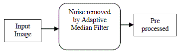
| Adaptive median filtering has been found to smooth the non repulsive noise from two-dimensional signals without blurring edges and preserve image particularly suitable for enhancing mammogram images. Therefore pre-processing is used in mammogram orientation, label and artefact removal, mammogram enhancement and mammogram segmentation. Pre-processing may also involve in creating mask for pixels with highest intensity to reduce resolutions and to segment the breast.The process flow of the proposed pre-processing using adaptive median filter is depicted in Fig.1. |
 |
V. PERFORMANCE EVALUATION |
| The following mathematical metrics are used for the evaluation of the quality of the image. |
| Peak Signal to Noise Ratio(PSNR) |
| Mean Squared Error (MSE) |
| Peak Signal to Noise Ratio (PSNR) |
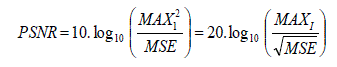
| The phrase peak signal-to-noise ratio often abbreviated PSNR, is an engineering term for the ratio between the maximum possible power of a signal and the power of corrupting noise that affects the fidelity of its representation. Because many signals have a very wide dynamic range, PSNR is usually expressed in terms of the logarithmic decibel scale. The PSNR is defined as |
 |
| Here, MAXI is the maximum pixel value of the image. When the pixels are represented using 8 bits per sample, this is 255. More generally, when samples are represented using linear PCM with B bits per sample, maximum possible value of MAXI is 2B-1. |
| Mean Squared Error (MSE) |
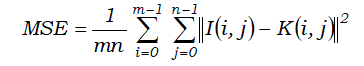
| The PSNR is most commonly used as a measure of quality of reconstruction in image compression etc. It is most easily defined via the Mean Squared Error (MSE) which for two m×n monochrome images I and K where one of the images is considered a noisy approximation of the other is defined as |
 |
| In general, a good reconstructed image is one with low MSE and high PSNR. That means that the image has low error and high image fidelity. |
VI. EXPERIMENTAL RESULTS |
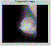
| The proposed methodology is experimented with Mammographic Image Analysis Society image databases and the results are presented separately. The images in the database have different sizes and are categorized in 10 classes as listed. In particular, a retrieved image is considered a match if and only if it is in the same category as the query. The pre-processing is done using adaptive median filter. The output image of the pre-processed image is a noise eliminated and enhanced one, which will be used for the image classification. The single mammogram image as input and the output of the pre-processed image as follows, |
 ' ' |
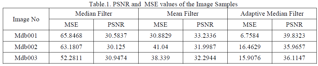
| The Table.1 shows the output of the preprocessed images PSNR and MSE values. From the experimental results it is concluded the adaptive median filter is best for face image noise removal and gives better performance by estimating the PSNR values. |
 |
VII. CONCLUSION |
| Pre-processing technique for enhancing the content of medical image based on removal of special markings and noise. Removal of special markings and noise existing in medical images will increase the quality of image segmentation. Here three types of filtering techniques for pre-processing of mammography images are considered. It compared to simulated output parameters such as image quality, mean square error, Peak signal to noise ratio. The comparisons of three types of filters are tested for mammogram images. From the experimental and results it is conclude that adaptive median filter is best for mammogram image noise removal gives better performance by estimating the PSNR values |
References |
|