ISSN ONLINE(2319-8753)PRINT(2347-6710)
ISSN ONLINE(2319-8753)PRINT(2347-6710)
| S.Anicham, C.Murukesh Dept Of Electronics & Instrumentation Engineering, Velammal Engineering College, Chennai-66, India |
| Related article at Pubmed, Scholar Google |
Visit for more related articles at International Journal of Innovative Research in Science, Engineering and Technology
Iris recognition is the most accurate and reliable biometric identification system used for security purposesThe iris recognition system consists of image acquisition, localization, normalization enhancement, feature extraction and classification. Segmentation is used for the localization of the correct iris region in an eye and it should be done to remove the reflection, eyelids, eyelashes, and pupil noises present in iris region. The proposed method uses Hough Transform segmentation method, then the iris and pupil boundary are detected from rest of the eye image in order to extract the noises. The segmented iris region is normalized to minimize the dimensional inconsistencies between the iris regions by using Daugman’s Rubber Sheet Model. The features of the normalized iris are extracted by contour let transform. LDA, SOM technique was chosen to classify the image. Iris Recognition is more efficient than using username and password technique and prevents the malicious action by the intruders. The above recognition experiment can be simulated using MATLAB.
Keywords |
| biometric, localization, normalization, LDA, SOM |
INTRODUCTION |
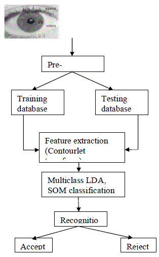
| Today, biometric recognition is a common and reliable way to authenticate and identify aPerson based on physiological or behavioral characteristics[1]. People cannot forget or lose their physical characteristics but they can lose passwords or their identity cards that is to be used for authentication. Among thesebiometric recognition, iris recognition iscurrently considered as one of the most reliable biometrics because of its unique texture. Moreover, iris is well protected from the external environment behind the cornea. Iris is relatively easy to acquire and stable all over the person’slife[2]. The image of a human iris thus constitutes a plausible biometric signature for establishing or confirming personal identity[3]. This research effort focuses on reducing the false rejectionby accurate iris detection and quality enhancement [4] irises are not identical even between the left and right eye of the same person[5]. Also irises are not alike for twins. Unlike other identifying characteristics that can change with age; the pattern of one's iris is fully formed by ten months of age and remains the same for the duration of their lifetime. Iris recognition technology is also accurate because it uses more than 240 points of reference in iris pattern as a basis for a match.[5],[6],[7]. In future, the use of passwords and PIN numbers for authentication becomes unnecessary. These methods are proved to be insecure and unsafe. Biometric identification utilizes many psychological and physical characteristics of an individual. Out of all physiological properties iris patterns are believed to be one of the most accurate. It has epigenetic formation and it is formed from the individual DNA. Eyes of the same individual seem to be very similar but contain unique patterns. Similarly, eyes of the identical twins also exhibit four different iris patterns [7]. This paper is organized as follows Section 1 describes about the introduction. Section 2 gives a full view about iris preprocessing techniques. Section 3 provides details about feature extraction. Section 4 gives a detail explanation about classification. |
 |
| A. Related Works |
| In [8], the phase component was used in 2D discrete Fourier transform of iris image with a simple matching strategy for phase based iris recognition technique. A 2D wavelet transform for the feature extraction and discriminate linear analysis for feature reduction with SVM techniques for iris pattern classifiers were used for iris recognition process in [9]. The Hough transform were applied for the iris localization, a Laplacian pyramid were used to represent the spatial characteristics of the human iris, with a modified normalized correlation were applied for the matching process [10][11]. With the development of neural networks algorithms, iris recognition systems may gain speed, accuracy and learning ability. In this paper iris segmentation algorithm for preprocessing, contourlet for feature extraction and Self organizing network for pattern classification were proposed. |
IRIS PRE-PROCESSING |
| The proposed iris recognition system is obtained fromthree major steps as follows: |
| a)Preprocessing includes image Acquisition, image iris localization, iris normalization, iris de-noising and enhancement. |
| b) Iris feature extraction. |
| c) Iris feature classification |
| A.Image Acquisition |
| It deals with capturing a high quality image of the iris. Images with sufficient resolution and sharpness are obtained. Good contrast in the iris pattern is obtained with proper illumination. Distance up to 3 meter. Near-infrared camera or LED. |
| B. Localization |
| The Hough transform is a standard computer vision algorithm that can be used to determine the parameters of simple geometric objects such as lines and circles present in an image. The circular Hough transform is used to deduce the radius and centre coordinates of the pupil and iris regions. Based on the circular Hough transform an automatic segmentation algorithm is employed by Wildes [12]. Firstly, an edge map is generated by calculating the first derivatives of intensity values in an eye image and then threshold the result. From the edge map, votes are cast in Hough space for the parameters of circle passing through each edge point. Those parameters are the centre coordinates of the circle say xc and yc, and theradius r, which are able to define any circle according to the equation. |
 |
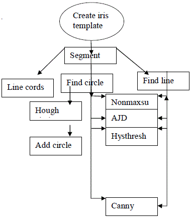 |
| Where (hj, kj) is the peak of the parabola and θj is the angle of rotation relative to the x-axis. To perform the preceding edge detection step, bias the derivatives in the horizontal direction for detecting the eyelids and in the vertical direction for detecting the outer circular boundary of the iris. The reason for this is that the eyelids are usually horizontally aligned and also the eyelid edge map will corrupt the circular iris boundary edge map if using all gradient data. A maximum point in the Hough space will correspond to the radius and centre coordinates of the circle best defined by the edge points. Wildes et al [14] and Kong and Zhang also make use of the parabolic Hough transform to detect the eyelids, approximating the upper and lower eyelids with parabolic arcs are represented perform the preceding edge detection. Consider only the vertical gradients for locating the iris boundary to reduce the influence of the eyelids when performing circular Hough transform, and not all the edge pixels defining the circle are required for successful localization. Performing this not only makes the circular localization more accurate [15], it also makes it more efficient since there are only less edge points to cast votes in the Hough space. There are number of problems associated with the Hough transform method. At first threshold values are to be chosen for edge detectionthis leads to the removal of critical edge points which fails to detect circles/arcs. Secondly, the Hough transform is computationally intensive due to its bruteforce approach and so Hough transform is notsuitable for the real time applications [16]. Segment iris- Performs automatic segmentation of the iris region from an eye image. Find circle - Returns the coordinates of a circle in an image using the Hough transform. Linecoords - Returns the x y coordinates of positions along a line.Find line - Returns the coordinates of a line in an image using the linear Hough transform and Canny edge detection to create the edge map. Hough circle - Takes an edge map image and performs the Hough transform for finding the circles in an image. Add circle – A circle generator for adding (drawing) weights into a Hough accumulator array.Nonmaxup – Function for performing non-maxima suppression on an image using an orientation image. Ajdgamma – Adjust image gamma. Image gamma value in the range 0-1 enhance contrast of bright regions, values > 1 enhance contrast in dark regions. Hysthresh - Function performs hysteresis threshold of an image. Canny - Function to perform canny edge detection. Upper and Lower Eyelid Detection Similar to iris outer boundary localization, the proposed method selects two search regions to detect upper and lower eyelids. The pupil centre, iris inner and outer boundaries are used as reference to select the two search regions. The search regions are confined within the inner and outer boundaries of the iris. The width of the two search regions is same with diameter of the pupil. Sobel edge detection is applied to the search regions to detect the eyelids. In order to reduce the false edges caused by eyelashes Sobel kernel is tuned to operate in the horizontal direction.After edge detection step, the edge image is generated. The eyelids are detected using linear Hough Transform method. The method calculates total number of edge points in every horizontal row inside the search regions. The horizontal row with maximum number of edge points is selected as eyelid boundary. In the proposed method, the eyelid boundaries are approximately modeled as straight lines. Edge detection cannot identify all pixels along the eyelid boundaries. The eyelid boundaries are normally occluded by the eyelashes. Therefore the eyelid boundaries are modeled with the straight line approximation. |
| C.Normalization |
| Once the iris region is successfully segmented from an eye image the segmented image is then transformed such that it has fixed dimensions in order to allow comparisons. The pupil dilation from various level of illumination causes stretching of the iris. This stretching is caused by the dimensional inconsistencies between eye images. The factors influencing the dimensional inconsistencies are the head tilt, varying imaging distance, rotation of eye within the eye socket and by rotation of the camera. The normalization process will produce iris regions with same constant dimensions. The act of normalizing makes the two photographs of the same iris working under different conditions to have their characteristic features at the same spatial location. Also it has been found that the pupil region is not always concentric within the iris region because it is slightly nasal. This must be taken into account while normalizing the “doughnutâÃâ¬ÃŸ shaped iris, it has been found that the undetected areas were small when compared with the size of the iris region. We will be using Daugman’s Rubber Sheet Model for normalization [17]. |
| D. Daugman’s Rubber Sheet Model |
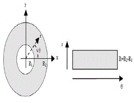
| Daugman suggested the transformation of Cartesian coordinates into polar coordinates which maps each pixel in the iris area into a pair of polar coordinates (r, θ), where r and θ lies in interval (0 1) and (0 2π). |
| This unwrapping can be formulated as |
| I(x(r, θ), y(r, θ)) I(r, θ) |
| Such that |
| X(r, θ) (1-r) xp(θ) + r x(θ) (3) |
| Y(r, θ) (1-r) yp(θ) + r y(θ) (4) |
| where I(x, y), (x, y), (r, θ), (xp, yp), (xi, yi) represent the iris region, Cartesian coordinates, polar coordinates, coordinates of the pupil and iris boundaries along the direction of θ respectively. Thus this representation often called as rubber sheet model. |
 |
| E. Image Enhancement UsingWeiner Filter |
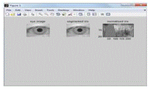
| The noise results while normalization process and while capturing the image some light illusion results were removed during enhancement. The contrast and brightness of image would increase and is used to represent the grayscale value of the image. |
 |
FEATURE EXTRACTION |
| The accurate recognition of individuals is obtained by extracting the most discriminating information present in an iris pattern. Only the significant features of the iris must be encoded.To create a biometric template most of the iris recognition systems makes use of a band pass decomposition of the iris image. The template generated by the feature encoding process requires a corresponding matching metric to provide a measure of similarity between two iris templates. This metric when compared with the templates generated from the same eye gives on range of values known as intraclass comparisons and another range of values is also obtained by comparing the same with the templates created from different irises which is referred to as inter-class comparisons. Both the range of values should be distinct and separate so as to identify whether the two templates are from same or from different irises. Feature extraction is one of the special form of dimensionality reduction. When the input data is found to be too large to be processed and notoriously redundant (much data, but not much information) then the input data will be transformed into a reduced representation set of features. |
| A.Extraction of Feature byContourlet |
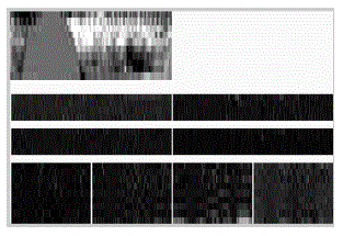
| The normalized iris image is used for feature extraction after preprocessing of an image. To extract feature from iris image the pyramidal and directional filter with level 2 subbands were used. Generally one dimensional transforms, such as Fourier transform and wavelet transforms such as Gabor wavelet were good at the catching point and only capture the edge of the image, due to this characteristics these transforms would not provide smooth contours in added to it starts to develop in continuous domain and then discrete for sampled data. So, in this proposed method feature extraction of an iris image were done by using contourlet transform. Contourlet transform is used for feature extraction in which the transforms starts with discrete domain and then continuous for sampled data by this smooth contours willbe obtained. The main advantage of contourlet transform is the multi resolution and multi directional expansion of discrete domain.[18]. By using the Laplacian pyramid and the directional filter bank the band pass image from LP are fed into DFB and the directional information from an image is captured. The main reason for combining the directional filter bank is to avoid the leak of low frequency in several directional subbands. |
| B. Filter Banks |
| Contourlet transform consists of two filter such as pyramidal filter and directional filter. Laplacian Pyramidal is used to obtain multiscale decomposition which will generate sub bands levels of an image and would obtain the band pass of image by taking difference between original and predication values. The process was iterated on the low pass signal and multidirectional filter bank for down sampled values . To obtain a directional filter bank (DFB) the binary level of tree decomposed into building blocks. The first block is divides into two directions as horizontal and vertical and the second block is for reordering the image samples using shearing operator. Contour let filter bank [19]. In which the Laplacian pyramid has the feature of generating only one band pass image and the frequency of this image would not scrambled and the original image can always be reconstructed from its Laplacian pyramid by reversing this process. |
 |
CLASSIFICATION |
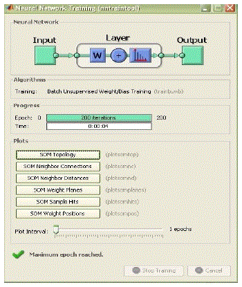
| SOM (self-organizing maps) is typically a kind of competitive learning that only one neuron will fire after mutual competition of neurons. The principal goal of self-organizing maps is to transform an incoming signal pattern of arbitrary dimension into a one or two-dimensional discrete map and to perform this transformation adaptively in a topologically ordered fashion. |
| A. Algorithm for Self-Organizing Map |
| a)Assume output nodes are connected in an array (usually 1 or 2 dimensional) |
| b)Assume that the network is fully connected-all nodes in input layer are connected to all nodes in output layer. |
| c)Randomly choose an input vector x. |
| d) Determine the “winning” output node i, wherewi is the weight vector connecting the inputs to output node i. |
 |
 |
 |
CONCLUSION |
| In this paper Iris recognition method is implemented using iris segmentation based on Hough transform and canny edge detection method. Iris normalization is performed using Daughman’s rubber sheet model, in order to reduce dimensional inconsistencies. Contourlet transform with multidirectional filter bank were used for efficient feature extraction of the iris pattern. Further it is classified using SOM. |
References |
|