ISSN ONLINE(2319-8753)PRINT(2347-6710)
ISSN ONLINE(2319-8753)PRINT(2347-6710)
Saravanan.M, Rajesh kumar.G.
|
| Related article at Pubmed, Scholar Google |
Visit for more related articles at International Journal of Innovative Research in Science, Engineering and Technology
Fractures are common in childhood. Incidence varies between geographical areas, and it has been proposed that the fractures in children are increasing. Repeated fractures, and especially vertebral fractures, in children may be a sign of impaired bone health, but it remains unestablished when and how fracture-prone children should be assessed. Bone mineral density (BMD) affects bone strength, and it can be measured with dual-energy X-ray absorptiometry (DXA). In this work, we studied epidemiology of fractures in children. Special attention was given to those children with frequent fractures or vertebral fractures; their bone health was thoroughly assessed. To evaluate the clinical use of two rarely used methods in children, we assessed the accuracy and advantages of vertebral fracture assessment (VFA) by densitometry, and histomorphometry from bone biopsy in children with suspected osteoporosis
Keywords |
| Bone fracture, Epidemiology, Material Properties, Stress variation |
INTRODUCTION |
| Fractures are common in children, and an increase in the incidence has been suspected. Underlying disease or life-style factors can influence bone health in children, and the risk for fractures might be modifiable. The diagnostic criteria of osteoporosis or the criteria for when to assess overall bone health in children without secondary causes have not been established. The present study was intended to obtain epidemiologic data on childhood fractures in Chennai; a 12- month survey was performed in all public health institutions in Chennai. All children with fractures were recorded for trauma and history of fractures; population-based incidence was obtained for children under 16 years. Further, a subgroup of apparently healthy children with frequent fractures or history of a vertebral fracture was assessed for bone health and risk factors for fractures. The usefulness of two methods that have not been widely used in children, vertebral fracture assessment from densitometerderived images and transiliac bone biopsy, were evaluated in children with suspected osteoporosis. |
II. PRESENTED WORK METHODOLOGY |
| The present work will focus on the ender nail fixation of fractured femoral bones (type of material and the size of nails). |
 |
| The present work will focus on the ender nail fixation of fractured femoral bones (type of material and the size of the nails). |
| Methodology: |
| ïÃâ÷ Getting the x-ray from hospital as jpg file. |
| ïÃâ÷ Importing the jpg file as raster image from Auto-Cad. |
| ïÃâ÷ Scaling and getting the actual dimensions and exporting the file asDXF file. |
| ïÃâ÷ Building the solid model using GID program. |
 |
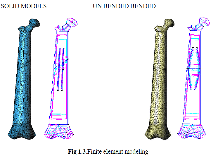
| Finite Element Modeling: |
 |
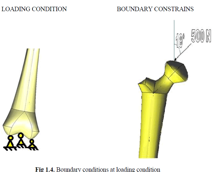
| Boundary Conditions: |
 |
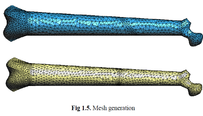
| Mesh Generation: |
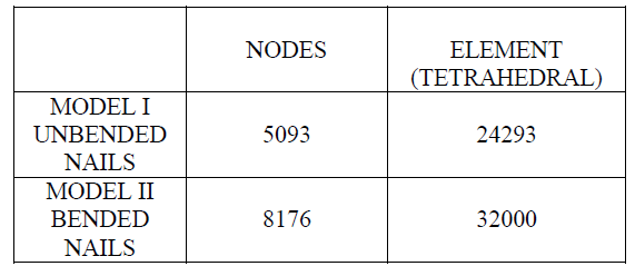 |
| Meshing was generated for two models .The table below shows the meshing data for the models. |
 |
| Material Properties: |
 |
III. RESULTS |
DIFFERENT CRANK ANGLES |
 |
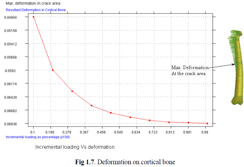
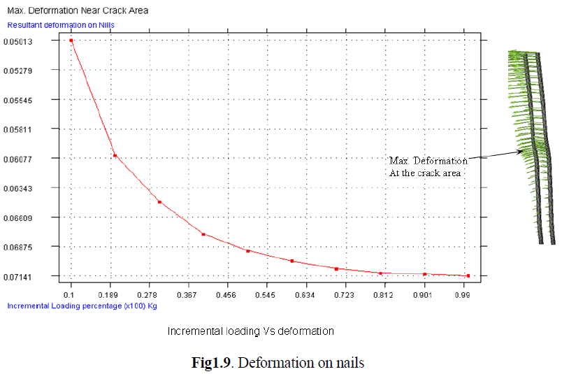
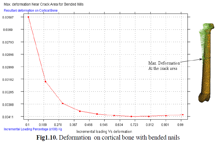
IV. DEFORMATION RESULTS |
DEFORMATION ON CORTICAL BONE |
 |
DEFORMATION ON CANCELOUSE BONE |
 |
DEFORMATION ON NAILS |
 |
DEFORMATION ON CORTICAL BONE WITH BENDED NAILS |
 |
DEFORMATION ON CANCELOUSE BONE FOR BENDED NAILS |
 |
DEFORMATION OF NAILS FOR BENDED NAILS |
 |
V. CONCLUSION |
| There are no established criteria for when and how to examine children with fractures. Repeated fractures or vertebral compressions are rare findings in healthy children; this group of patients is at risk of having impaired bone health and requires thorough evaluation. Based on the present findings, these patients can be identified from the children with newly diagnosed fracture, and screening those with frequent low-energy fractures from the emergency cohort is valuable. Lifestyle factors, biochemical parameters, including vitamin D, and DXA should be assessed. Mineralized mass is clearly not the only factor relevant to bone strength. Attention to bone mass, as measured by DXA, however, is justified, in part because it does play a substantial role, and in part, because it is to some extent controllable. Histomorphometric findings in this work underscore the difficulties in diagnosing pediatric osteoporosis. |
References |
|