Research & Reviews in Pharmacy and Pharmaceutical Sciences
e-ISSN:2320-1215 p-ISSN: 2322-0112
e-ISSN:2320-1215 p-ISSN: 2322-0112
1Research Scholar, Karpagam University, Coimbatore – 2, Tamil Nadu, India.
2Department of Pharmaceutical Chemistry, SRM College of Pharmacy, SRM University, Kattankulathur – 603203, Tamil Nadu, India.
Received: 27/03/2013 Accepted: 14/04/2013
Visit for more related articles at Research & Reviews in Pharmacy and Pharmaceutical Sciences
The effect of chloroform and methanol extract of the aerial parts of Tephrosia spinosa against Dalton’s Ascitic Lymphoma(DAL) have been evaluated in Swiss albino mice .A significant increase in the life span and a decrease in the cancer cell number and tumor weight were noted in the tumor –induced mice after treatment with chloroform and methanol extract of Tephrosia spinosa (L.f) Pers.The hematological parameters were also measured in tumor-induced mice .5-Fluorouracil was used as the standard .These observations are suggestive of the protective effect of Tephrosia spinosa against DAL.
Anticancer activity, Chloroform extract, Methanol extract, Tephrosia spinosa (L.f) pers.
Cancer is class of disease or disorder characterized by uncontrolled division of cells and the ability of these to spread, either by direct growth into adjacent tissue through invasion or by implantation into distant sites by metastasis [1]. Modern man is confronted with an increasing incidence of cancer and cancer deaths annually statistics indicate that men are largely plagued by lung ,colon, rectal and prostrate cancer, while women increasingly suffer from breast ,colon, rectal and stomach cancer [2] .The literature indicates that many natural products are available as chemo protective agents against commonly occurring cancer types [3].
Tephrosia spinosa (L.f) pers belongs to the family papilionaceae [4] and it is a stiffy throny shrub, known as mullukolinji commonly found in South India on dry barren lands on the coast and inland to the hills of Coimbatore, Madurai and Tirunelveli districts [5]. The phytochemical studies revealed the presence of flavanoids [6,7]. It is used in traditional system of medicine for anti-rheumatic, antipyretic, indigestion, anti-diarrheal, anti inflammatory, anthelminitc and to control excessive thirst [8]. No systematic studies on anticancer activity have been reported on Tephrosia spinosa. Hence an effort has been made to establish the anticancer activity.
The aerial parts Tephrosia spinosa was collected from Madurai district in 2009 and authenticated by Dr.D.Stephen who is the taxonomist in American College, Madurai.A voucher Specimen (KMCP/RXA/CC-0280) was deposited in the department of Pharmacognosy, KM College of Pharmacy, Uthangudi,Madurai for future reference. The air dried aerial parts of plant material was ground into coarse powder using cutter mill and then stored in an air tight container for further use.
The coarsely powdered plant material was defatted with hexane using cold maceration process and further subjected to extraction with chloroform followed by methanol successively by cold maceration for five days until complete extraction was effected. It was then concentrated under reduced pressure at 50oc and finally dried in desiccators. The chloroform and methanol extracts were used for anticancer activity.
The experimental protocol was approved by the institutional animal care and use committee of K.M. College of Pharmacy Uthangudi, madurai.RXA/08/Oct/2008/P.T/Ph.D with registration number 661/02/CPCSEA and date of registration 19/07/2002 and approved dated on 17/10/2010.As per the standard practice, the mice were segregated based on their gender and quarantined for 15 days before the commencement of the experiment. They were fed on healthy diet and maintained in hygienic environment in our animal house.
Male Swiss albino mice (20-25 gm) were produced from animal experimental laboratory, and used throughout the study [9]. They were housed in micro nylon boxes in a control environment (temp 25±2°C) and 12 hrs dark /light cycle with standard laboratory diet and water ad libitum.
Dalton’s ascitic Lymphoma (DAL) was supplied by Amala cancer research center, Trissur, Kerala, India. The cells maintained in vivo in Swiss albino mice by intraperitoneally transplantation. While transforming the tumor cells to the grouped animal the DAL cells were aspirated from peritoneal cavity of the mice using saline. The cell counts were done and further dilution were made so that total cell should be 1 x 10⁶, this dilution was given intraperitonealy. Let the tumor grow in the mice for minimum seven days before starting treatments.
Swiss Albino mice were divided into five group of six each. All the animals in four groups were injected with DAL cells (1 x 10⁶ cells per mouse) intraperitoneally, and the remaining one group is normal control group [10].
Group 1 The normal control.
Group 2 The tumor control. Group 1 and 2 receives normal diet and water.
Group 3 The positive control, was treated with 5-fluorouracil at 20 mg/kg body weight, Intraperitoneally.
Group 4 Treatment control group and was administered chloroform extract of Tephrosia spinosa(CETS) in a dose of 200mg/kg orally.
Group 5 Treatment control group and was administered methanol extract of Tephrosia spinosa(METS) in a dose of 200mg/kg orally.
For the study against DAL cell lines the chloroform extract and methanol extract of Tephrosia spinosa suspended in 2ml of sterile water for oral use.
In this study, drug treatment was given after the 24 hrs of inoculation, once daily for 14 days. On the 14th day after the last dose, all mice from each group were sacrificed by Bell jar euthanasia method (Painless killing). Blood was withdrawn from each mouse by retro orbital plexus method and the following parameters like tumor volume ,tumor cell count, increase in life span ,body weight, serum enzymes ,lipid profiles and hematological parameters were checked [11-14].
The mice were dissected and the ascitic fluid was collected from the peritoneal cavity.The volume was measured by taking it in a graduated centrifuge tube and packed cell volume was determined by centrifuging at 1000 rpm for five minutes [15].
The effect of extracts of Tephrosia spinosa tumor growth was monitored by recording the mortality daily and percentage increase in the life span (%ILS) was calculated [16].
Red blood cell count (RBC) hemoglobin content and White blood (WBC) count and platelets were studied in the mice of all groups .Blood was collected from all the mice in the groupings puncturing retro-orbital plexus method and counted for RBC, WBC, Hemoglobin content and platelets [17].
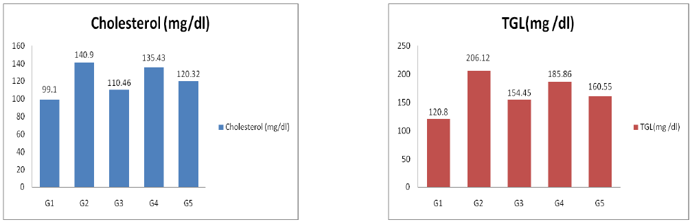
The serum was analyzed for the following parameters like AST, ALT, ALP, Total cholesterol level (TC) and triglycerides (TGL) [18-20].
The ascitic fluid was taken in a heamatocrit (micro) tube and diluted 1000 times .Then a drop of the diluted cell suspension was placed on the neubauer counting chamber and the cells in 64 small squares were counted.


Table 1: Effect of CETS and METS on the life span, body weight and cancer cell count of tumor induced mice.
G1 – Normal Control, G2 – Cancer Control, G3 – Positive control, G4 –Treatment control (CETS),G5– Treatment control (METS). All values are expressed as mean ± SEM for 6 animals in each group, **a: Values are significantly different from control (G1) at P < 0.001, *b: Values are significantly different from cancer control (G2) at P < 0.01, **b: Values are significantly different from cancer control (G2) at P < 0.001, CETS: Chloroform extract of Tephrosia spinosa, METS: Methanol extract of Tephrosia spinosa

Table 2: Effect of CETS and METS on Hematological parameters
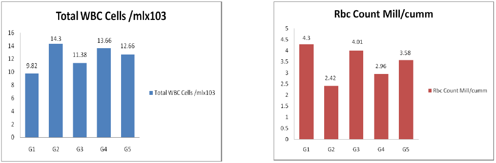

Table 3: Effect of CETS and METS on serum Enzymes and lipid proteins
In the DAL tumor control group, the average life span of animal was found to be 48% where as CETS and METS at the dose of 200 mg/kg body weight increase the life span to 66% and 72% respectively and these values were significant. How ever the average life span of 5- Fluorouracil treatment was found to be 92%, indicating its potent antitumor nature. The antitumor nature of CETS and METS was evidenced by the significant reduction in percent increase in body weight of animal treated with CETS and METS at the dose of 200 mg/kg body weight when compared to DAL tumor bearing mice.
It was also supported by the significant reduction in packed cell volume and viable Tumor cell count in both the extent of treatment when compared to the DAL tumor control. (Table No .1)
As shown in (Table No.2) RBC, Hemoglobin content, Platelets were decreased and WBC count was significantly increased in the DAL control group compared to the normal control group. Treatment with CETS and METS at the dose (200 mg/kg) significantly increases the Hemoglobin content, RBC, Platelets and significantly decrease the WBC count to about normal level. All these results suggest the anticancer nature of the both extract. However, the standard 5-Fluorouracil at the dose of 20 mg/kg body weight produced better result in all these parameters.
The inoculation of DAL cells caused significantly increase in the level of Total Cholesterol, Aspartate amino Transferase, Alanine amino Transferase, Alkaline Phosphatase in the tumor control animals(G2), when compared to the normal group. The treatment with CETS and METS at the dose of 200 mg/kg body weight reversed these changes towards the normal level. (Table No. 3) All the value was found to be significant. The treatment with standard 5- Fluorouracil also gave similar results.
The alternative system of medicines like Ayurveda, siddha, unani and other tribal folklore medicines have significantly contributed to the health care of the population of India. Today these systems are not only complementary but also competitive in the treatment of various diseases. Plants have served as a good source of antitumor agents. Several studies have been conducted on large number of plants possessing anticancer properties have been documented [22-27].
Plants of Tephrosia spinosa was traditionally used in the treatment of tumors. The present investigation was carried out to evaluate the antitumor activity of chloroform and methanol extracts of Tephrosia spinosa in DAL bearing mice. The CETS and METS treated animals at the doses of 200 mg/kg significantly inhibited the tumor volume, packed cell volume, tumor (viable) cell count and brought back the hematological parameters to more or less normal levels.
Many studies have reported the useful effects of plant products against DAL.when DAL is induced in animals the cancer cell count in the peritoneal fluid has been used as the marker to confirm the proliferation of cells .For a similar observation, in this study a cancer control, group was used. The increased cell count after 14 days confirmed the proliferation of cells in this group. A decrease in cancer cell count as a confirmatory evidence for protection against DAL has been reported [28]. In this study also a similar decrease was observed following the administration of the extracts .Consequently increased life span was observed with extract treated mice .Hematological parameters also enable to conclude on the protective effect .A decrease in RBC count, increase in WBC count and increase in cholesterol ,and TGL following cancer cell proliferation .In the same study an increase in RBC count and decrease in elevated WBC count were reported as confirmatory markers for the protection against DAL [29].