ISSN ONLINE(2319-8753)PRINT(2347-6710)
ISSN ONLINE(2319-8753)PRINT(2347-6710)
L. Ashok Kumar1, K.Karthika2, S.Kavitha2, T. Vinoth Krishnan2
|
| Related article at Pubmed, Scholar Google |
Visit for more related articles at International Journal of Innovative Research in Science, Engineering and Technology
Stroke is one of the leading threats to human health. Stroke and heart attack would seriously cause human morbidity and mortality. The carotid artery of intima-media thickness is a key indicator to the disease and is one of the major criteria for early screening and diagnosis of stroke. The intima-media thickness can be measured by segmenting the ultrasound images of carotid artery. First, the ultrasound image is loaded and the image is enhanced by reducing the speckle noise by using wavelet filter. Then the enhanced image is segmented into contours using level set method in which Chan-Vese algorithm is used. The level set formulation by Chan and Vese using random initialization provides a segmentation of the common carotid artery ultrasound images into different distinct regions. Finally, the resulting segmented image is classified into several classes which is used for IMT measurement of carotid artery. The resulting smoothed lumen intima boundary combined with anatomical information providean excellent initialization for parametric active contours that provide the final IMC segmentation which is used for intima-media thickness measurement.
Keywords |
| Carotid artery, Intima-media thickness, Active contours, wavelet filter, Image segmentation, Chan-Vesealgorithm, Ultrasound imaging, Level sets |
INTRODUCTION |
| According to recent data of World Health Organization cardiovascular diseases represent the third leading causeof death in the world. Therefore, the individuation of earlyprevention increased risk of cardiovascular disease which is of uppermost importance in clinical practice. Carotid intima media thickness is the distance between the lumen-intima and themedia-adventitia interfaces.It is a measure of early atherosclerosis. Intima media thickness can be measured through the segmentationof the intima media complex which corresponds to theintima and media layers of the arterial wall. One of the most widely used indicator of cardiovascular and cerebrovascular risk is the common carotid artery intima-media thickness. Several studies have showed that the IMT had an important diagnostic and predictive value for incident myocardial infraction. Ultrasound is the most used diagnostic tools for assessing CVDs. Ultrasound scanning could offer several paramount advantages in IMT clinical practice such as realtime quick examination, low-cost or economic, repeatable and reliable and non-radiation and non-invasiveness The technique used is the active contours without edges. Active contourswithout edges with the incorporation of anatomical information are used to establish intensity information for theUS images normalization. Following image normalization, activecontours without edges are again used to identify an initialintima-media boundary. This boundary provides an excellent, completely automatic initialization for the snake segmentationalgorithm that gives the final IMC segmentation. |
IMAGE SEGMENTATION |
| Image segmentation is the process of partitioning an image into multiple segments. The goal of segmentation is to simplify or change the representation of an image into more meaningful image. It is the process of assigning a label to every pixel in an image such that pixels with the same label share certain visual characteristics. The result of image segmentation is a set of segments that collectively cover the entire image. |
SPECKLE NOISE REMOVAL |
| Speckle is a form of granular multiplicative noise causedwhen the surface imaged appears rough to the scale of thewavelength used. Speckle noise has a significant impact on thecorrectness of boundary detection. The edges of the adventitia are also affected by this noise. This noise can be removed by using wavelet filtering. In this filtering, first the image is converted into a signal. Then, noise is reduced using wavelet filter. After the noise is removed, the signal is converted into a two dimensional image. This filter allows speckle noise to be reduced completely. |
LEVEL SET METHOD |
| The level set method is a numerical technique for tracking interfaces and shapes. The advantage of level set method is that one can perform numerical computations involving curves and surfaces on a fixed cartesian grid without having to parameterize these objects. This method makes it easier to follow shapes that change topology. Thus, level set method is a great tool for modelling time varying objects . |
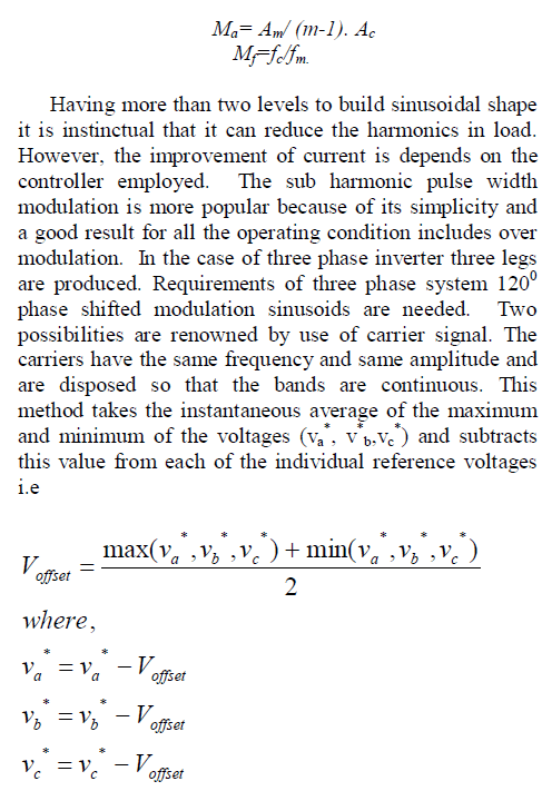 |
| This is a partial diferential equation and it can be solved numerically. |
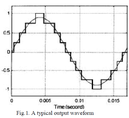 |
CHAN-VESE ALGORITHM |
| The Chan-Vese algorithm is an example of a geometric active contour model. Such models begin with a contour in the image plane defining an initial segmentation and then evolve this contour according to some evolution equation. The goal is to evolve the contour in such a way that it stops on the boundaries of the foreground region. This model is based on the Mumford-Shah functional for segmentation, and is used widely in the medical imaging field, especially for the segmentation of the brain, heart and trachea. The model is based on an energy minimization problem, which can be reformulated in the level set formulation, leading to an easier way to solve the problem. |
POLYLINE DISTANCE METRIC |
| The basic idea is to measure the distance of each vertex of a boundary to the segments of the other boundary. The measured distance becomes very robust because it is independent on the number of points of each boundary. This measure is very important since it reflects theaverage distance between two computer-generated boundariesand the corresponding ground truth profiles. |
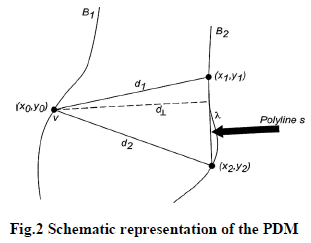 |
EXISTING SYSTEM |
CLINICAL SIGNIFICATION OF IMT MEASUREMENT |
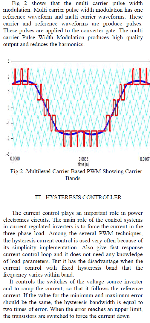
| Both CCA wall segmentation and IMT measurement are themost important two steps in computer-aided clinicalapplications.IMT measurement is the key to perform measurementsof further clinical use.Thestudies confirmed that the increase of the IMT value is oftenrelated to a change of the arterial wall properties, thus exposingthe patients to increased CVD risks. There is a high correlationbetween CVD and IMT value, therefore, it is important tomeasure and monitor IMT of vessels with a progression orregression plaque on the wall.This review will focus on the techniques that have beendeveloped to perform CCA wall segmentation and IMT measurement in 2D B-Mode ultrasound longitudinal images. |
 |
DYNAMIC PROGRAMMING TECHNIQUES |
| Dynamic programming method is used to segment the vessel walls. It was applied in an iterative progress, starting from the imagein a coarse representation and then slowly applying step-by steprefinement iteration. Hence, the global position of theartery in the image was estimated on a coarse scale, whileprecise position of the wall layers was estimated in a finescale. Therefore, the dynamic programming method couldbalance reducing computational cost and ensuring accurate performance. |
| This techniquecombinemultiple measurements of echo intensity, intensitygradient, and boundary continuity. The system also includedoptional interactive modification by the human operator. Aninitial set of ultrasound images was manually segmented byexpert operators and served as reference. Theestimated values of the three boundary featureswere linearlycombined in order to create a cost function. Each image pointwas associated with a specific cost that in turn correlated withthe likelihood of that point being located at the echo interface.Therefore, points being located at the interfaces betweendifferent artery layers were expected to be associated with alow-cost, whereas points located far from the interfaces wasassociated with a high cost. The weights of the linear combinationoriginating the cost function were determined bytraining the system, considering the ground truthasreference.Dynamic programming was used in order to reduce the computationalcost given by the need of inspecting all the points ofthe image. The major limitation relies in the need fortraining of the system. Therefore, retraining is required when scanneris changed. |
 |
DRAWBACKS |
| • Speckle noise is not completely removed |
| • Requires trained physician for segmentation |
| • Sensitive to change in topology structure |
| • No accuracy in monitoring carotid wall evolution |
THE PROPOSED SYSTEM |
| The proposed methodology allows segmenting the common carotid artery for measuring the intima-media thickness by active contours methodology in which level set technique is used with Chan-Vese algorithm.The proposedsystem is described as in the following stages: |
Stage 1 |
Loading the image and image enhancement: |
| The input image is loaded and the speckle noise present in the image is removed by using median filter. Then the denoised image is subjected to histogram equalization and the contrast of the image is adjusted to get better visualization. |
Stage 2 |
Segmentation of the enhanced image: |
| Level set method is used to segment the image by using Chan-Vese algorithm. The method tries to separate the image into regions based on pixel intensities. The most important step is to actually determine a level set function that segments the image in a meaningful way. |
Stage 3 |
Classification of the segmented image: |
| The intent of classification is to categorize all pixels into several classes. Unsupervised classification is a method which examines a large number of unknown pixels and divides it into number of classes. It does not require manual intervention. |
Stage 4 |
Measurement of intima-media thickness: |
| The intima-media thickness can be measured using polyline distance metric. Then, the measurement is used to detect the occurrence of cardiovascular disease. |
| All stages of the proposed system are illustrated in the given diagram |
 |
ADVANTAGES |
| • More robust to speckle noise |
| • It is fully automatic |
| • It also includes a smoothness constraint for robustness against noise |
| • Not depend on edges of images |
| • Handle topological changes naturally |
CONCLUSION |
| Thus, the presented segmentation of the IMT and evaluation of the IMT provide an objective and reproducible technique. The method is fully automatic, achieves lower absolute mean IMT error compared to interobserver variability in the evaluated dataset which has comparable results to both automatic and semiautomatic methods .The methodmay be used in clinical practice for the reliable segmentation ofthe carotid artery and the estimation of features of the arterial wall with physiological significance for the evaluation of earlystages of atherosclerosis and corresponding risks. It may represent a generalized and standard methodology towards completely automated and accurate IMT measurement. |
References |
|