ISSN ONLINE(2319-8753)PRINT(2347-6710)
ISSN ONLINE(2319-8753)PRINT(2347-6710)
Dr.DhairyashilNamdeoEdake1, Dr. Shaikh Shoeb Yakub2, Dr.AmitHemrajPatil3 and Dr.IshanAtulMota1
|
| Related article at Pubmed, Scholar Google |
Visit for more related articles at International Journal of Innovative Research in Science, Engineering and Technology
Every clinician who has performed root canal treatment has experienced a variety of emotions ranging from a perfect fill till the root apex to endodontic instrument breakage in the root canal. Non-surgical removal of these fractured or separated instruments is a real challenge for the clinician. Today, with the advancement of newer techniques in magnification(dental loupes and surgical operating microscope), access, instrument removal kits(Masseran kit), ultrasonic instrumentation and studies on tooth fractures after using these newer techniques, a clinician is able to make the right choices for separated instrument retrieval from the root canal with which this article deals with.
Keywords |
| Fractured instrument, Masserann kit, Ultrasonics, Vertical root fracture. |
INTRODUCTION |
| Vertical root fracture (VRF) is a longitudinal fracture of the root that extends through the entire thickness of the dentine from the root canal to the periodontium[1]. Many factors and procedures can cause VRF such as the placement of posts, anatomical characteristics of the teeth, masticatory forces and the removal of fractured instruments from the canal [2]. Instruments, such as the Masserann kit, the Canal Finder System, the Ruddle IRS and ultrasonic systems, have been used in the removal of metallic objects from root canals [3]. Among the instruments mentioned, ultrasonic tips and the Masserann kit are the two used most frequently [4]. In both techniques, the root canal must be enlarged sufficiently coronally, and a staging platform at the coronal part of the instrument should be prepared for better visualization and easier handling of the instruments [5]. Removal of a fractured instrument from the middle third of the root decreased the force required to fracture the root vertically, regardless of the technique used for instrument removal (Masserann technique and ultrasonic tips) [6]. Endo-surgical microscopes and magnifying loupes are essential for the removal of fractured instruments. However, there is no available literature about the use of enhanced vision and the effect of instrument retrieval different techniques on the force that is required to fracture the root vertically. |
| The aim of the study was to evaluate the force required to fracture roots vertically after ultrasonic and Masserann kit for removal of broken instruments using Magnifying loupe and Endo-surgical microscope. |
MATERIALS AND METHODS |
Group 1 Ultrasonic Removal using Endo-Surgical Microscope |
| A staging platform was prepared to improve the visibility of the fractured instrument using Gates Glidden (GG) drills. This was done by cutting off the guiding tip using a diamond bur, and a flattened end was prepared at the maximal cross-sectional diameter of the GG. All the procedures for removal of the instruments were performed under a surgical dental microscope at 10x magnification. The tips were activated without coolant to allow visualization of the energized tip around the fractured instrument, and the dentine around the fragment was trephined. The tip was inserted between the fractured instrument and the dentine and used around the fractured instrument in a counter-clockwise direction. |
Group 2 Ultrasonic using Magnifying loupe |
| All the procedures for removal of the instruments were same as previous group and performed using a magnifying loupe at 2.5x magnification. |
Group 3 Masserann kit using Endo-surgical microscope |
| Radicular access to the coronal end of the fragment was created with GG drills as in the ultrasonic group. Fan‐shaped gauge was used to measure outer diameter of broken fragment which facilitate correct choice of trepan. After ensuring that the fragment could be visualized, a gutter was prepared around it with a trepan bur. The extractor tube was engaged in the prepared space, and the plunger rod was turned manually in a clockwise direction to grip the fragment. When the tightest grip was felt manually, the entire assembly was rotated in an anticlockwise direction to unscrew the fragment. |
Group 4 Masserann using Magnifying loupe |
| All the procedures for removal of the instruments were same as previous group and performed under a magnifying loupe at 2.5x magnification. |
Sample Preparation |
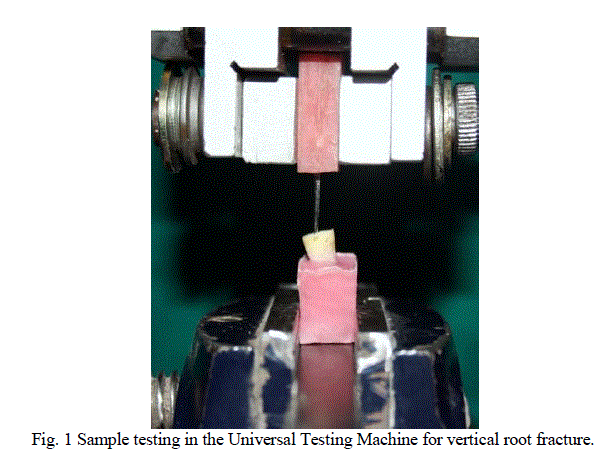
| The samples were embedded in acrylic resin such that, for each root, 3 mm of the apical part was embedded in the acrylic resin. A universal testing machine (Instron 3345; InstronCorp, Canton, MA, USA) was used to evaluate the force required to fracture the roots. One-way ANOVA test was used to determine the differences among the groups, and the Tukey HSD test was used to detect the group that caused the difference. The level of significance was set at P < 0.05. |
 |
RESULTS |
| Ten teeth were included in each group. The means and standard deviations of the force required for vertical fracture of the roots in each group are presented in Table 1. There were significant differences in relation to the force required for vertical fracture among the groups (P < 0.05, one-way ANOVA). The force required to fracture the roots in the control group was significantly higher (P < 0.05) than that required to fracture the roots in all the other groups. Roots from which the fractured instruments had been removed with the Masserann kit using dental loupes was the weakest followed by the group in which ultrasonic tips with dental loupes are used, followed by the group in which Masserann kit was used with surgical operating microscope followed by the group in which ultrasonic tips and surgical operating microscope was used which was found to be the strongest; however, this difference was not statistically significant (P > 0.05). |
 |
DISCUSSION |
| The loss of dentine increases the chances of teeth to fracture [7].Excessive instrumentation of the root canal, excessive pressure during canal filling, dehydration of dentine and preparation of post space lead to fracture of the teeth[7],[8]. In this regard, when an attempt is made to remove a fractured instrument, the potential loss of dentine must be taken into consideration. |
| In the present study, mandibular 2nd Premolars that had roots of similar length and similar buccolingual diameters were selected to obtain standardized groups. In addition, the coronal parts of the roots were cut at the same length. Also, 3 mm of the apical region was embedded in acrylic resin to compensate for differences in length. In the experimental groups, all instruments were fractured at almost the same level in the canal. However, despite all standardization procedures, uniform fracture strength was not obtained which can be explained by the characteristics of dentine, which vary as a result of age, dentine sclerosis and the physiological variations found in extracted teeth. In the present study, the force was applied vertically to the long axis of the teeth. Force is transmitted uniformly by this method according to many studies. |
| Also, many studies on the effectiveness of the Masserann kit and ultrasonic devices in the removal of fractured instruments have been published [3], [4], [9]. The force that is required to fracture roots vertically after the removal of broken instruments using ultrasonic tips has been investigated in many studies[2],[10].In the same study, it was also reported that the removal of fractured instruments from the apical third of the root canal caused the greatest loss of root dentine, followed by the middle and coronal areas.The Masserann kit is another instrument that was designed to remove metallic objects from the root canals. It is limited in its application because it uses rigid and relatively large trepan burs and extractors.Thus establishment of straight-line access to the target object often requires removal of considerable amounts of root dentine, which can lead to failure [11]. Although some studies have reported relatively good rates of success in removing fractured instruments, a recent survey showed that 61.8% of dentists had experienced complications during or after the removal of fractured files [10]. The most common complication reported was the excessive removal of tooth structure [2],[8],[10]. This process can reduce root strength by 30–40% and may predispose the tooth to VRF [2],[8]. This can lead to the extraction of single rooted teeth and amputation or hemisection of multirooted teeth [1]. In the present study, only one tooth was perforated during the use of the Masserann kit and was eliminated from the study. |
| According to some studies on magnification methods for locating the MB2 canal in maxillary molars, dental loupes provide magnification of as much as 10X whereas surgical operating microscope has a magnification of as much as 30X. Thus, in the present study, the use of the dental loupes and the surgical operating microscope have been compared for retrieval of the broken endodontic instrument in the root canal. |
| The results of this study show that the use of Endo-Surgical Microscope minimize the damage to the surrounding dentin, is that of magnifying loupe. |
CONCLUSION |
| According to the results of the present study, the difference between the force required to fracture roots vertically after removal of a broken instrument with the Masserann kit and with an ultrasonic device was statistically significant. |
| The main reason for the decrease in the force required to fracture roots might be attributed to the preparation of a staging platform under microscope and loupe. |
| In addition to clinical benefits associated with the use of the microscope in instrument retrieval, can be done in less time because of the greater visibility. |
References |
|