International Journal of Innovative Research in Science, Engineering and Technology
ISSN ONLINE(2319-8753)PRINT(2347-6710)
ISSN ONLINE(2319-8753)PRINT(2347-6710)
| Johndevadoss Gobinath, and Ravichandran Ramanibai* Unit of Aquatic Biodiversity, Department of Zoology, University of Madras Guindy Campus, Chennai-600 025, Tamil Nadu, India |
| Related article at Pubmed, Scholar Google |
Visit for more related articles at International Journal of Innovative Research in Science, Engineering and Technology
The present study documents the histopatological effects of gram positive probiotic bacteria Lactobacillus rhamnosus in the freshwater fish Labeo rohita fingerlings challenged with Vibrio cholerae. Fingerlings were infected by V. cholerae were fed with 10-4, 10-5, 10-6 and 10-7 CFU/g of L. rhamnosus supplemented diet for a period of 30 days. Fingerlings without infection were considered as control and diet without probiotic was taken as control infected. The results revealed that the gill, liver and kidney of control infected fingerlings showed pronounced difference in their structure and completely damaged. On the other hand, these tissues in the experimental fingerlings showed less pronounced damage. However, the fingerlings fed with 10-6 CFU/g (diet 3) probiotic feed showed intact structure as in control. Therefore, the present study suggested that L. rhamnosus (at 10-6 CFU/g) can be used as an effective probiotic against V. cholerae for L. rohita fingerlings.
Keywords |
| Probiotic, formulated feed, disease, vital tissues, aquaculture |
INTRODUCTION |
| India is primarily an agro-based country with more than 60–70% of its population dependent on agriculture. However, 30% of its agricultural production is lost owing to pest infestation. In the absence of a better alternative deployment of pesticides becomes inevitable despite their known hazardous effects. Application of pesticides in India contributed 3% of the total worlds consumption and is increasing at the rate of 2–5% per annum (Bhadbhade et al., 2002). One of the most economically exploited fishery resources worldwide has experienced an intense global expansion since 1955. Histopathological studies reveal the impact of toxicants on fish as it provides direct translation of toxic xenobiotics effects on vital anatomical functions. Histological analysis appears to be a very sensitive parameter and is crucial in determining cellular changes that may occur in target organs such as the gills, muscle, liver and kidney (Dutta, 1996). Histological investigation may therefore prove to be a cost effective tool to determine the health of organisms hence reflecting the health of an entire aquatic ecosystem. Histology of fish liver could therefore serve as a model for studying the interactions between stress factors which include bio-toxins, parasites, infectious germs, physicochemical parameters and pollutants (Brusle and Anadon, 1996). Pathogens produce pathological changes in fish such as necrosis in the liver, tubular damage of kidney and gill lamellar abnormalities (Ramalingam, 1985). Therefore, histopathological studies are necessary for the description and evaluation of potential lesions in aquatic animals exposed to various infections and toxicants in aquaculture (Mayers and Hendricks, 1985). |
| The degree of contamination in aquatic environment is frequently assessed by comparing contaminant concentration in associated biota (Yang and Chen, 1996). Since bioconcentration of compounds have been determined in the environment, it has been observed that there are many quantitative relationships between structure and biological activity of chemicals established in aquatic system. Developing an accurate knowledge of the stress responses in fish is a crucial element for better understanding of the problems related to the well-being and survival of fish when exposed to forceful physical and chemical stimuli. A variety of blood-borne parameters have been recognized as reliable tools in determining the relative severity of stress in fish ( Brown et al 1990; Waring et al 1992; AlKindi et al 1996). The recent study clearly documented in the impact of probiotic effect against the pathogen Vibrio cholerae. on different tissues studied. |
II.MATERIALS AND METHODS |
ANIMAL COLLECTION AND MAINTENANCE |
| Fingerlings were collected from Porur lake Chennai Tamilnadu, India, were brought to the laboratory in plastic bags with oxygenated habitat water then acclimatized in to the laboratory conditions for 10 days in disinfected 1000 L circular FRP tanks. During acclimatization the Labeo rohita fingerlings were fed with formulated diet. Healthy and disease free fingerlings weighing average body Weight and length 22 ± 2g and 6 ± 0.3 cm were selected for further experiments. |
| For histological studies different tissues such as gill, liver, muscle and kidney were dissected from both control and infected fingerlings of Labeo rohita fingerlings. The isolated tissue samples were fixed in Bouin’s fixative for 24 hrs and washed with distilled water. The samples were dehydrated in different grades of alcohol series and processed further. Sections of 5-6 μm thickness were taken using a microtome and stained using haematoxylin and eosin. Respectively mounted using DPX and observed under a compound microscope. |
III.RESULTS |
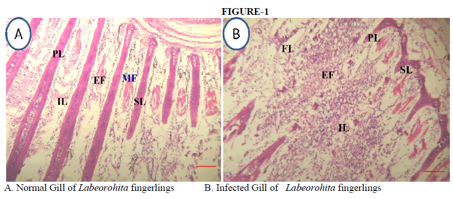
GILLS |
| Histological observations on gills of fingerlings fed with a control diet showed the normal architecture of gill filaments such as primary lamellae, secondary lamellae with mucus cells lying scattered on both sides. Whereas the gills of infected fingerlings showed proliferation of filamentary epithelium, lamellar fusion, loss of secondary lamellae, swelling of inter lamellae and excessive secretion of mucus on the surface of filaments. However the fingerlings fed with group 3 probiotic diet for a period of 30 days and challenged with Vibrio cholerae did not showed any deformities in the gills. Fingerlings of the gill supported the normal architecture as seen in the control fingerlings. The gills of both studied fish showed degenerative, necrotic and proliferative changes in gill filaments and secondary lamellae, edema in gill filaments and secondary lamellae and congestion in blood vessels of gill filaments. These pathological changes may be a reaction to toxicants intake or an adaptive response to prevent the entry of the pollutants thorough the gill surface. The observed alterations like proliferation of the epithelial cells, partial fusion of some secondary lamellae and epithelial lifting are defense mechanisms, since, in general, these result in the increase of the distance between the external environment and the blood and thus serve as a barrier to the entrance of contaminants . The cellular damage observed in the gills in terms of epithelial proliferation, separation of the epithelial layer from supportive tissues and necrosis can adversely affect the gas exchange and ionic regulation (FAS Mohamed - |
 |
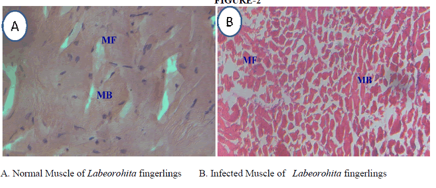
MUSCLE |
| Muscles of fingerlings (control) showed the normal arrangements of muscle fibers and muscle bundles . However, infected fingerlings showed many deformities such as degenerated muscle fibers and edema in the muscle tissu gradual infiltration of macrophages, muscular necrosis and fragmentation of muscle fibers . However the pre groups group 3 probiotic did not show any marked deformities in the muscles. Histology of the muscles showed the normal architecture as seen in the control fingerlings |
 |
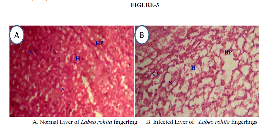
LIVER |
| The liver of Labeo rohita fingerlings fed with control diet showed large hepatic cells, polygonal in shape with almost centrally placed nuclei. Hepatic plate, hepatocytes with prominent central vein, sinusoids and nuclei were also observed. Liver obtained from infected fingerlings hepatic plate, granulation in cytoplasm, necrosis, vacuolization and disruption of hepatocytes . However the fingerlings fed with D3 probiotic diet and challenged with of control fingerlings. |
 |
KIDNEY |
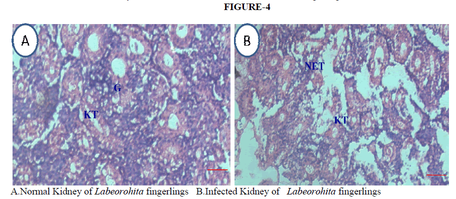
| Fingerlings challenged with Vibrio prominent tubular epithelium, glomerulus with Bowman’s capsule and inter bacterial treatment showed invading lymphocytes, hyperplasia of interregnal ce in tubular cells, vacuolation of kidney tubules, necrosis of epithelial lining and depletion of lymphoid cells in kidney. However the pre-treated groups 3 probiotic did not show any marked deformities in the kidney tubules, observation on kidney showed the normal architecture as seen in fingerlings fed with control diet. |
 |
DISCUSSION |
| In the present study histological alterations were documented for different tissues namely gills, muscle, liver and kidney of Vibrio cholerae infected rohu fish Labeo rohita fingerlings, fed with probiotic supplemented diets. Histological observations on gills of control and probiotic supplemented diets fed fingerlings showed normal architecture of primary and secondary lamellae. Whereas fingerlings infected with Vibrio cholerae in gills showed fusion and loss of secondary lamellar epithelium. The pathological effects included an increase in the thickness and the damage to the mucosa. The parasitic infection in turn disturbed the metabolic pathways (Esch et al.,1977). Similarly muscle of Labeo rohita fingerlings, infected with Vibrio cholerae showed fragmentation of muscle fibers and necrosis. The histopathology of muscle duplicated progressive damage in the structure of muscle with increasing concentrations of the sago effluent. Similar observations were reported by Nagarajan and Suresh (2005) in the muscle tissue of the fish Cirrhinus mrigala with increasing concentrations of sago effluent. Sakr and Gabr (1991;) Das and Mukherjee (2000) have studied the effect of different pollutants on fish muscles. |
| The other vital organ the liver also showed changes and damage to the hepatic cells due to bacterial infection. Histophathological changes such as vaculation, necrosis and nuclear condensation in hepatocytes were evident in the fingerlings infected with Vibrio cholerae. Fish liver can be regarded as the body’s detoxification organ and hence a target organ of various xenobiotic substances. A first level screening to identify potential pollutant exposure and effect can be accomplished on the basis of overt and relatively simple measures of condition. Such measures may serve to identify the most sensitive members of a fish population. In addition, they may provide information on energy reserves and possibly the ability of animals to tolerate toxicant challenges along with other environmental stresses (Mayer et al., 1992). |
| Histology of kidney showed a normal architecture in the fingerlings fed with supplemented diets. Whereas, control infected fingerlings showed dark granule accumulation in tubular cells, vaculation in kidney tubules and necrosis. The kidney is one of the first organ to be affected by contaminants in the water (Thophon et al.,2003). Most common alterations found in the kidney of fishes exposed to water contamination are tubule degeneration (cloudy swelling and hyaline droplets) and changes in the corpuscle, such as dilation of capillaries in the glomerulus and reduction of Bowman´s space (Takashima and Hibiya, 1995). |
V.ACKNOWLEDGEMENT |
| I like to thank UGC-BSR fellowship Delhi for providing financial support to carry out the work. |