International Journal of Innovative Research in Science, Engineering and Technology
ISSN ONLINE(2319-8753)PRINT(2347-6710)
ISSN ONLINE(2319-8753)PRINT(2347-6710)
Dr. Rajendra D. Kanphade1, Devesh D. Nawgaje2
|
| Related article at Pubmed, Scholar Google |
Visit for more related articles at International Journal of Innovative Research in Science, Engineering and Technology
This paper presents a novel approach to implement Evolutionary Computing (EI) techniques like Fuzzy Logic, Genetic Algorithm, Neural Network, and Adoptive Neuro-Fuzzy Inference System (ANFIS) for diagnosis of cancer using TMS320C6713 (Texas Instruments) DSP (Digital Signal Processor). The simulator has been developed using MATLAB and Neurosolution, while implementation has been done using code composer studio for TMS320C6713 DSP. Performance is compared by considering the metrics like accuracy of diagnosis and mean square error. The simulation and implementation result show that this EI approach can be effectively used for cancer detection to help oncologist to enhance the survival rates significantly.
Keywords |
| Evolutionary Computing, Fuzzy Logic, Genetic Algorithm, Neural Network, Adaptive Neuro Fuzzy Inference System, Digital Signal Processor. |
INTRODUCTION |
| Evolutionary computing system focus on systems that use knowledge based techniques to support human decisionmaking, learning and action. Such systems are capable of cooperating with human user and so the quality of support given and the manner of its presentation are important issue [1,2]. Evolutionary Computing is dedicated to system solutions based on soft computing techniques. It provides rapid dissemination of important results in evolutionary computing technologies, a fusion of research in genetic programming, neural science, neural net systems, fuzzy set theory, fuzzy systems, chaos theory and chaotic systems. Evolutionary computing encourages the integration of soft computing techniques and tools into both everyday and advanced applications. [6] Cancer is a group of diseases in which cells in the body grow, change, and multiply out of control. Usually, cancer is named after the body part in which it originated; thus, breast cancer refers to the erratic growth and proliferation of cells that originate in the breast tissue. A group of rapidly dividing cells may form a lump or mass of extra tissue. These masses are called tumors. Tumors can either be cancerous (malignant) or non-cancerous (benign). Malignant tumors penetrate and destroy healthy body tissues. A group of cells within a tumor may also break away and spread to other parts of the body. Cells that spread from one region of the body into another are called metastases.[4][5] A major class of problem in medical science involves the diagnosis of disease, based upon various tests performed upon the patient. When several tests are involved, the ultimate diagnosis may be difficult to obtain, even for a medical expert. This has given rise, over the past few decades, to computerized diagnostic tools [3], intended to aid the physician in making sense out of the confusion of data. |
| A. Related Work |
| Fuzzy logic, Neural Network, Genetic algorithm and adaptive neuro fuzzy inference system approaches have been used earlier in the computerized diagnostic system for cancer [5][7-14]. After experiments with Fuzzy logic, ANN and ANFIS they achieved a classification accuracies ranging from 50% to 90%. The work in this paper is aimed to find not only a better accuracy and usage of Evolutionary computing approach in diagnosis of cancer but also its implementation on TMS320C6713 digital signal processor. |
II. TMS320C6713 DSP |
| The C6713 device is based on the high-performance, advanced very-long-instruction-word (VLIW) architecture developed by Texas Instruments. The TMS320C62x is a 16-bit fixed point processor and the „67x is a floating point processor, with 32-bit integer support. The C6713 DSK is a low-cost standalone development platform that enables users to evaluate and develop applications for the TI C67xx DSP family. The DSK also serves as a hardware reference design for the TMS320C6713 DSP [20]. In our work we have develop a program of segmentation using c-code. After writing the code, the next step is to compile the code to machine language. The Build command will compile all the files that are included in this project and make an executable file for the DSP. Finally, to run the program, load the executable file (.out) that the compiler generated into the DSP and run the file loaded into DSP. The generated output of DSP is then open code composer studio. |
III. IMPLEMENTATION OF FUZZY LOGIC FOR CANCER DIAGNOSIS |
| The algorithm described in this paper is based on the subjection of a set of four pixels, part of a 2x2 window of an image to a set of fuzzy conditions which help to highlight all the edges that are associated with an image. The fuzzy conditions help to test the relative values of pixels which can be present in case of presence on an edge. So the relative pixel values are instrumental in extracting all the edges associated to an image. The system implementation is carried out considering that the input image and the output image obtained after defuzzification are both 8-bit quantized; this way, their gray levels are always between 0 and 255. The fuzzy sets are created to represent each variable‟s intensities; these sets are associated to the linguistic variables “Black”, Edge and “White”. The adopted membership functions for the fuzzy sets associated to the input are trapezoidal and to the output are triangular. The functions adopted to implement the “and” and “or” operations are the minimum and maximum functions, respectively. The Mamdani method is chosen as the defuzzification procedure, which means that the fuzzy sets obtained by applying each inference rule to the input data are joined through the add function; the output of the system is then computed using weighted average method of the resulting membership function. The values of the three membership functions of the output are designed to separate the values of the blacks, whites and edges of the image. The inference rules depend on the weights of the four neighbour gray level pixels, if the neighbours‟ weights are degree of blacks or degree of whites. The powerful of these rules is the ability to extract all edges in the processed image directly. This study is assaying all the pixels of the processed image by studying the situation of each neighbour of each pixel. The condition of each pixel is decided by using the floating 2x2 mask which can be scanning the all grays. In this location, some of the desired rules are explained. The proposed system is tested with different images. The algorithm to find the edges associated with cancer cells had been introduced which has been instrumental to abridge the concepts of fuzzy logic and digital image processing. The FIS algorithm has tremendous scope of application in the area of cancer image processing. The image edge detection and segmentation using FIS algorithm has been successful in obtaining the edges of the cancer affected cells that are present in medical images. FIS is implemented on TMS320c6713 DSP kit. Outputs have been shown to make to understand the accuracy of the algorithm and which helps to detect exact shape of cancer cells.[5][19] |
| This paper adopts OSTU Principle as the image segmentation standard. In this method, we can directly calculate the threshold without pretreatment to histogram. This algorithm is simple and is a remarkable method for selecting the threshold. Here‟s the fundamental principle. The gray value of a grey-scale map is 0~255.The total number of pixels is defined as N, ni is the number of pixels which‟s gray value is i. By normalizing the histogram, the following equations could be obtained. |
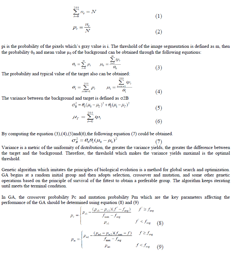 |
| fmax the maximum fitness; favg the average of the fitness; f‟ the larger fitness of the two individual which will take the cross-operation. f the fitness of the individual. GA gives better result for the segmentation of mammogram image which is helpful for the diagnosis of cancer treatment. Simulation is done on CCS whereas implementation is done on TMS320C6713 digital signal processor on number of images. The average computing time for simulation of GA using Intel core I3, 2.13GHz processor is 8.39 minutes, whereas computing time using TMS320C6713, 225MHz DSP is 24 seconds which shows the implementation using DSP decreases run time greatly.[20] |
V. ARTIFICIAL NEURAL NETWORK |
| ANNs are massively distributed parallel processing systems made up of highly interconnected neural computing elements that have the ability to learn and thereby acquire knowledge and make it available for use [15,16,17]. Neural networks are applicable in virtually every situation in which a relationship between the predictor variables (independents, inputs) and predicted variables (dependents, outputs) exists, even when that relationship is very complex and not easy to articulate in the usual terms of “correlations” or “differences between groups”[21,22]. In this paper Jordan and Elman network is used. |
| A. Simulation models |
| Jordan and Elman network and ANFIS were evaluated on the same data set. The data set consist of 125 clinical cases. All cases are first segmented based on fuzzy logic and after feature extraction database was generated. Out of 125 clinical cases 90 cases are used for training the network, 20 cases are used for cross validation and 15 cases are used for testing. Each case has to be categorized into either of the two categories: Benign or malignant |
| B. Architecture of Network Used Jordan and Elman network is train using I/P PEs 10, O/P PEs 2, Hidden Layers = 2, for Both Hidden Layers & O/P Layer Transfer Function TanhAxon, Learning Rule Momentum, Momentum = 0.7, Maximum Epoch 2000 C. Jordan and Elman networks Jordan and Elman networks extend the multilayer perceptron with context units, which are processing elements (PEs) that remember past activity. Context units provide the network with the ability to extract temporal information from the data. In the Elman network, the activity of the first hidden PEs are copied to the context units, while the Jordan network copies the output of the network. Networks which feed the input and the last hidden layer to the context units are also available. Figure 1 shows the training MSE versus Epoch graph of Jordan and Elman network |
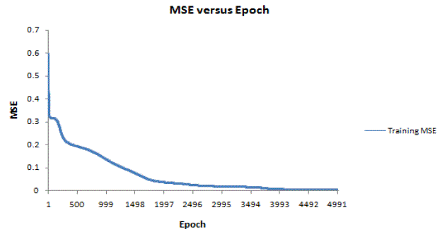 |
| Figure 1: Training MSE versus Epoch graph of Jordan and Elman networks |
VI. NEURO FUZZY SYSTEM |
| Neuro Fuzzy (NF) computing is a popular framework for solving complex problems and is an integrated system combining the concept of FIS (Fuzzy Inference System) and ANNs. ANFIS is a fuzzy inference system implemented in the framework of adaptive networks, and hence has the advantages of both FIS and NNs.[23,24] A. ANFIS Structure Figure 2 shows the structure of ANFIS. The first and fourth layers in the ANFIS are learning rules, used to tune the model adaptive layers. The first layer contains premise parameters which characterize the shape of the input membership functions. These parameters are associated with the IF part of the rule. The fourth layer also has consequent parameters, which are associated with the THEN part of the rule. Suitable learning rules are used to tune the model parameters. A hybrid learning algorithm used in ANFIS combines the gradient descent and the least squares method for an effective search for the optimal parameters. In this procedure, an ANFIS system can learn from the existing data with correct output and possibly predict a result with new input set by tuning its internal node values |
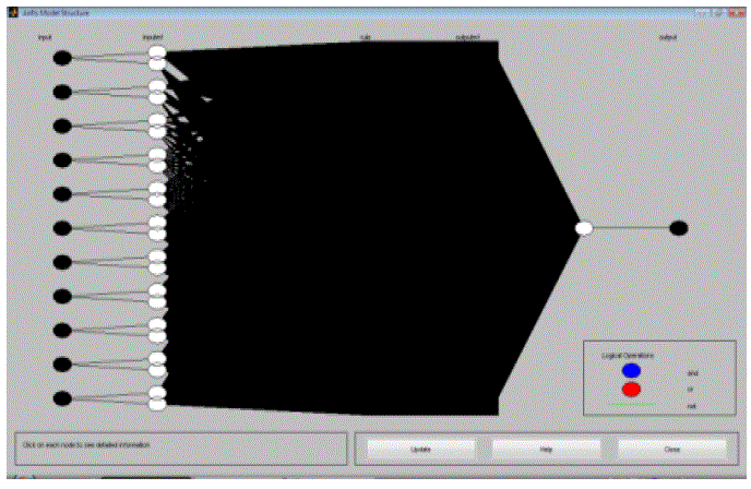 |
| Figure 2 ANFIS structure |
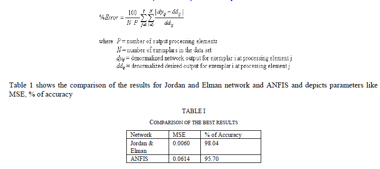
VII. PERFORMANCE COMPARISON |
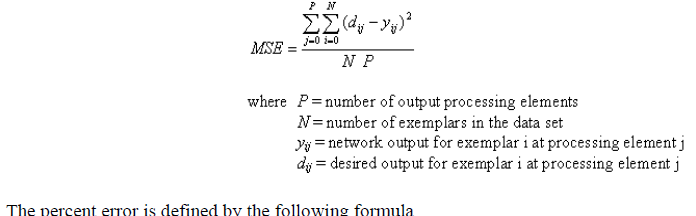
| The Performance Measures component provides two values that can be used to measure the performance of the network for a data set The mean squared error is simply two times the average cost. The formula for the mean squared error is |
 |
 |
VIII. CONCLUSION |
| The success of an image analysis system depends on the quality of segmentation. In the analysis of medical images for computer-aided diagnosis and therapy, segmentation is often required as a preliminary processing task. In this paper fuzzy logic and genetic algorithm is used to select the threshold in cancer image segmentation. Experimental data demonstrate the accuracy of segmentation which helps doctor for diagnosis purpose. Moreover the hardware implementation of both algorithm on DSP TMS320C6713 is achieved successfully. Whereas Jordan and Elman network and ANFIS were develop for differentiating the malignant and benign cancer and run on Matlab for the medical images obtained from the radiologist. Outputs have been shown to make to understand the accuracy of the algorithm and which helps us to detect the cancer. After the training of Jordan and Elman network and ANFIS, an unknown images was taken and tested for malignant or benign. After working, we find Jordan and Elman network has achieved a top result of accuracy 98.04%. |