International Journal of Innovative Research in Science, Engineering and Technology
ISSN ONLINE(2319-8753)PRINT(2347-6710)
ISSN ONLINE(2319-8753)PRINT(2347-6710)
Raman Gupta1, Sandeep Singh1, Kashish Garg1 and Shruti Jain2
|
| Related article at Pubmed, Scholar Google |
Visit for more related articles at International Journal of Innovative Research in Science, Engineering and Technology
This paper provides electronic implementation of electrocardiograph (ECG) circuit by using instrumentation amplifier (IA) as bio-potential amplifier in such a manner which reduces noise, common voltage, DC offset value and RF interference from the existing circuit.Noise and common voltage can be removed from ECG using driven right leg circuit or by using isolator circuit. DC offset can be removed by using integrator as feedback. In the differential amplifier part of IA, we can add single resistance, T-network or inverter circuit with integrator to improve impulse response. By using filters, we can reduce RF interference. In this paper, we have used instrumentation amplifier as a bio-potential amplifier
Keywords |
| ECG, Bio-potential amplifier, Driven right leg circuit, DC offset, Common mode rejection Ratio, Filter. |
INTRODUCTION |
| An electrocardiogram (ECG or EKG) is the measurement and graphical representation of electrical signals associated with the human heart. Applications of an ECG range from monitoring heart rate, heartbeat, heart rhythm to the diagnosis of specific heart conditions. The basics of ECG measurement are the same for all applications, but there can be variation in the methods and representation of the circuit. |
| All ECGs pick up heart signals through electrodes connected externally to specific locations on the body i.e. arms and legs. Then the body generates the heart signals which are of few mill volts amplitudes. The specific locations of the electrodes allow the heart's electrical activity to be viewed from different angles, each of which is displayed as a channel on the ECG printout. The channels are commonly referred to as "leads" and the number of leads varies from 1 to 12 depending on the application [1]. 12-leadECG are recorded using right arm (RA), left arm (LA), left leg (LL), right leg (RL), and chest (C) electrodes. Standard lead system can be divided into two planes i.e. frontal and transverse plane. They comprise a combination of electrodes taking measurements from different regions and can be further divided into bipolar limb leads, unipolar leads and the chest leads. Bipolar limb leads derive signals from electrodes on the limbs, and are designated as leads I (RA to LA), II (RA to LL), and III (LA to LL). Unipolar leads are designated as aVR, aVL, and aVF, and can be designed by connecting RA, LA and LLrespectively to non-inverting terminal and remaining two electrodes to inverting terminal of IA. The remaining six leads, V1, V2,…V6, are chest leads [2]. In this paper we are using lead I ECG system. |
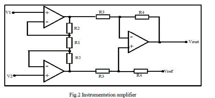
| The basic design of a bio-potential amplifier consists of an instrumentation amplifier. The amplifier should possess several characteristics, including high amplification, high input impedance [3], high common mode rejection ratio (CMRR) and the ability to reject electrical interference, all of which are needed for the measurement of these biopotentials [4]. Our aim in this paper is to reduce the electronic circuit of ECG as simple as possible. |
RELATED WORK |
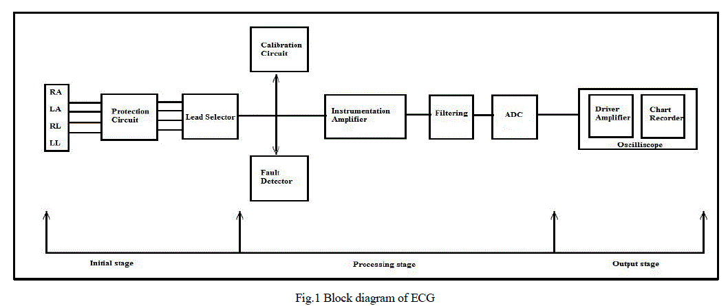
| In this section we will discuss the existing functional blocks of ECG as shown in fig. 1. |
| a. Protection circuit: This circuit includes protection devices so that the high voltages that may appear across the input to the electrocardiograph under certain conditions do not damage it. |
| b. Lead selector: Each electrode connected to the patient is attached to the lead selector of the electrocardiograph. The function of this block is to determine which electrodes are necessary for a particular lead and to connect them to the remainder of the circuit. It selects one or more leads to be recorded. |
 |
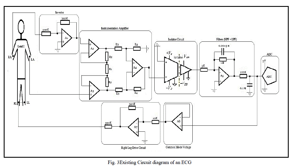
| On the basis of block discussed in fig. 1, internal circuit diagram is shown in fig. 3 which is already discussed in several papers[5] [6]. But this circuit diagram has problems associated with it. They are mentioned below: |
| 1. No need of inverter at RA. |
| 2. Presence of noise and common mode values. |
| 3. DC offset and suppression. |
| 4. Separate filters at the end are making circuit complex. |
| All these problems are explained and resolved further in this paper. |
 |
MATERIALS AND METHODS |
| Removal of inverter: As for lead I, RA (right arm) is at negative terminal and LA (left arm) is at positive terminal so, instead of using inverter after RA we put this point at the negative terminal of instrumentation amplifier. |
| Removal of noise and common mode values: Followings are the methods to remove noise and common mode values from ECG waveform to increase CMRR. |
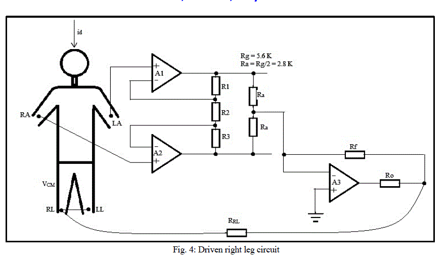
| a) Driven right leg system (Feedback loop to reduce noise): This circuit provides a reference point on the patient that normally is at ground potential [7]. This connection is made to an electrode on the patient’s right leg as shown in Fig.4. Right leg driver circuit is used in a feedback configuration to reduce 60 Hz noise and drive noise on patient to a lower level. VCM (common mode voltage) is given by eq. 2. |
 |
 |
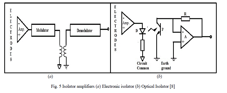
| b) Isolation amplifier: It is used to isolate patient from high voltages and currents to prevent electric shock where there is specifically a barrier between passage of current from the power line to the patient. It can be done by two ways i.e. electrical isolation and optical isolation. Electrical isolationcan be done by inserting a transformer in the signal path (Fig. 5(a)). It limits the possibility of the passage of any leakage current from the instrument in useto the patient. Optical isolation can be done by introducing an opticalcoupler (Fig. 5(b)).The electric signal from the amplifier is first converted to light by a light-emitting diode (LED).This optical signal is modulated in proportion to the electric signal, and transmitted to the detector.[8] |
 |
| This method is bit costly so we prefer to use driven right leg circuit for removal of noise and common mode values. |
| Removal of DC offset and DC suppression: DC-offset and DC-suppression are key parameters in bioelectric amplifiers. Bioelectric amplifiers require a high gain level, a low density of equivalent input noise, a high common mode rejection ratio (CMRR) [9] and a high-impedance input. Most of these features can be achieved by using an instrumentation amplifier (IA) as a front stage. But some DC voltage levels appear at the output of the IA. These levels are produced by several factors such as impedance imbalance from the input electrodes, electrode contact potentials and input bias currents. These DC levels must be removed; otherwise, they would produce output saturation phenomena when amplified in the subsequent stages. Several techniques have been developed to remove the DC levels. These are explained below: |
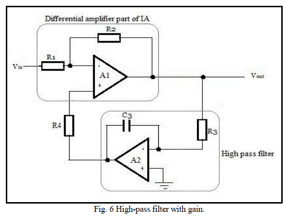
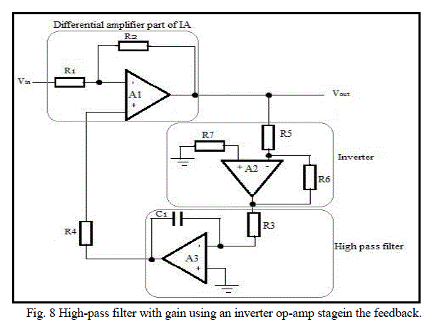
| a) As shown in Fig.6, the stage is a high-pass filter with gain, cascaded after the instrumentation amplifier [10].This circuit consists of feedback integrator which acts as low pass filter and used to eliminate DC level. Hence when feedback is applied, the DC component is eliminated at output voltage and whole stage thus behaves as a HPF with gain G. As input voltage is given on inverting terminal so this behaves like an inverter circuit which will amplify the input. Eq. 3 shows transfer function for the circuit shown in fig. 6. |
 |
 |
| Filters: Sometimes there is RF interference in circuit so to reduce it we need to add LPF [11]. Filtering should be included in the front end of the instrumentation amplifier. As shown in Fig.9, low pass filter is adjusted with instrumentation amplifier. Now with this there is no need of filtering after IA. |
 |
PROPOSED CIRCUIT DIAGRAM OF AN ECG |
| Fig. 10 shows the proposed circuit diagram of ECG system. This circuit diagram comprises of instrumentation amplifier with pre-amplifier circuit and right leg drive circuit to reduce noise and common mode value from circuit, high-pass filter with gain using an inverter op-amp stage in the feedback to block DC offset value. The final circuit diagram is small and compact in comparison with the existing circuit. |
CONCLUSION |
| This paper proposes the electronic implementation of ECG circuit by using instrumentation amplifier as bio-potential amplifier. This paper also explains the several techniques to reduce noise and to increase CMRR by using driven right leg circuit. With the help of high pass filter with gain G, we can reduce DC offset.RF interference can be reduced by filtering. At the end we have combined all the parts and made the existing circuit compact in size. In future we will check all the electrical parameters with the help of this circuit. |
 |
References |
|