Osteopoikilosis is an asymptomatic osteosclerotic dysplasia initially described by Albers Schönberg in 1915. Osteopoikilosis is a rare disease with an estimated incidence of 1/50,000 and with an unknown etiology. Osteopoikilosis can have a very typical radiographic appearance and distribution; however, diverse clinical associated pathologies can lead to diagnostic uncertainty and the need for further investigation. We emphasize on the radiological characteristics of this condition with clinical presentations and associated dysplasias. The extensive search was conducted using an indexed search database (Pubmed and Cochrane) using MeSH terms, the relevant articles were chosen by title, abstract, language and publication date. The importance of its differential diagnosis is stressed with relevant review of the literature. When uniform multiple radio-dense round or oval sclerotic lesions in a periarticular distribution are found on radiographic examination, osteopoikilosis must be in the differential diagnosis. Osteopoikilosis should not be considered a coincidental radiographic finding, but rather part of a systemic disorder, as several developmental dysplasias coexisting with this disorder has been reported.Radiologically, the differential diagnosis are Enostosis, dysplasias of endochondral bone formation including osteopetrosis (Albers-Schönberg disease), pycnodysostosis and osteopathia striata (Voorhoeve disease), each with varying radiologic appearance. Although osteopoikilosis is generally considered an incidental finding, several developmental dysplasias coexisting with this disorder must be considered and excluded. In patients with a known or suspected malignancy, radionuclide bone scan has a critical role in distinguishing osteopoikilosis from osteoblastic bone metastases.
Keywords |
| Osteopoikilosis, dysplasias, malignancy |
Introduction |
| Osteopoikilosis (osteopathia condensans disseminata, spotted bones) is an asymptomatic osteosclerotic dysplasia initially described by Albers-Schönberg in 1915 [1-3]. Osteopoikilosis associated with benign conditions can have a very typical radiographic appearance and distribution; however, with diverse clinical associated pathology can lead to diagnostic uncertainty and the need for further investigation. |
METHODS |
| The extensive search was conducted using an indexed search database (Pubmed and Cochrane) using MeSH terms, the relevant articles were chosen by title, abstract, language and publication date. We review the literature available on osteopoikilosis with focus on characteristics, associated benign conditions, relevant differential diagnosis and diagnostic workup. |
DISCUSSION |
| Osteopoikilosis is a rare disease with an estimated inci-dence of 1/50,000 [1-3] and with an unknown etiology [1, 2]. Actual incidence is not known[1] since the disease is asymptomatic, actual incidence may be under-reported). It appears with variable onset in both sexes regardless the age but preferably adult males. The lesions have been described in all age groups, and although prevalence studies have shown a higher frequency among men, the apparently unequal sex distribution may be a result of referral bias in the literature (men are more likely than women to present to hospital with traumatic injuries requiring radiologic investigation [4, 5] |
Pathogenesis |
| A hereditary failure (abnormality in endochondral bone maturation process and collagen regulation) to form normal trabeculae along lines of stress is blamed in the pathogenesis [6]. Furthermore, an altered osteogenesis may be responsible for the lesions [7]. Benli et al. studied epidemiological, clinical and radiological features of the disease in 53 patients from four families to evaluate its genetic transmission [1]. |
| Osteopoikilosis exists in hereditary (autosomal dominant transmission) and sporadic forms [7-9] and is one of several bone dysplasia’s characterized by defective endochondral bone formation [10]. It is more frequent in self-contained communities were consanguineous marriages are frequent [8, 11]. Buschke-Ollendorff syndrome is an autosomal dominant, generally benign, combination of osteopoikilosis and connective tissue nevi (dermatofibrosis lenticularis disseminata) or juvenile elastoma [12, 13]. |
| Familial clustering suggests a dominant inheritance, associated with LEMD-3 gene, responsible for the disease, featured by abnormality in enchondral bone maturation process [12, 14-19]. The LEMD-3 gene codes for a protein of the inner nuclear membrane that inhibits both the BMP and TGF-b signaling pathways [14, 20]. |
| Endochondral ossification refers to the formation of the long and flat bones, which begins from a primitive hyaline cartilaginous model. This process is in contrast to intramembranous ossification, which refers to direct transformation of condensed mesenchymal cells into cortical bone without a cartilaginous phase, as is typically seen in the formation of the skull bones [10]. The condensation of cancellous bone in osteopoikilosis consists of a peripheral area of trabeculae in which osteocysts are scant and there are no osteoblasts and osteoclasts, together with a central core of irregular trabeculae, in which both osteoblasts and osteoclasts are present. The lesions appear to be metabolically active, and they become denser with time, but later their size may change or they may even disappear. The precise origin of these abnormal areas remains debatable, but they appear to represent foci of deranged differentiation in cancellous bone [21, 22]. |
Clinical and radiographic presentation |
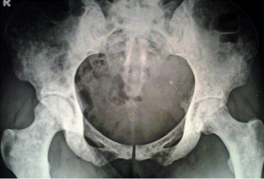
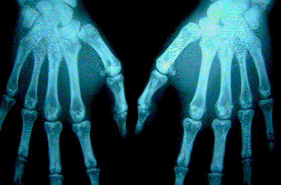
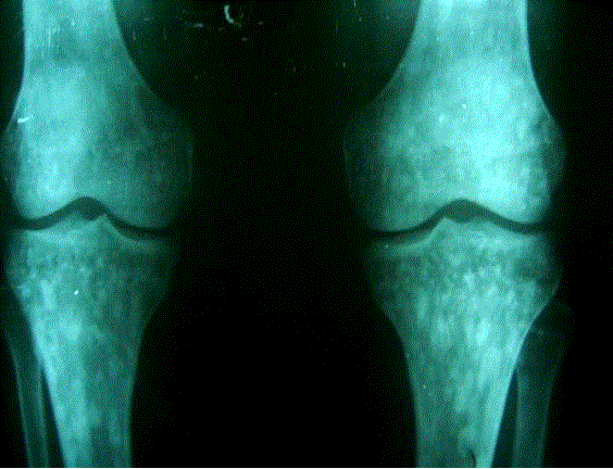
| This rare hereditary condition is usually noticed radiologically as an incidental finding of diagnostic sclerotic lesions identified during the investigation of unrelated problems in which there is no clinical history suggestive of either malignant or systemic disease [23, 24]. These lesions are symmetric, numerous, small, well-defined, homogeneous and circular or ovoid [25]. The most frequently involved sites are the epiphyses and metaphyses of long tubular bones and the carpi, tarsi, pelvis and scapulae [1]. |
| According to an epidemiologic study on 53 patients with osteopoikilosis members of four families, Benli et al[1], noticed that the most frequent sites for op appearance were the phalanges (100%), carpal bones (97.4%), metacarpals (92.3%), phalanges of the foot (87.2%), metatarsals (84.4%), tarsal bones (84.6%), pelvis (74.4%), femur (74.4%), radius (66.7%), ulna (66.7%), sacrum (58.9%), humerus (28.2%), tibia (20.5%) and fibula (12.8%). It is generally accepted in the literature that it is more frequently located in long bones and pelvis and male to female ratio is 3:2 [2, 3]. |
| Although osteopoikilosis is generally considered an incidental finding, several developmental dysplasias coexisting with this disorder have been reported [26]. Izge Gunal et al. reported a family in whom various members had osteopoikilosis with 5 different associated lesions and suggest that osteopoikilosis is a bone manifestation of a generalized fibroproliferative or stenosing disease [27]. |
Diagnostic work up of radiographic lesions |
| When Osteopathia condensans disseminata or spotted bones presenting with sclerotic lesions of bone are areas of increased bone density seen on plain radio graphs, it is necessary to consider the following three elements: the appearance of the spots, their number, and their location and distribution. |
| As mentioned previously, the characteristic radiologic feature in osteopoikilosis is multiple, punctate, sclerotic, rounded or oval foci. The multiple benign lesions of osteopoikilosis tend to have a characteristically symmetrical periarticular distribution within the epiphyseal and metaphyseal regions of the axial and appendicular skeleton that is virtually diagnostic of the condition. Osteopoikilosis has not been described in the skull. |
| Assessment of the radiograph as well the patient’s clinical history is highly relevant to the workup of bone lesions. The diagnosis of osteopoikilosis is clear when the characteristically benign-looking periarticular spots are found incidentally in an otherwise healthy person or in a patient presenting with a traumatic injury. Occasionally, OP can be painful without causing any deformity or dysfunction at the location site [74]. Although a benign condition, it may lead to diagnostic problems when the patient undergoes diagnostic imaging of the skeletal system due to various reasons like malignancy [28]. |
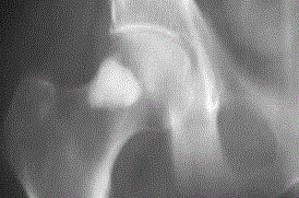
| In benign Enostosis, or bone islands, which also have sclerotic rounded lesions, the peripheral margins tend to blend or merge with the underlying normal trabeculae (Figure 3). These lesions are typically small (< 1 cm) and frequently isolated. Although the lesions in Enostosis are usually round or oval, their shape is not specific. If the lesions are larger than 1 cm, other sclerotic bone conditions or lesions must be considered, despite the characteristic appearance of the peripheral margins. If sclerotic lesions are large or numerous, the diagnosis of osteopoikilosis or Enostosis is questionable. In addition, bone islands should be blandly homogeneous, as they represent a hamartoma (i.e., cortical bone where medullary bone should be). |
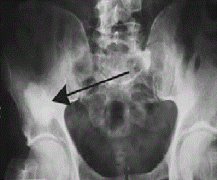
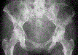
| Nonhomogeneous or multiple lesions are cause for concern, as they may indicate osteoblastic activity related to an underlying marrow-replacing disease such as metastasis (Figure 4). Metastases tend to be more frequent in the pelvis and spine. Metastatic disease can affect any bone, but it predominates in the axial skeleton, is rarely seen below the knee or elbow and tends not to follow a periarticular distribution. In these situations, a review of previous imaging is essential to show that the lesion has remained stable over time. In patients with a known or suspected primary malignancy, radionuclide bone scan has a critical role in distinguishing osteopoikilosis from osteoblastic bone metastases when previous images are not available [29, 30]. Although osteopoikilosis may have the appearance of sclerotic metastatic disease to the bone on plain films and CT, the lesions are classically inactive on scintigraphy [31]. There have been a few cases reported in literature of abnormal bone scans in patients with osteopoikilosis [32-34]. In such cases, the pattern of increased uptake is usually symmetric and localized to the distal ends of tubular, carpal, and tarsal bones. This may perhaps correspond to active bone remodeling of these multiple foci, which has also been reported in pathologic specimens of osteopoikilosis [6]. In addition, fluoro-deoxyglucose positron emission tomography scan is normal and can further support the case for benign bone lesions [31]. |
| Although uptake of 99mTc is unusual in osteopoikilosis and bone islands, it is commonly observed in connection with metastasis, sarcoidosis, mastocytosis and tuberous sclerosis [33, 35-38]. Such conditions are associated with systemic illness in addition to their radiologic manifestations, and their appearance is usually clinically overt by the time skeletal changes occur [36-38]. However, Mungovan et al. suggest that an abnormal bone scan does not exclude the diagnosis of osteopoikilosis in young patient if the roentgenographic findings are characteristic of that entity [39]. Although there have been reports describing uptake of 99mTc in patients with osteopoikilosis and Enostosis, such false-positive results are uncommon and are usually seen only in patients with larger lesions [1-3, 28]. The uptake may be due to active remodeling of the bone, i.e., a process similar to the formation of callus [2]. |
Differential Diagnosis (Table 1) |
| Radiologically, the differential diagnosis are enostosis, dysplasias of endochondral bone formation include osteopetrosis (Albers-Schönberg disease), pycnodysostosis and osteopathia striata (Voorhoeve disease), each with varying radiologic appearances [10, 14]. |
| Lagier et al. documented that the radiological appearance of rounded and linear densities in osteopoikilosis corresponded to old and inactive remodeling of spongy trabeculae in epiphyseal and metaphyseal locations [6]. On histological examination, these lesions were found to be composed of lamellar osseous tissue containing haversian systems. It was suggested that the lesions were not probably formed through enchondral ossification of cartilage rests. |
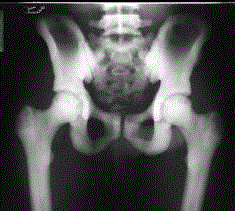
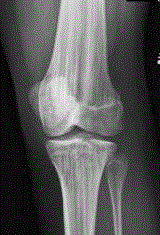
| Enostosis histopathologically and radiographicaly most closely resembles osteopoikilosis and the difference is that bone islands can be isolated and small. If multiple, they are usually scattered and do not display a characteristic periarticular distribution. Histopathologically, the lesions consist of compact, markedly hypertrophied trabeculae composed of lamellar osseous tissue [40]. Osteons within a bone island are not regularly oriented and contain well-vascularized canals surrounding narrow rings of lamellae, but which are empty of osteoclasts and osteoblasts [40]. Both osteopetrosis (Figure 5) and pycnodysostosis have a very different radio graphic appearance from osteopoikilosis; the lesions appear in a more diffuse pattern of sclerosis, rather than spotted or rounded [8]. These patients also have severe systemic manifestations. Osteopathia striata (Figure 6) also has a different appearance. The sclerotic areas within the bone are neither round nor oval. Instead, they are linearly striated and periarticular in distribution [40-42]. Clinical manifestations of this disorder are subtle or nonexistent. Histopathologic and biochemical studies are able to differentiate osteopetrosis and pycnodysostosis from osteopoikilosis, but little pathologic data are available on osteopathia striata [14, 33]. Fortunately, all of these disorders can be diagnosed and differentiated readily by their radiologic features.[43] |
Associated Syndromes (Table 2) |
| Osteopoikilosis is usually incidentally found on radiographic examination, but it can be rather part of a systemic disorder. In approximately in 25% of cases, whitish fibrocollagenous infiltrations (Buschke-Ollendorf Syndrome) are found [12, 14, 16, 44-53]. Hasan et al. reported case of synovial chondromatosis associated with osteopoikilosis [54]. If cellular activity exists in the foci of osteopoikilosis, it is conceivable that a malignant lesion could develop [55, 56]. This is also true of synovial chondromatosis [55, 56]. By taking into account the similarities, we can speculate that chondromatosis is the synovial manifestation of osteopoikilosis (synosteopoikilosis) and increasing evidence suggests that, when osteopoikilosis is detected in a case, other associated disorder, especially lesions of fibroproliferative origin should be sought. Tander et al reported a case where etiology of the symptom was related to an osteopoikilosis and the MRI findings was confusing, which can mimic bone metastases and concluded that, in patients with similar findings of metastases of MRI should be carefully re-evaluated for the presence of osteopoikilosis [57-59]. |
| Spinal stenosis, dwarfism, dystocia and mild articular pain with or without joint effusion have been reported in patients with osteopoikilosis [60]. This disorder may resemble Osteoblastic metastases, mastocytosis and tuberous sclerosis [76]. Günal et al. have reported five members of a family with dacryocystitis with osteopoikilosis [61]. Overlap syndromes, formerly known as mixed sclerosing dystrophies, in which osteopoikilosis and other osteosclerotic bone disorders (such as fibrous dysplasia, osteopathia striata, and melorheostosis) are combined, must be borne in mind in the differential diagnosis. A combination of melorheostosis, OPK, and osteopathia striata (type I) is the most frequent among the other overlap syndromes [10, 62]. |
CONCLUSIONS |
| The discovery of bone spots on a radiograph is often disturbing, and benign conditions need to be differentiated from indicators of serious disease. Osteopoikilosis is one of several uncommon benign variants of bone formation that must be distinguished from more worrisome disorders, most notably osteoblastic metastases on the basis of the lack of internal architecture, irregular margins, the periarticular distribution and relative sparing of the skull. Although the natural course of this condition is benign and requires no treatment, and coexisting pathologic conditions require medical attention. Therefore, it is important that an accurate diagnosis be made. Despite the fact that osteopoikilosis is a very rare asymptomatic condition that most physicians are not familiar with, it is valuable to take it into consideration, particularly when diagnostic issues on bone radiography occur and severe pain at the adjacent joints co-exists. |
Key Message |
| • When uniform multiple radiodens round or oval sclerotic lesions in a periarticular distribution lesions are found on radiographic examination, osteopoikilosis must be in the differential diagnosis before invasive diagnostic procedures and dangerous unnecessary treatments are planned. |
| • No further investigation is required if the patient is otherwise healthy and there is no suspicion of systemic or metastatic disease. |
| • Although osteopoikilosis is generally considered an incidental finding, several developmental dysplasias coexisting with this disorder must be considered and excluded. |
| • Technetium 99m Scintigraphy is helpful in ruling out disorders that are of greater concern like bone metastasis. |
References |
- Benli IT, et al., Epidemiological, clinical and radiological aspects of osteopoikilosis. J Bone Joint Surg Br. 1992;74(4):504-6.
- Borman P, et al., Osteopoikilosis: report of a clinical case and review of the literature. Joint Bone Spine. 2002;69(2):230-3.
- Bicer A, et al., Coexistence of osteopoikilosis and discoid lupus erythematosus: a case report. Clin Rheumatol. 2002; 21(5):405-7.
- Szabo AD. Osteopoikilosis in a twin. Clin OrthopRelat Res. 1971;79:156-63.
- Ikonomow GK. [Osteopoikilosis in twins]. PrzeglLek. 1972;29(2):333-5.
- Lagier, R., A. Mbakop, and A. Bigler, Osteopoikilosis: a radiological and pathological study. Skeletal Radiol, 1984;11(3):161-8.
- Mindell ER, CS Northup, and HO Douglass Jr. Osteosarcoma associated with osteopoikilosis. J Bone Joint Surg Am. 1978;60(3):406-8.
- Sarralde A, et al. Osteopoikilosis: report of a familial case. Genet Couns. 1994;5(4):373-5.
- Agostinelli JR. Osteopoikilosis. A case report. J Am Podiatry Assoc. 1983;73(10):529-31.
- Vanhoenacker FM. et al., Sclerosing bone dysplasias: genetic and radioclinical features. Eur Radiol. 2000;10(9):1423-33.
- Butkus CE, et al. Melorheostosis in a patient with familial osteopoikilosis. Am J Med Genet. 1997;72(1):43-6.
- Mumm S, et al. Deactivating germline mutations in LEMD3 cause osteopoikilosis and Buschke-Ollendorff syndrome, but not sporadic melorheostosis. J Bone Miner Res. 2007;22(2):243-50.
- Ehrig T and CJ Cockerell. Buschke-ollendorff syndrome: report of a case and interpretation of the clinical phenotype as a type 2 segmental manifestation of an autosomal dominant skin disease. J American AcadDermatol. 2003;49(6):1163-1166.
- Hellemans J, et al. Loss-of-function mutations in LEMD3 result in osteopoikilosis, Buschke-Ollendorff syndrome and melorheostosis. Nat Genet. 2004;36(11):1213-8.
- Couto AR, et al. A novel LEMD3 mutation common to patients with osteopoikilosis with and without melorheostosis. Calcif Tissue Int. 2007; 81(2):81-4.
- Zhang Y, et al. Novel and recurrent germline LEMD3 mutations causing Buschke-Ollendorff syndrome and osteopoikilosis but not isolated melorheostosis. Clin Genet. 2009; 75(6):556-61.
- Baasanjav S, et al. Osteopoikilosis and multiple exostoses caused by novel mutations in LEMD3 and EXT1 genes respectively--coincidence within one family. BMC Med Genet. 2010;11:110.
- Lynch SA, et al. The 12q14 microdeletion syndrome: six new cases confirming the role of HMGA2 in growth. Eur J Hum Genet. 2011;19(5):534-9.
- Gurcay E, et al. Osteopoikilosis. A case report with a family screening. Saudi Med J. 2006; 27(12):1917-8.
- Hellemans J, et al. Germline LEMD3 mutations are rare in sporadic patients with isolated melorheostosis. Hum Mutat. 2006;27(3):290.
- Verbov J. Buschke--Ollendorff syndrome (disseminated dermatofibrosis with osteopoikilosis). Br J Dermatol. 1977;96(1):87-90.
- Ayling RM and PE Evans. Giant cell tumor in a patient with osteopoikilosis. ActaOrthop Scand. 1988;59(1):74-6.
- Padin-Paz EM and JA Diaz-Peromingo. Osteopoikilosis in the knee. CMAJ. 2011. 183(4): p. 461.
- Hii CH and CK Tan. Osteopoikilosis in the pelvic region. CMAJ. 2011;183(4):460.
- Young LW, I Gershman and PR Simon. Radiological case of the month. Osteopoikilosis: familial documentation. Am J Dis Child. 1980;134(4):415-6.
- Carpintero P, et al. Clinical features of ten cases of osteopoikilosis. Clin Rheumatol. 2004;23(6): 505-8.
- Gunal I and E Kiter. Disorders associated with osteopoikilosis: 5 different lesions in a family. ActaOrthop Scand. 2003;74(4):497-9.
- Tuncel M and B Caner. Osteopoikilosis: a major diagnostic problem solved by bone scintigraphy. Rev Esp Med NuclImagen Mol. 2012;31(2):93-6.
- Kennedy JG, et al. Metastatic breast carcinoma to bone disguised by osteopoikilosis. Skeletal Radiol. 2003;32(4):240-3.
- Dahan S, et al. Iconography of BuschkeOllendorff syndrome: x ray computed tomography and nuclear magnetic resonance of osteopoikilosis. Ann DermatolVenereol. 1989;116(3):225-30.
- Ng C, et al. Osteopoikilosis: A Benign Condition With the Appearance of Metastatic Bone Disease. J Clin Oncol. 2014.
- An YS, et al. Abnormal bone scan in an adult with osteopoikilosis. Clin Nucl Med. 2004;29(12): 856-8.
- Wadhwa SS and R Mansberg. Abnormal bone scan in osteopoikilosis. Clin Nucl Med. 1999; 24(1):71-2.
- Appenzeller S, GR Castro, and IB Coimbra. Osteopoikilosis with abnormal bone scan: long-term follow-up. J Clin Rheumatol. 2007;13(5):291-2.
- Canade A, et al. Reasoned approach to multiple osteosclerotic lesions. Combined diagnostic imaging in a case of osteopoikilosis. Rays. 2005;30(3):273-7.
- Delsignore JL, et al. Mastocytosis presenting as a skeletal disorder. Iowa Orthop J. 1996;16: 126-34.
- Nijjar SS and WD Leslie. A case of skeletal sarcoidosis imitating skeletal metastases on bone scintigraphy. CMAJ. 2008;178(2):153-4.
- Bell DG, et al. Imaging characteristics of tuberous sclerosis. AJR Am J Roentgenol. 1991;156(5):1081-6.
- Mungovan JA, et al. Tc-99m MDP uptake in osteopoikilosis. Clin Nucl Med. 1994;19(1):6-8.
- Caballes RL and RA Caballes. Polyostotic giant enostoses with strongly positive radionuclide bone scan. Ann DiagPathol. 2004;8(4):247-251.
- Calvo Romero JM, et al. Osteopoikilosis: report of 3 cases and review of the literature. An Med Interna. 2000;17(1):29-31.
- Bernard C, et al. A case of osteopathiastriata. Sem Hop. 1984;60(8):573-6.
- Ayling RM and PE Evans, Grimer RJ. Giant cell tumor in a patient with osteopoikilosis. ActaOrthop Scand. 1988;59(1):74-6.
- Roberts NM, et al. Case report: osteopoikilosis and the Buschke-Ollendorff syndrome. Br J Radiol. 1993;66(785):468-70.
- Czerwienska B, A Wystrychowski and AWiecek. Osteopoikilosis--rare, hereditary determined, benign bone dysplasia. case report. Pol Arch Med Wewn. 2005;113(5):466-70.
- Pastinszky J and Z Csato. On skin variations in osteopoikilia (Buschke-Ollendorff syndrome). Z Haut Geschlechtskr. 1968;43(8):313-23.
- teigleder GK and K Jung. Disseminated lenticular dermatofibrosis with osteopoikilia. Buschke--Ollendorff syndrome]. Z Hautkr. 1974;49(16):697-702.
- Morrison JG, EW Jones, and DM MacDonald. Juvenile elastoma and osteopoikilosis (the Buschke--Ollendorff syndrome). Br J Dermatol. 1977;97(4):417-22.
- Grasa MP, et al. Buschke-Ollendorff Syndrome (connective tissue nevus and osteopoikilosis). Study of 7 familial cases. ActasDermosifiliogr. 1981;72(11-12):565-70.
- Lippelt C and H Petzel. Dermatofibrosislenticularisdisseminata with osteopoikilosis (Buschke-Ollendorff syndrome). Radiologe. 1982;22(12):553-61.
- Loreck D, I Tausch, and H Albrecht-Nebe. Buschke-Ollendorff syndrome--combination of dermatofibrosislenticularisdisseminata with osteopoikilosis. RadiolDiagn (Berl). 1984; 25(3): 283-91.
- Tausch I., et al. Dermatofibrosislenticularisdisseminata with osteopoikilosis (Buschke-Ollendorff syndrome). DermatolMonatsschr. 1984;170(5):322-31.
- Bonde CT and L Vielfreund. Buschke-Ollendorff syndrome. Connective tissue nevi in osteopoikilosis. UgeskrLaeger. 2001;163(2):170-1.
- Havitcioglu H, I Gunal, and S Gocen. Synovial chondromatosis associated with osteopoikilosis--a case report. ActaOrthop Scand. 1998;69(6):649-50.
- Chatterjee P, et al. Intraosseus and extraosseusjuxtaarticular calcification: Osteopoikilosis with synovial osteochondromatosis - an association. J Radiol Case Rep. 2009;3(3):1-5.
- Ono H, et al. Locking wrist with synovial chondromatosis: report of two cases. J Hand Surg Am. 1994;19(5):797-9.
- Ghandur-Mnaymneh L, LE Broder, and WA Mnaymneh. Lobular carcinoma of the breast metastatic to bone with unusual clinical, radiologic, and pathologic features mimicking osteopoikilosis. Cancer. 1984;53(8):1801-3.
- Paraskevas G, et al. Osteopoikilosis: a case report of a symptomatic patient. J Radiol Case Rep. 2009;3(12):38-43.
- Ozdemirel AE, et al. A rare benign disorder mimicking metastasis on radiographic examination: a case report of osteopoikilosis. Rheumatol Int. 2011;31(8):1113-6.
- Weisz GM. Lumbar spinal canal stenosis in osteopoikilosis. Clin OrthopRelat Res. 1982;166: 89-92.
- Gunal I, et al. Dacryocystitis associated with osteopoikilosis. Clin Genet. 1993;44(4):211-3.
- Ihde LL, et al. Sclerosing bone dysplasias: review and differentiation from other causes of osteosclerosis. Radiographics. 2011;31(7):1865-82.
- Hasan SA, et al. Osteopoikilosis. Mymensingh Med J. 2010;19(2):290-3.
- Zajdel J and R Zajdel. Rheumatoid arthritis in a patient with osteopoikilosis. WiadLek. 1996; 49(7-12):143-6.
- Sari I, et al. Osteopoikilosis coexistent with ankylosing spondylitis and familial Mediterranean fever. Rheumatol Int. 2009;29(3):321-3.
- Kavukcu S, et al. A case of osteopoikilosis coexisting with amyloidosis of familial Mediterranean fever. PediatrNephrol. 2003;18(12):1313-4.
- Grimer RJ, et al, Chondrosarcoma in a patient with osteopoikilosis. Apropos of a case. Rev ChirOrthopReparatriceAppar Mot. 1989;75(3):188-90.
- Marshall J. Osteopoikilosis and connective tissue naevi: a syndrome of hereditary polyfibromatosis. S Afr Med J. 1970;44(27):775-7.
- Zajac J, A Soltys, and M Wilimowska. Osteopoikilosis and Guillain-Barre syndrome. Pol TygLek. 1967;22(42):1621-2.
- Kaparov A, et al. De Quervain's syndrome associated with osteopoikilosis: a case report and review of the literature. Rheumatol Int. 2011;31(6):809-13.
- Zhang XT, et al. Familial osteopoikilosis in the pelvic region combined with bilateral gluteal muscle contracture: a case report. ZhongguoGu Shang. 2013;26(3):246-7.
- Brandt K, et al. Acute lymphoblastic leukaemia in a patient with osteopoikilosis. Br J Haematol. 2011;155(5):535.
- Sanyal K and B Stuart. Osteopoikilosis: spotted bone disease in a patient with psoriatic arthritis. J Clin Rheumatol. 2010;16(5):251.
- Reznick D, Niwayama G. Enostosis, hyperostosis and periostitis. Reznick D, Niwayama G. Diagnosis of Bone and Joint Disorders. W.B Saunders Company Philadelphia 1988, pp. 4084-4088.
- Wynne-Davies R, Fairbank TJ. Fairbanks Atlas of general affections of the skeleton. 2nd edition, Edinburgh: Churchill Livingstone 1976; pp: 142.
- Strewler GJ. Diseases of Bone and Bone Mineral Metabolism: Wyngaarden JB, Smith LH. Cecil Textbook of Medicine 17th ed. Tokyo: Igaku-Shoin/Saunders International; 1985.
|
