Research & Reviews: Journal of Medical and Health Sciences
ISSN: 2319-9865
ISSN: 2319-9865
C Lavanya Varma1*, PK Raju2 and T Rajeshwari1
Dept of Anatomy, Bangalore Medical College & RI, Bangalore-560002, Karnataka, India.
Dept of Orthopaedics, Victoria Hospital, Bangalore Medical College & RI, Bangalore-560002, Karnataka, India.
Received: 18 June 2013 Accepted: 02 July 2013
Visit for more related articles at Research & Reviews: Journal of Medical and Health Sciences
In 60 human cadavers which were embalmed, with the soft tissue in situ, the vertical diameter of head of femur, diameter of acetabulum and the depth of acetabulum were studied. The mean vertical diameter of head of femur was found to be 44.49 mm on right side and 44.92 mm on left side in males . The same was found to be 39.65mm on right side and 39.28 mm on left side in females. This parameter is useful in sex determination. The mean diameter of acetabulum was found to be 45.38 mm on right side and 45.54 mm on left side in males. While in females it was found to be 40.47mm on right side and 40.18 mm on left side. The measurements were found to be significantly greater in males than females. The vertical diameter of head of femur was lesser than the diameter of acetabulum in all cadavers hence accounting for the rarity of osteoarthritis of hip joint in Indians. The mean depth of acetabulum was 28.09 mm on right side and 28.49 mm on left side in males .The same was found to be 27.12 mm on right side and 26.77 mm on left side in females. The present parameters will not only assist forensic experts for sex determination but will also assist biomedical engineers to construct suitable prostheses of hip joint. Knowledge of these parameters will also helping better understanding of aetiopathogenesis of osteoarthritis of hip joint.
hip joint, cadaver
Hip joint is one of the major joints in human body. Body proportions and absolute dimensions widely vary in respect to age and sex of individual and again within and between racial groups. Such variations are partly due to variability in muscularity and adiposity, but chiefly due to skeletal variation. Physical anthropology is making a an important medicolegal contribution through careful examination of skeletal remains. The margin of sex identification varies with the parts available for examination.
In the present study measurements of upper end of femur and acetabulum have been carried out.
Hip replacement is a common surgery performed these days. As this a cadaveric study various dimensions of hip joint have been taken with soft tissue in situ. This gives average values of various parameters to near normal situations as encountered on operation table. Hence these dimensions studied will assist Biomedical Engineers and Orthopaedic Surgeons to construct suitable prostheses.The Anatomy and Biomechanics tend to produce certain pathological conditions, which can lead to osteoarthritis of hip joint. The knowledge of parameters of bony components of hip joint is very essential in understanding the aetiopathogenesis of osteoarthritis of hip joint.
The study provides useful information to biomedical engineers to construct suitable hip joint prostheses. It also aids the forensic experts for early detection of disputed sex. The present study also helps to understand the aetiopathogenesis of osteoarthritis of hip joint. With the soft tissue in situ vertical diameter of head of femur, diameter of acetabulum and the depth of acetabulum have been measured.
The parameters have been compared between males and females
The right side parameters have been compared with left side in both sexes.
Transverse diameter of acetabulum has been correlated with average vertical diameter of head of femur in males and females.
The above measurements have been carried out in 40 male and 20 female cadavers aged 40 to 70 years, available in department of Anatomy, Bangalore Medical College.
The hip joints were dissected and measurements have been carried out on these cadavers with no osteoarthritic changes . Following measurements were taking using Vernier Calipers 1/50mm accuracy and a metallic strip:
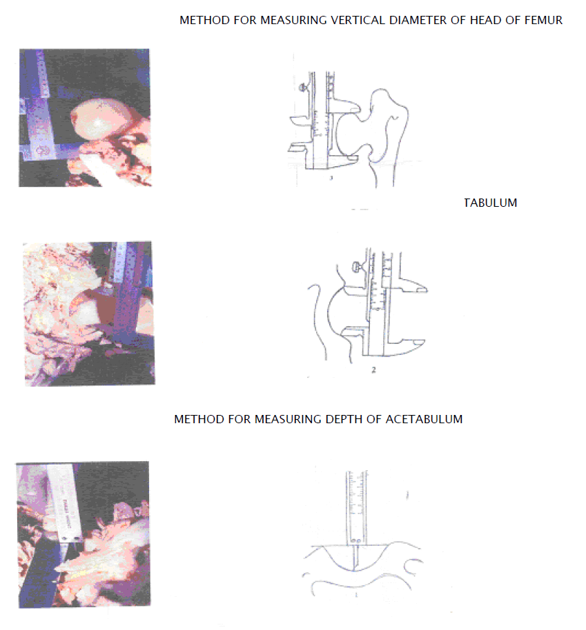
1. Vertical diameter of head of femur: was taken as the distance between the most superior and most inferior point on head of femur.
2. Diameter of acetabulum : was recorded as the maximum transverse diameter of acetabulum
3. Depth of acetabulum: the meatallic strip was placed across the diameter of acetabulum . Depth of acetabulum was measured from centre of acetabulum to the metallic strip.
All measurements were taken three times and the mean of three measurements was recorded.
From the measurements recorded the following observations were made.
In case of males
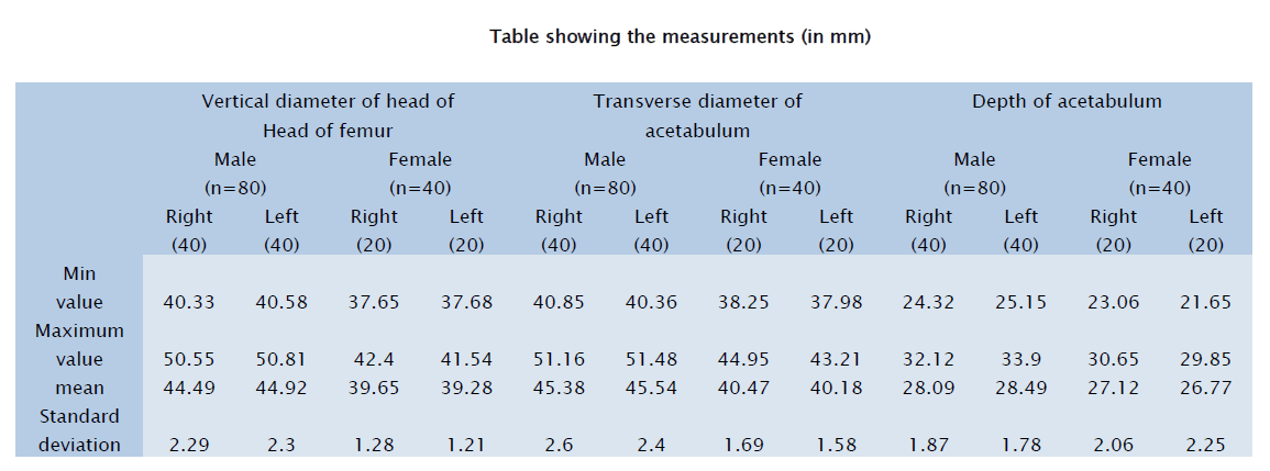
1. The mean vertical diameter of head of femur on right side was 44.49mm and 44.92mm on left side with a range of 40.33mm to 50.55mm on right side and 40.58mm to 50.81mm on left side. (TableI)
2. The mean diameter of acetabulum was 45.38 mm on right side and 45.54 mm on left side with a range of 40.85mm to 51.16mm on right side and 40.36mm to 51.48mm on left side. (Table II)
3. The mean depth of acetabulum was 28.09mm on right side and 28.49 on left side with a range of 24.32mm to 32.12mm on right side and 25.15mm to 33.9mm on left side. (Table III)
The difference was no greater than 2mm for all three parameters between right and left side.
In case of females
1. The mean vertical diameter of head of femur on right side was 39.65mm and 39.28 mm on left side with a range of 37.65mm to 42.40mm on right side and 37.68mm to 41.54mm on left side. (Table I)
2. The mean diameter of acetabulum was 40.47mm on right side and 40.18 on left side with a range of 38.25mm to 44.95mm on right side and 37.98mm to 43.21mm on left side. (Table II)
3. The mean depth of acetabulum was 27.12mm on right side and 26.77 on left side with a range of 23.06mm to 30.65mm on right side and 21.65mm to 29.85mm on left side. (Table III)
On comparing the measurements between males and females the mean of three parameters was more in males . The difference was statistically significant for vertical diameter of head of femur and diameter of acetabulum . In either sex the diameter of acetabulum was greater than the vertical diameter of head of femur again with no statistical significance again.
The above three parameters i.e vertical diameter of head of femur, diameter of acetabulum and depth of acetabulum have been carried out to assist Orthopaedic Surgeons, Biomedical Engineers to make suitable prostheses ,detection of disputed sex by Forensic Experts and to understand aetiopathogenesis of diseases like primary osteoarthritis of hip joint.
The vertical diameter of head of femur has been recorded by many workers from different countries including India. Workers like Davivong [3] , Rajendra Prasad et al [6], Gonzalo et al [4], Asala [1] and Ruma Purkait and Heeresh Chandra [7] show statistically significant difference in vertical diameter of head of femur. In this study the mean vertical diameter of head of femur on right side was 44.49mm and 44.92mm on left side in case of male cadavers and the same is right side was 39.65mm on right side and 39.28 mm on left side of female cadavers. As in other studies, statistically significant difference is noted for this measurement between males and females(p<0.01)in this study too. This difference is based on the fact that the male skeleton is more robust than the average female, although the magnitude of difference varies from population to population. The initial impact of this weight is borne by the the femur in transmission of body weight. The modification of female pelvis with respect to specialized function of reproduction also bears its indentation on femur.
This measurement alone can help Forensic experts to determine sex from skeletal remains. If the vertical diameter of femoral head is above 44mm it can can safely be said to belong to a male where as if it is less than 39 mm it can be sexed as a female.
The mean diameter of acetabulum is 45.38 mm on right side and 45.54 mm on left side in case of males. The same is found to be 40.47mm on right side and 40.18 on left side in case of female cadavers. The difference of measuremants between male and female is statistically significant (p<0.01). The diameter of acetabulum was found to be greater than the vertical diameter of head of femur in all cadavers accounting to rarity of osteoarthritis of hip joint in Indians. Rarity of osteoarthritis of hip joint can be explained on the basis of meachanical perfection, absence of undue stress and strains due to social habits of Indians.
The average depth of acetabulum is found to be 28.09mm on right side and 28.49 on left side. The mean depth of acetabulum was 27.12mm on right side and 26.77 on left side in case of female cadavers. On comparison to the study by Chauhan et al [2], these values are greater suggesting a deeper acetabulum in South Indians compared to North Indians. According to Lloyd Roberts [5] a shallow acetabulum is a most common cause of osteoarthritis among idiopathic causes. The deeper acetabulum noted in South Indian cadavers suggests rarity of osteoarthritis in and congenital subluxation of hip joint in this population .
Comparison of right and left sided measurements showed no significant difference for all the three parameters. In all cadavers, the difference between measurements of right and left side was not more than 2mm. In the present study the mean values for all the three parameters was greater on left side for males , but for females the mean values of all the three parameters was greater on right side.
In conclusion, for a hip joint replacement, knowledge of the dimensions of depth of acetabulum and femoral head in both sexes will assist the Biomedical Engineers to construct suitable prostheses. As all these measurements are take with soft tissue in situ, the recordings made of parameters are as very near to normal situation as encountered in patients at operation table. Knowledge of the anatomical parameters of bony components of hip joint are also very essential to better understand the aetiopathogenesis of primary osteoarthritis and will help in early detection of disputed sex by Forensic experts.