ISSN ONLINE(2319-8753)PRINT(2347-6710)
ISSN ONLINE(2319-8753)PRINT(2347-6710)
Naidile.S1, Shrividya.G2
|
| Related article at Pubmed, Scholar Google |
Visit for more related articles at International Journal of Innovative Research in Science, Engineering and Technology
Rapid growth in the use of e-commerce applications requires reliable user identification for secure access control. Hand vein biometrics is a recent technology that offers secure system for identification/authentication. Just like any other identification system, this has four steps- image acquisition, pre-processing, feature extraction and classification/matching. This paper presents a simulation to identify the individuals using dorsal hand vein images. In the proposed system an infrared camera is used to capture the hand image. After pre-processing, method known as maximum curvature is used to find the centerlines of veins. For matching, a simple correlation method is used. Maximum curvature method is robustous against any temporal fluctuations in brightness and vein width.
Keywords |
| Vein, Biometric, Maximum curvature, matched filter. |
INTRODUCTION |
| Biometrics is a type of automated method that recognizes a person based on physiological or behavioural characteristic. They have become the framework of highly secure identification and personal verification solutions. The number of security breaches and fraud has increased over the years. As a result there is an urgent need for highly secure identification and verification technologies. Biometric based recognition system has many applications like secure electronic banking, investing, financial transactions, law enforcement and health and social services. It consists of two phases- enrolment and recognition phase. The enrolment phase includes initial profile measurement for database storage and average and acceptable range is calculated. It is generally slow and considers multiple measurements. In the recognition phase the matching of the presented profile with stored profile is done. Hence this phase is very quick and needs only few attempts. Some of the examples of biometric system include face, fingerprint, iris, retinal scan etc. These systems have their own advantages and disadvantages. But recently, vein pattern recognition has become a new branch of biometric identification and has been attracting much attention from researchers for its convenience and security. The pattern of veins is unique even in identical twins. The vein technology has the following advantages which makes it a better biometric system compared to the already existing systems. |
| • Living body recognition: Vein pattern can be obtained from a person who is alive. • Internal: As the veins are hidden inside the body it is not damageable and non-wearable. • Highly secure: It is extremely difficult to counterfeit or duplicate. • Universality: It can be obtained from anyone. • Constant: Vein patterns do not change with time. • Unique: Vein patterns are different for different individuals. Even identical twins have different pattern. |
| Paper is organized as follows. Section II describes the prior work done in the field of hand vein biometrics. Section III describes the proposed architecture of the hand vein recognition system. The experimental results obtained using this system is described in section IV. Finally the conclusion and the future work are described in the section V. |
II. RELATED WORK |
| The first biometric system based on vein pattern using dorsal region of the hand was developed by P. Macgregor and R. Welford in 1991. This system was called as “Veincheck”. They used tungsten filament to provide illumination. The vein structure information was displayed on VDU and plotted manually onto the acetate sheet. The image was traced to enable feature extraction and further analysis took place away from the graphic display. Vein patterns were determined by viewing IR images using the intensity and contrast controls on VDU. This method was useful for comparing the images but was not adequate for a practical system [3].C. L. Smith and J. M. Cross developed a system where an array of LED’s was used to illuminate the back hand. This approach automatically segmented the pattern of veins from a digitized image and the medial axis representation of the vein pattern was obtained. This offered a better representation of the vein pattern. But some improvement was needed with respect to the image acquisition system [4]. Im et al. tried to reduce the pre-processing time and improve the recognition time by developing a system using DSP. They also designed a filter with a shift and add architecture because of which the pre-processing time and the recognition time greatly improved. A reliability of 99.45% and a verification speed of 150 ms/person were obtained which demonstrated enhanced performance. Maximum FAR was 0.001%. However some improvement in the performance of vein pattern extracting algorithm was needed [5]. Wang et al. designed a new vein recognition system. Unlike the other verification systems which compared the vein patterns based on a predefined set of features extracted, this system recognized the shape of the vein patterns by calculating their line segment Hausdorff distances. FAR of 0% was achieved. However since it was applied on a very small database it casted a shadow over the validity of their experiments. Also the factors such as ambient temperature etc affected the quality of the image [6]. Kumar et al. presented a new approach which could be applied over a large database. Their database included different images from 100 persons. To represent multiple features, box and branch point approach was used. In this method, ROI extracted was convolved with the Gabor filter. Multiple features were extracted from the real part of the convolved images using feature extraction techniques based on the branch point. These multiple features were then combined at the decision level. AND and OR fusion rules were used to combine the decisions taken by the individual matcher. For a FAR of 0.1%, GAR was found to be 99.6%[7]. Shahin et al. developed an authentication system using fast spatial correlation of vein patterns. In order to evaluate the system performance a prototype was designed and a dataset of 50 persons was acquired. The recognition ratio of 94.5% was obtained [8]. Hu had defined seven moment invariants based on normalized central moment feature recognition and classification. The seven moment invariants compared to the other moments were better in regard to translation, rotation and scale. But the recognition result was not satisfactory when they were used and their values kept varying widely [9]. Therefore Li et al developed an algorithm for the improvement of recognition ratio. This algorithm acquired a one pixel width vein skeleton from the original image. Then the seven corrected moment invariants of the vein pattern were extracted as feature vector. This was given as input into the SVM for training and recognition. A recognition ratio of 95.5 % was obtained under the same conditions used by Shahin et al. [10]. To further increase the recognition ratio Wang et al. proposed a method on Partition Local Binary Pattern (PLBP). According to this method, after preprocessing, the image is divided into sub-images. LBP uniform pattern features are extracted from all the sub images were combined to form the feature vector. It was tested on 102 individuals. The results showed PLBP performs better than other features. Furthermore, when the image was resized to 256x256, a recognition rate of 98.14% was obtained[11]. |
III. THE PROPOSED SYSTEM ARCHITECTURE |
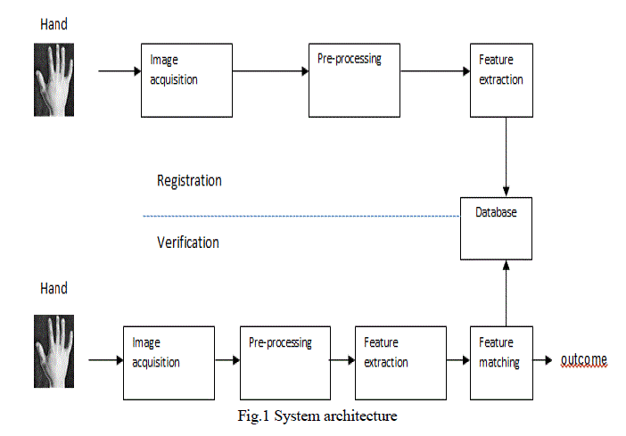
| The overall system architecture of the proposed biometric identification system is shown in Fig.1. Each of the constituent modules is described in the following sections. |
 |
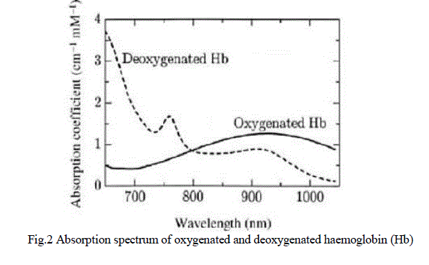
| A. Image Acquisition Veins are present under the body surface. They are not visible to the naked eye. Most of the popular methods of vein imaging are based on infrared (IR) light absorption. The primary transporter of oxygen in human beings is the haemoglobin molecule. Arteries carry oxygenated blood and veins carry deoxygenated blood and blood contains haemoglobin. Oxygenated and deoxygenated haemoglobin have different absorption spectra as shown in Fig.2. It is found that near infrared (NIR) light with a wavelength of 700nm- 900nm can transmit through a body tissue. Deoxygenated haemoglobin absorbs the light having wavelength of about 760nm. When these rays pass through the hand, veins appear darker than the other parts because they absorb IR rays better than surrounding tissue. The absorption coefficients of blood and other tissue are different for NIR illumination. Higher absorption coefficient of blood means the amount of NIR illumination absorbed by blood is greater compared to the surrounding tissue. In this work a lost cost infra-red sensitive camera is used. It consists of IR LED’s socket to provide the illumination and an USB module to transfer the captured images to PC. In order to capture the image the person has to place his clenched fists in front of the camera |
 |
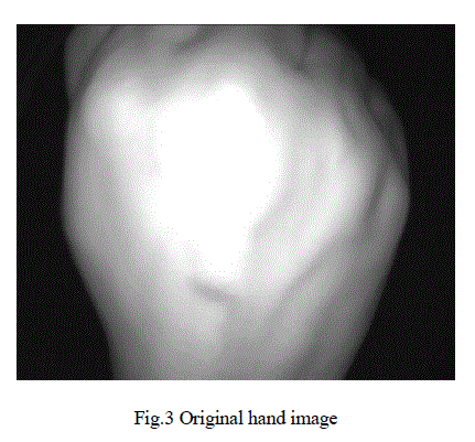
| The hand image acquired using the infrared camera is shown in Fig.3. |
 |
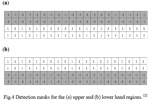
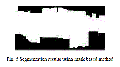
| B. Preprocessing After capturing the hand image it is converted to a grayscale image. This conversion is practical due the ability to manipulate the image easily for further stages. Then global thresholding is applied to the converted image. In order to remove small or extra particles from the thresholded image, morphological processing is done. Later the image is resized and the region of interest (ROI) is determined. In order to localize the hand region, masks were used. Fig.4 shows the masking values used to determine the upper and lower boundaries of the hand region. |
 |
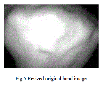
| Fig.5 shows the original image after resizing. It is resized in order to increase the processing speed. Fig.6 shows binary image of the segmentation after applying the mask. The white region represents the region of interest. |
 |
 |
| C. Feature Extraction There are several methods to extract the vein pattern. In the proposed system matched filter is used. The matched filter method uses two-dimensional filter. It consists of four different filter kernels with each filter kernel rotated by 45º in order to obtain the vein pattern in different directions. Each filter is then convoluted to the captured image independently and all convolution values are added together. Hence a Gaussian shaped filter can be used to match the vessel. Another advantage of Gaussian shaped filter is that it reduces the noise. The 2-D Gaussian function is given as, |
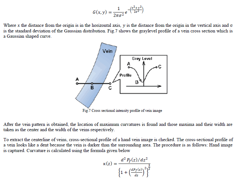 |
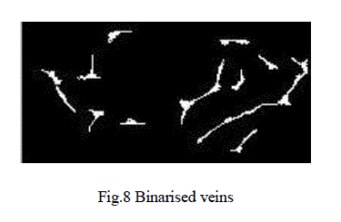
| whereïÿýïÿýïÿýïÿý ïÿýïÿý is a cross-sectional profile of the hand image. After obtaining the curvature, profile is classified as concave or convex depending on whether ïÿýïÿý (ïÿýïÿý) is positive. Local maximums of ïÿýïÿý ïÿýïÿý in each concave area are calculated. These points indicate the center position. Next, scores are assigned to each center position. Scores are assigned to a plane, V. To connect the centres of veins and eliminate noise, the following filtering operation is conducted. First, the two neighbouring pixels on the right side and two neighbouring pixels on the left side of pixel (ïÿýïÿý, ïÿýïÿý) are checked. If (ïÿýïÿý, ïÿýïÿý) and the pixels on both sides have large values, a line is drawn horizontally. If ïÿýïÿý, ïÿýïÿý has a small value and the pixels on both sides have large values, a line is drawn with a gap at ïÿýïÿý, ïÿýïÿý . If (ïÿýïÿý, ïÿýïÿý) has a large value and the pixels on both sides of (ïÿýïÿý, ïÿýïÿý) have small values, a dot of noise is at (ïÿýïÿý, ïÿýïÿý) .The operation is applied to all pixels in all four directions. Finally, a final image ïÿýïÿý (ïÿýïÿý, ïÿýïÿý) is obtained by selecting the maximum curvature for each pixel.The vein pattern ïÿýïÿý (ïÿýïÿý, ïÿýïÿý) is binarized by using a threshold. Pixels with values smaller than the threshold are labelled as parts of the background, and those with values greater than or equal to the threshold are labelled as parts of the vein region. Fig.8 shows the binarised vein obtained after applying the maximum curvature method. The white lines represent the vein pattern. |
 |
| D. Matching After the enrolment stage, matching is done. This process compares the captured image with the image from the database. Two methods are commonly used for pattern matching. They are –structural matching and template matching. Here template matching is used which is based on comparison of pixel values unlike the structural matching that requires additional extraction feature points like line endings and bifurcations. An image ïÿýïÿý(ïÿýïÿý, ïÿýïÿý) is correlated with a template image ïÿýïÿý ïÿýïÿý, ïÿýïÿý to find all the possible matching positions and the matching score is calculated. |
IV. EXPERIMENTAL RESULTS |
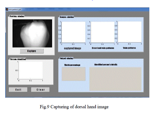
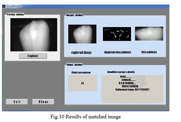
| The performance of the proposed recognition system was evaluated in real time on hand vein database. This database consists of the right hand and left hand images of the volunteers. The images of both male and female subjects are taken for the experiment.The acquisition setup is explained in the section II. The size of the acquired image is 720x480 pixels. The acquired image is subjected to some rotational changes and scaling changes as it’s likely that the user may present his/her hand at different distances from the camera during the experiment. The ROI extracted is from the resized image of size 390x189 pixels. Matlab 7.8 was used to obtain the above results. The main reason of using GUI is to provide a clear picture of the proposed system thereby reducing the complexity of understanding. The proposed GUI system first captures the dorsal hand image. Next, the original hand image and the vein pattern extracted along with the overlaying of the extracted vein pattern over the original image is displayed. In addition to these images, the matching score and the details of the identified person are displayed. The proposed system GUI snapshot is shown below as an example for one person. If there is a reference image it will be displayed as well. Fig.9 shows the capturing of dorsal hand vein image. |
 |
| Fig.10 shows the results of a matched image along with the matching.As there is no reference image for this person, it has not been displayed. |
 |
V. CONCLUSION |
| Experimental results show a positive detection rate of 75% and negative detection rate of 25%. Hence this method can be successfully used for identification. Future work may involve working with a large database system, applying additional/ alternative pattern recognition algorithms or turning it into a multimodal system where other additional biometrics traits are considered and making the system more invariant to illumination conditions. |
References |
|