International Journal of Innovative Research in Science, Engineering and Technology
ISSN ONLINE(2319-8753)PRINT(2347-6710)
ISSN ONLINE(2319-8753)PRINT(2347-6710)
Pohekar P.G.1,Rathod S.M.2, Sinha A.3
|
| Related article at Pubmed, Scholar Google |
Visit for more related articles at International Journal of Innovative Research in Science, Engineering and Technology
Microbiology is the science which deals with study of microorganisms. Some of them are beneficial and important with respect to industrial production. However most of the microorganisms are pathogen and cause the disease in human. There are several ways to control the population of microorganism including the use of antibiotics at the top; however the LASER (Light Amplification by Stimulated Emission of Radiation) irradiation also provides the effective way to control the microbial population. The bacteria selected were include both Gram positive i.e. Streptococcus pyogenes and Gram negative i.e. Proteus mirabilis. These bacteria are exposed to the 632.8nm He-Ne laser beam in order to study the effect. The increase in the exposure time of the radiation resulted in the reduction of the bacterial count due to their destruction because of the radiation. For Streptococcus pyogenes, the best results shows at 17.5 mW and 150 seconds, where as for Proteus mirabilis, the reduction occur at 17.5 mW for 150 seconds. Today LASER use in many field such as skin treatment, ulcerous would, the diabetic wounds. So once this method approve, this could be the invaluable tool to control bacteria
Keywords |
| He-Ne laser, Streptococcus pyogenes, Proteus mirabilis. |
INTRODUCTION |
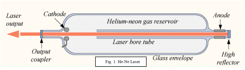
| This wonder beam has penetrated into every aspect of life like medicine [1-2], communication, industry, defence almost everything. A helium-neon laser (He-Ne) is a type of gas laser consisting of mixture of helium and neon gas inside the small bore capillary tube. The best known and most widely used He-Ne laser operates at a wavelength of 632.8 nm in the red part of visible spectrum. The He-Ne laser is highly compact, reliable. The mechanism responsible causing bacterial death has been reported to involve the formation of singlet oxygen and free radicals [3-4]. He-Ne laser has spatial characteristics such as 632.8 nm of wavelength, good directivity, high intensity, good monochromatic and coherence is a variable low-level laser [5-8]. The low-level laser has some biology effects such as cell vitality [9-11], phagocytosis [12-13], but as far as its usage for the treatment of digestive disease, there were only few reports. Streptococcus pyogenes (Group A streptococcus) is a Gram-positive, non motile, non spore forming coccus that occurs in chains or in pairs of cells. S. pyogenes is the cause of many important human diseases, ranging from mild superficial skin infections to life-threatening systemic diseases. Infections typically begin in the throat or skin. Examples of mild S. pyogenes infections include pharyngitis ("strep throat") and localized skin infection ("impetigo"). Erysipelas and cellulitis are characterized by multiplication and lateral spread of S. pyogenes in deep layers of the skin. S. pyogenes invasion and multiplication in the fascia can lead to necrotizing fasciitis, a potentially life-threatening condition requiring surgical treatment. Proteus mirabilis is part of the Enterobacteriaceae family. It is a small Gram-negative bacillus and a facultative anaerobe. Proteus mirabilis is characterized by its swarming motility, its ability to ferment maltose and its inability to ferment lactose. When this organism, however, enters the urinary tract, wounds or the lungs it can become pathogenic. Proteus mirabilis commonly causes urinary tract infections and the formation of stones. Proteus can also cause wound infections, septicemia and pneumonias, mostly in hospitalized patients. Infections caused by P. mirabilis are seen most often in nursing home patients. These infections are commonly caused by infected medical equipment including catheters, nebulizers (responsible for inhalation), and examination gloves (responsible for wound infections). The length of catherization is directly related to incidence of infection. Each day of catherization gives an infection rate of 3-5%. The effect of He-Ne laser on Bacillus subtilis and Escherichia coli was previously studied, shows that decrease in viability at effective combination of 7.5 mW and 90 seconds [14]. A present experimental study we found that two different dosages of He-Ne laser with power 7.5 mW and 17.5 mW at different time 60, 90, 120 and 150 seconds, resulted in different effects. The best effect was the dosage 17.5 mW, 120 seconds of He-Ne laser in case of Proteus mirabilis, and 17.5 mW, 150 seconds in case of Streptococcus pyogenes which showed the dependence on intensity and dosage. The purpose of our project was to identify possible bactericidal or bacteriostatic effects of He-Ne laser on the Proteus mirabilis and Streptococcus pyogenes. |
II. MATERIALS AND METHODS |
| 1. A suitable strain of bacteria such as Proteus mirabilis and Streptococcus pyogenes were used. 2. He-Ne Laser of varying power (7.5 mW, 17.5 mW) 3. Standard plate count method was used. |
METHODOLOGY |
| A pure culture of each species of bacteria and sterile saline was used as diluents for both the bacteria. The medium used were nutrient agar for Proteus mirabilis and MRS medium [16] (De MAN, ROGOSA, SHARPE) for Streptococcus pyogenes. Serial dilution used was 10-6 for irradiation purpose by using He-Ne laser (As shown in the figure 1) and spread plate method was performed. Plates were incubated at 370 C for 24 hours and number of colonies was measured. Once reproducible results were obtained next phase of exposure was started. Bacterial suspensions were irradiated by passing the laser beam in the test tube vertically. Irradiation is done using different Laser power and different time, like power of laser is varied of 7.5 mW and 17.5 mW and time 30, 60, 90 ,120 and 150 seconds. After irradiation procedure each group, bacterial suspension was spread over surface of medium and plates were incubated at 370 C for 24 hours, after that the bacterial colonies were counted. A control plate of each bacterial species was made for every time exposure dose. Every time we tried to maintain relatively similar laboratory conditions during experiments. |
 |
III RESULTS |
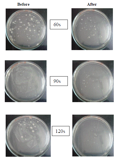
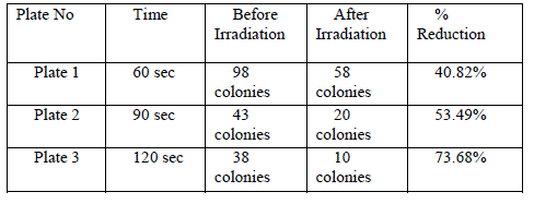
| From table 1 it was observed that the 632.8nm light produced bactericidal effect on P. mirabilis for all the exposure time and the growth was decreasing with respect to time, where the power of the laser beam was 17.5 mW. At 120 seconds we had got almost 73.68% inhibition (Figure 1). It is clear that the along with the along with power of the laser, the exposure time also play an important role in decreasing the viability. |
 |
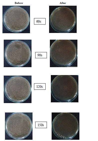
| Similarly from table 2 which shows the effect of 17.5 mW He-Ne laser at different time on Streptococcus pyogenes for different time exposure. It is evident that there was no growth on the plate for 17.5 mW at 150 seconds. It means that complete inhibition of Streptococcus pyogenes takes place at the power mentioned above (Figure 2). The results of this study show that exposure of bacterial cultures to He-Ne laser light results in a decrease in viability. However the most-effective combination was 17.5mW, 120 seconds for Proteus mirabilis and 17.5 mW, 150 second for Streptococcus pyogenes suggesting that power of laser and time of exposure plays an important role in its efficacy. However further studies are needed to determine its clinical suitability, once this method of killing bacteria is approved it could be valuable tool to control the growth of these bacteria. |
 |
| Table2: Effect of 17.5 mW He-Ne laser at different time exposure on Streptococcus pyogenes |
 |
IV DISCUSSION |
| The properties of laser make it highly applicable source of light because laser beam can be selectively focused into beam of very small size. And therefore beam can be irradiated on a very small portion of material where the effect is to be produced. The laser photons are highly distinguishable from other photons and hence they can be easily separated from other unwanted photons with the help of simple spectrum analyzers. The above discussion shows that we may find numerous fields where lasers can act as a source of radiation. Infact laser is applied in infinite number of fields. Medical science is one of the important fields where laser has been successfully utilized. |
| Several researchers have reported that laser irradiation of various bacteria result in destruction of the organisms. The mechanism of laser-induced cell destruction has important implications in clinical therapy. According to Karu [15] exposing a cell to laser light causes acceleration of electron transfer in some areas of the respiratory chain. We have performed the experiments in which we studied effect of He-Ne laser on Proteus mirabilis and Streptococcus pyogenes. As shown in Tables 1 and 2 the number of colonies decreases with respect to exposure time, particularly for the 17.5 mW He-Ne laser. Each exposure time represents a radiant energy dose; the longer the time of irradiation, the higher the dose. In case of Streptococcus pyogenes up to 120 seconds there is no effect may be indicating that, mentioned bacteria are resistant to the dose and time employed. However it will no longer tolerate more dose 17.5 mW He-Ne laser at 150 seconds, seemed to be a effective dose results in its inhibition or killing. |
V CONCLUSION |
| With respect to the known methods of control of microorganism this method emerged as a physical control of microorganism. The method is seemed effective in case of burnt patients and other skin infections where other control methods not work. Therefore once this method approve, it could be the valuable tool in many field. |
References |
|