
Research & Reviews: Journal of Medical and Health Sciences
 Menu
MenuISSN: 2319-9865
ISSN: 2319-9865
Muhammad Asim, Hamza Ishfaq, Ume Habiba*
Department of Eastern Medicine, The Islamia University of Bahawalpur, Bahawalpur, Pakistan
Received: 18-Oct-2024, Manuscript No. JMAHS-24-150399; Editor assigned: 21-Oct-2024, Pre QC No. JMAHS-24-150399 (PQ); Reviewed: 04-Nov-2024, QC No. JMAHS-24-150399; Revised: 17-Mar-2026, Manuscript No. JMAHS-24-150399 (R); Published: 24-Mar-2026, DOI: 10.4172/2319-9865.15.1.001
Citation: Habiba U, et al. Preliminary Steps to Prevent Nipah Virus Outbreak in the United States of America and Other Countries. RRJ Med Health Sci. 2026;15:001.
Copyright: © 2024 Habiba U, et al. This is an open-access article distributed under the terms of the Creative Commons Attribution License, which permits unrestricted use, distribution and reproduction in any medium, provided the original author and source are credited.
Visit for more related articles at Research & Reviews: Journal of Medical and Health Sciences
Nipah Virus (NiV) outbreaks in different countries throughout the world have created fear and panic situation in the international community with the most recent outbreak in India during September 2023 with a fatality rate of 34% (2 deaths out of 6 cases). Overall, 25 outbreaks (from 1998 to 2023) of this virus have been recorded with fatality rates ranging from 38%-100%. NiV is a serious threat to human beings. It presents with high mental and respiratory problems. Still, no vaccine is available against the protection from this virus. Researchers and healthcare professionals from different countries worldwide are working on preparing vaccines and antiviral medicine for NiV. Observing the recent situation of NiV outbreaks worldwide may also come in the United States of America. So, to protect the international community from Nipah virus infection, it is necessary to study the emerging zoonotic disease. The priority should be to control the natural reservoirs of the Nipah virus and prevent its transmission. This review is about the NiV breakout and its current status. It highlights the preventive and monitoring procedures that should be applied at both government and nongovernment levels to ensure no occurrences of this outbreak in the world.
Nipha virus; Nipah virus; Infectious disease; Government
From 12 to 15 September 2023, the Ministry of Health, Govt. of India claimed 6 (six) NiV cases with 2 deaths in district Kerala [1]. The origin of the initial case of NiV was unknown. It was initially misidentified as a Japanese encephalitis virus [2]. In five years, that was the 4th outbreak of NiV in India (Kerala). However, these outbreaks affected small geographical areas but their fatality rate was too high. Again in 2019, the Nipah virus was seen in Bangladesh in which 8 cases were reported out of which 7 people died [3-5].
NiV belongs to the family Paramyxoviridae and the genus Henipavirus. It is a negative sense RNA virus. Hendra Virus (HeV) and Cedar virus also contain henipavirus [6]. Its genome encodes six structural proteins, which are large protein (L), glycoprotein (G), matrix protein (M), phosphoprotein (P), nucleocapsid protein (N), fusion protein (F), or maybe RNA polymerase in order 3'-L-G-M-P-N-F-5'. C, V, and W are three non-structural proteins that are encoded by the P gene (Figure 1).
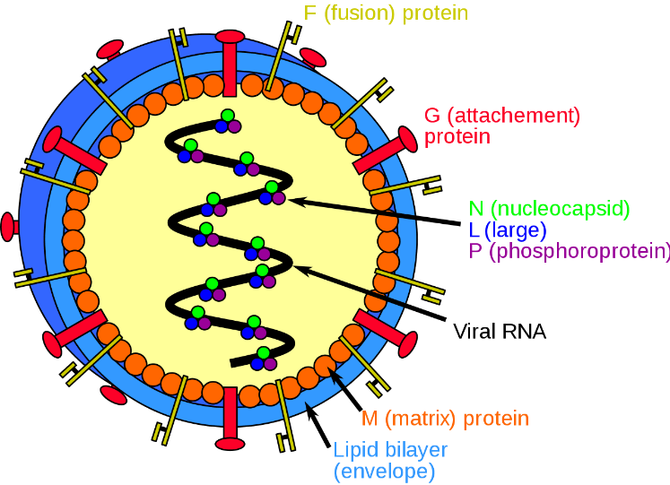
Figure 1. Structure of Henipavirus.
NiV has different strains. Bangladeshi virus genome is indicated by NiV-B while the Malaysian virus genome is indicated by NiV-M. The origin of NiV-B was the utilization of raw date palm sap on the other hand NiV-M was pigs. NiV-B outbreak was more severe than NiV-M. In NiV-M; the mortality rate was 39% while NiV-B was 79% [7]. In 2014, NiV infection was detected in the Philippines. The fatality rate was 42%. The Philippines NiV infection strain was similar to the strain of Malaysia but spread in the Philippines was happen by person-to-person contact [8].
In May 2018, they identified 23 cases of NiV infection in Kerala state India. The death rate of these cases was 91%, with 21 patients dying and only 2 surviving. While the outbreak continued for more than 1 month, the situation was very panicky there. The main factors in overcoming the NiV outbreak were its early diagnosis, prevention, and measurement to control its transmission [9]. According to the classification, NiV is a Biosafety level 4 pathogen. It causes small outbreaks in humans throughout the world but with a fatal outcome. The small size of the outbreak is due to the low transmission of the virus from one individual to another. Transmission may occur from human to human. NiV was 1st time detected in Malaysia from 1998 to 1999. During its 1st outbreak, 283 cases of acute encephalitis appeared with 109 deaths. The fatality rate of NiV infection ranges from 38%-100%. In its severe form, it affects the key organs of the body like the brain, central nervous system, lungs, and kidneys. Therefore, it is necessary to prevent Pakistan from NiV infection and to prepare our healthcare system to fight against this virus. Here in this article, we have discussed the NIV genome, causes, signs symptoms, outbreaks, diagnosis, transmission, and its prevention [10].
Methods
We concluded this literature by using digital platforms like Google Scholar and Pubmed. The following terms were used epidemiology of Nipah virus, Nipah virus infection, clinical features of Nipah virus, Vaccine of Nipah virus, surveillance of Nipah virus, diagnosis of Nipah virus, Nipah virus infection in India, Niv in Malaysia, Niv Philippines, Niv in Bangladesh, treatment and vaccine of Nipah virus, uses of herbal antibiotics. All literature reviews, original article papers and all cases, and reports regarding aspects of Nipah virus origin, diagnosis, clinical presentation, mood of transmission, and management were reviewed. The cross references from all these papers and case reports were also included. Additionally, epidemiological reports from the National Center for Disease Control of India, WHO, and other public health organizations were assessed for this paper. The purpose of this review article was to research strategies for the prevention of the Nipah virus-transmission of this virus control strategies, and diagnosis of infection.
Disease
Nipah virus causes encephalitis, which is a severe condition for normal survivors. Because of this disease, the mortality rate of the Nipah virus infection is huge [11]. Encephalitis appears within a week to ten days. Other symptoms are mental status retarded, hypotonia, colonic spasm of muscles, and limb weakness [12]. The period of incubation ranged from 4 days to 2 months in 19% of patients after the last exposure to pigs and subsequent onset of illness. Most patients gave a history of 2 weeks [13]. In some cases, encephalitis may appear later. There may be relapsing NiV encephalitis [14]. Respiratory illness is also caused by the Nipah virus. Both diseases are highly fatal [15]. Patients may present with headache, fever, muscle stiffness, fatigue, confusion agitation, speech seizure, and many more issues. In infants, it may cause poor feeding, nausea, and irritability [16].
Causes
The main causes of the Nipah virus are domestic animals like dogs, chickens, cats, goats, rodents, birds, wild boar, bats, and pigs. Bats are emphasized depending upon the findings that fruit bats of Pteropid species were the reservoir of the Hendra virus [17]. The Nipah virus and the Hendra virus have a phylogenetic relationship. Some serological evidence proved that domestic animals were infected with viruses and believed that the origin of infection for these domestic species was infected pigs that are effectively “dead-end” hosts.
Transmission
Still, the exact way of transmission of the Nipah virus is unknown. Initially, it was thought that it was transmitted through nosocomial and oropharyngeal routes. Until 2014, due to an earlier outbreak in Bangladesh, it was thought that the Nipah virus was transmitted through date palm trees [18].
Fruit bats: In Malaysia, it has been seen that the natural reservoir of NiV infection is Pteropus bats [19]. Antibodies against NiV were seen in blood and NiV RNA was seen in serum, saliva, and urine of Pteropus in many areas of Asia [20-22]. NiV human infection was seen in Kerala in May 2018. All these patients were showing acute respiratory syndrome and mental retardation transmitted through fruit bats [23,24].
Pigs: In Malaysia, Pigs were infected with NiV infection by eating food that was contaminated with the saliva and urine of bats and after that, they infected the humans. These patients showed the symptoms of respiratory disease with a high lethality rate. All those workers were infected who were in direct contact with these animals [25]. Among slaughterhouses of pig workers, an outbreak was seen in Malaysia, and Singapore in 1999. They controlled this epidemic by prohibiting the importation of pigs from the infected areas.
Diagnosis
Indian laboratories prescribe throat swabs, blood, urine, and Cerebrospinal Fluid for diagnosis. Samples must be transported at 2-8c in a triple-coated box. Storage must be at the -20C recommended level after 48 hours of sample collection. In the BSL-2 laboratory, virus inactivation by radiation was a good technique for making the sample safe for use [26].
PCR: The US centers for disease control and prevention developed conventional PCR targeting the N gene. Because PCR was considered as the 1st method of choice for the observation of the virus, it can diagnose any infection within a few hours as we have seen NiV has an incubation period ranging from 4 to 17 days and may be longer [27].
Chemistry
For immunohistochemistry, tissues are used by formalin. In the vascular endothelium, viral replication occurs in a lot of tissues, like the brain, kidney, lymph nodes, spleen, and lungs. In pregnant animals, the products of conception, placenta, and uterus are also examined [28].
In the BSL-4 laboratory, the virus is isolated from urine, respiratory secretion, CSF, and other tissue specimens. The Vero cell line is the line of choice for NiV and HeV [29].
Antibody: For diagnosis, IgM antibodies in serum or CSF were used. During epidemiological investigation, it has been seen that the examination of IgG antibodies gives better results for surveillance in humans and identification in reservoir animals. During outbreaks, it has been used for human investigation [30].
ELISA: ELISA is used most commonly for serological diagnosis because of it is high responsiveness, ease, quickness, and welfare in use. In Malaysia, ELISA is used for the examination of IgG and IgM which are prepared by the CDC for accurate diagnosis. Also, it has been used for monitoring in Bangladesh during the NiV outbreak [31].
Outbreak
Malaysia: In Malaysia and Singapore, the first outbreak of NiV in humans was recognized from September 1998 to June 1999. In this outbreak 283 people were infected, mostly pig farm and stockyard workers, through getting in touch with sick pigs. In this outbreak, 39% (Figure 2) of people died. So, this was an alarming situation for national and international societies [32,33].
India: The worldwide second outbreak of NiV occurred in India from January through February 2001 in Siliguri, near Bangladesh. The fatality rate was sixty-eight percent. All these patients were presented with both encephalitis and respiratory system disease [34]. In 2007, In India, individual-to-individual transmission was reported. This outbreak was observed from a person who contracted the disease because of the ingestion of alcohol. That alcohol was extracted from date palms. This infection affected the health workers and family members. The worker who collected the blood from the patient of NiV was affected [35]. A 3rd outbreak of the Nipah virus was observed in India, in May 2018. The death rate was increased up to 91% (Figure 2). 21 out of 23 patients died. This outbreak lasted for more than one month.
A 4th outbreak of NiV appeared on the 4th of September 2021 in Kerala, the state of India. In August 2021, a boy showed mild pyrexia and was admitted to several hospitals but his condition was not under control. After a few days, on 4th September, NiV was seen in his plasma, serum, and CSF. Samples were confirmed by the test as RT/PCR and IgM antibodies also were seen in the plasma sample on the ELISA report. The patient died the next day. Then NiV infection was seen in different areas of Kerala state and 20 people died because of this virus (Figure 2).
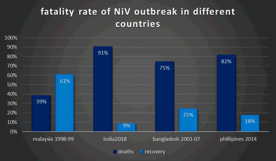
Figure 2. Fatality rate of NiV outbreak in different countries.
Bangladesh: A third outbreak had been seen in Bangladesh from 2001–2007. 2001–2007 outbreak of Nipah virus infection, here it was seen that this virus is transmitted by the usage of sick cows, sap of fresh date palm, and individual-to-individual exposure [36,37].
The mortality rate was 75% (Figure 2) in this area. All these outbreaks were observed between the month of 1 and 5. In Bangladesh, there is evidence of individual-to-individual exposure to the Nipah virus. In April 2001, in the Meherpur district of Bangladesh, 30 cases of Encephalitis were seen with 9 deaths. In this outbreak, the death ratio was 69%. At that time, testing facilities were limited, so the United States center for disease control and prevention in 2003 was acknowledged as the 1st outbreak of human NiV infection in Bangladesh [38,39].
In the past 25 years, Nipah virus monitoring activities have evolved substantially in Bangladesh. So, with the help of the establishment of national surveillance, almost all NiV cases in the country have been detected [40,41]. The data of this surveillance showed that from 2007-2014, about 50% of the cases were not reported because patients lived in rural areas that were away from the surveillance hospitals [42].
From April 2001 to December 2021, a total of 322 human cases appeared in Bangladesh, including 81 (25%) probable cases and 241 confirmed cases. Still, some cases of Nipah virus are seen in 33 districts of Bangladesh.
Philippines: In 2014, a Nipah virus infection outbreak appeared in the Philippines. Here, 17 cases were reported. The death ratio was eighty-two percent (Figure 2). Ten patients have a history of association with horses and horse meat ingestion. In the Philippines, 10 deaths were confirmed. 9 out of 10 patients showed neurological symptoms. Healthcare personnel get this disease through individual-to-individual exposure (Table 1). The Philippines strain was similar to the Malaysian strain. One individual to another’s exposure had not been observe before [43].
| S. No. | Country | Year | Place | \Cases | \Deaths |
| 1 | Malaysia | 1998 | 283 | 109 | |
| 2 | Malysia | 1999 | Perak, Negeri, Sembilan, Selangor | 265 | 105 |
| 3 | Singapore | 1999 | Singapore | 11 | 1 |
| 4 | India | 2001 | Siliguri | 66 | 50 |
| 5 | Bangladesh | 2001 | Meherpur | 13 | 9 |
| 6 | Bangladesh | 2003 | Naogaon | 12 | 8 |
| 7 | Bangladesh | 2004 | Manikganj and rajbari | 42 | 14 |
| 8 | Bangladesh | 2004 | Fareed pur | 36 | 27 |
| 9 | Bangladesh | 2005 | Tangail | 12 | 11 |
| 10 | India | 2007 | Nadia | 50 | 3-5 |
| 11 | Bangladesh | 2007 | Kushtia and Thakurgaon | 15 | 8 |
| 12 | Bangladesh | 2008 | Manikgang and Rajbari | 9 | 8 |
| 13 | Bangladesh | 2010 | Fareed pore | 8 | 7 |
| 14 | Bangladesh | 2011 | Cumilla Dinajpur, Fareedpore,Lalmohirhat,Nilphamari | 44 | 40 |
| 15 | Bangladesh | 2012 | Joypurhat | 12 | 10 |
| 16 | Bangladesh | 2013 | Gaibandha, Manikgani, Naogaon, Natoeri, Pabna | 24 | 21 |
| 17 | Philippines | 2014 | 17 | 9 | |
| 18 | Bangladesh | 2015 | Gaibandha, Manikgani, Nao gaon, Natori, Pabna | 261 | 199 |
| 19 | Bangladesh | 2017 | Fareed pore | 3 | 2 |
| 20 | India | 2018 | Kerala | 23 | 21 |
| 21 | India | 2017 | Kozhikode and Malappuram | 18 | 17 |
| 22 | Bangladesh | 2019 | 8 | 7 | |
| 23 | Bangladesh | 2020 | 6 | 4 | |
| 24 | India | 2021 | Kerala | 2 | 1 |
| 25 | India | 2023 | Kerala | 6 | 2 |
Table 1. Yearly occurrence of Nipah virus outbreaks in different countries.
Prevention
Prevention of this outbreak in the United States of America and other countries should be the government's priority. Here are the following steps that should be immediately considered to save the international community from the fatal outcome of the NiV.
Control of natural reservoirs of NiV
By staying away from such places where the chances of Nipah virus breeding are high, people should also use masks and wash their hands well. In this way, they can be protected from this virus. People should distance themselves from all the things that may cause this virus to spread. Similarly, travellers coming from countries where this virus has arrived should be fully tested. If anyone is ordering food items from those countries where the Nipah virus outbreak occurred, they should be banned. This way international communities can be protected from this virus [44].
Awareness among population
The international community can be protected from the Nipah virus by telling everyone about the thread of consuming sap of date palms and stopping individual-to-individual exposure. Covering the sap-producing area of the date palm tree with skirts that have been found to effectively prevent contact with bats, date palm sap should be prevented from contamination [45]. Like the government of Bangladesh, the international community should also conduct an awareness campaign among the people. Healthcare professionals should warn people about its spread and prevention through social media, newspapers, conferences, press releases, radio, talk shows, and national public and private channels. It should be told and through all these platforms people should be told in which places this virus spreads more and people should be told that it can spread from November to May [46].
Control of transmission
The government of any country can prevent individual to individual transmission by isolating patients by using personal protective equipment and good hygiene practices [47]. Hospital surfaces are contaminated by NiV around patients, by cleaning the hospital surfaces, the international community can prevent its spread. Implementation of standard infection control practices can also help individual-to-individual exposure in hospital settings.
Improvement of the healthcare system
By thoroughly studying the Nipah virus and treating it accordingly, the Nipah virus should be avoided by providing financial assistance to poor countries. If the health systems of poor countries are improved, the spread of the Nipah virus can be stopped. Similarly, by prohibiting transmission of this virus from one individual to another individual, the Nipah virus should be controlled [48].
Vaccine and antibiotics: A vaccine is essential to control any virus, but no vaccine has yet been developed for the Nipah virus. Patients can only be cured by taking precautions, supporting them, and maintaining their body's salt and water levels. Apart from this, there is a wide range of antibiotics available that are given to these patients. These antibiotics have been used to control outbreaks of the Nipah virus in Malaysia and Singapore but have not been effective.
Throughout the world, researchers and healthcare professionals are working on the preparation of vaccines and antiviral drugs for NiV. HeV-sG evokes a protective immune response against both HeV and NiV. In the prevention of the Nipah virus, several strategies have been developed based on the G glycoproteins (SG) of NiV and HeV, to develop a successful vaccination against the Nipah virus, but no successful outcome was observed. All these vaccines have been experienced in animal models. The use of monoclonal antibody m 102.4, is also used to stop the speed of outbreaks in different areas of the world [49]. Further research in this field can result in the development of an appropriate effective vaccine that can effectively prevent the disease from occurring [50,51].
There are few trials of vaccines conducted on different types and methods of preparation of vaccine on a lot of candidates.
Here we will discuss about advantages and disadvantages of these types of vaccines [52,53].
Because there are no curative measures for NiV so, the virus spreads easily, there is a considerable health risk. Creating vaccinations to stop NiV infection could stop it from spreading, especially in more susceptible communities. However, because handling live infections has additional dangers related to biohazard, developing vaccinations for bats the virus's major reservoir presents practical obstacles.
Inactivated pathogens or their proteins are frequently used as antigens in conventional vaccinations; however, researchers prefer different kinds of antigens to minimize the risk of biohazard. Subunit vaccines, which employ pieces of a pathogen's protein or glycoprotein to elicit a defense response in the immune system, are one potential strategy. For example, when given subcutaneously to cats, soluble G glycoprotein alone can stimulate the formation of serum-neutralizing antibodies. For up to two months, vaccinated cats had noticeably greater antibody levels (titer ~20,000) [54]. HeVsG administration protected against NiV infection in AGMs. A serum-neutralizing antibody titer of 2560 was seen on day 14 post-inoculation; this titer decreased after 28 days. When challenged, none of the immunized animals displayed any symptoms of disease, viral replication, or pathology. The recombinant subunit vaccine's effectiveness in non-human primates encourages its continued preclinical study for possible use in humans [55]. Prior immunization with subunit vaccine candidates targets either the NiV F or NiV G protein, or both, depending on the kind of NiV infection. Below are specifics on the most recent advancements in viral vector-based vaccinations against NiV [56].
Recombinant measles virus-based NiV vaccine
To fight NiV, researchers are looking into the possibility of employing recombinant Measles Virus-based (MV) vaccinations. There were worries that the generation of NiV antigenic proteins would be hampered by widespread measles vaccination. On the other hand, Yoneda et al. [57] have shown that a recombinant MV expressing the NiV envelope proteins (F, G) generated an antibody response that was specific to NiV. African green monkey and hamster models were used to test this vaccine candidate, which produced total protection against fatal NiV challenges [58].
Recombinant vaccinia virus-based vaccine candidate
The highly attenuated Modified Vaccinia Ankara (MVA) strain is a frequently utilized viral vector strain for preclinical and clinical investigations. Recombinant VV expressing NiV glycoproteins (G and F) was used to inoculate hamsters, and the outcome was an immunological response that protected the animals against a deadly NiV challenge (1000 PFU/animal). By giving an Intramuscular (IM) dosage of 108 PFU on days 0 and 21, the immunogenicity of the MVA-NiV-G/NiVsG candidate vaccines was assessed. The results demonstrated a considerable generation of NiV-G-specific IgG antibodies in mice following a single injection [59].
Vesicular stomatitis virus-based candidates
The recombinant Vesicular Stomatitis Virus (rVSV) serves as a platform for the production of antibodies against NiV and has been utilized to investigate the biology of negative-strand RNA viruses. A single dosage of rVSV-G-NiVBG, which expresses NiV G, completely protected monkeys from the NiV challenge [60]. The generation of NiV-specific antigens is still necessary for protection against NiV, even if the VSV vector system seems to speed up the activation of innate and adaptive immunity. For pseudotyping NiV glycoprotein, researchers investigated a glycoprotein-deleted (VSV-ΔG) vector [61].
Rabies virus-based vaccine candidates
Because the Rabies Virus (RABV) can multiply and express foreign proteins in target cells, it presents a promising option for use as a major viral vaccine delivery platform. In particular, it has been confirmed that the rERAG333E strain of the oral rabies vaccine is safe for dogs to receive orally [62]. The low level of anti-RABV serum antibodies in humans makes it possible for a recombinant RABV vector to express NiV structural proteins [63].
Adeno-associated virus-based vaccines
The development of vaccines against certain lethal viral diseases appears to be greatly promising with the Adeno-Associated Virus (AAV) platform. Notably, when given at a dose of 104 particle-forming units of NiV or HeV, the AAV-expressing NiV G vaccine candidate is successful in shielding hamsters from fatal NiV challenges.
ChAdOx1-vectored vaccine
Researchers have successfully performed challenge investigations in a Syrian hamster model and integrated NiVB glycoprotein (ChAdOx1-NiVG). It's interesting to note that giving ChAdOx1-NiVG produced a defense-oriented immunological reaction. Immunization against NiV challenges was effective with both prime and booster doses of the vaccine [64].
ChAdOx1-vectored vaccine
A promising platform in the search for vaccines against NiV has been the simian adenoviral vector or ChAdOx1. Researchers have successfully performed challenge investigations in a Syrian hamster model and integrated NiVB glycoprotein (ChAdOx1-NiVG). It's interesting to note that giving ChAdOx1-NiVG produced a defense-oriented immunological reaction.
Newcastle disease virus-based vaccine
Research has demonstrated that in both mice and pigs, recombinant NDV expressing NiV G/F elicits a neutralizing antibody response [65]. Through CD8+ T cells, the NDV-NiVF vaccine candidate stimulates humoral immunity and cell-mediated immunity [66]. These results highlight the vaccine candidate's encouraging potential for use against Nipah in humans.
Canarypox virus-based vaccine
For swine vaccinations, the live attenuated canarypox virus vector (ALVAC) vaccine has proven to be successful. By producing the Nipah F/G subunit, ALVAC prevents viral shedding in feces by inducing a high production of NiV-specific antibodies in pigs [67].
Virus-like particles
Using particular viral structural proteins to self-assemble into Virus-Like Particles (VLPs) is the basis of the Virus-Like Particle (VLP)-based vaccination approach. Research has demonstrated that the NiV Matrix protein (NiV-M) may efficiently combine to create VLPs, either by itself or in conjunction with the viral fusion protein (NiV-F) or receptor-binding protein (NiV-G) [68].
mRNA
The mRNA vaccine has shown promise as a vaccination approach to tackle the COVID-19 pandemic because of its many benefits. This sort of vaccine is easily adaptable and can be produced quickly. Both humoral and cellular immunity are successfully elicited by it [69,70]. Up to 70% of Syrian hamsters were protected against a deadly NiV challenge by administering a single-dose mRNA vaccination expressing HeV glycoprotein, which also produced neutralizing antibodies. With a restricted CMI response, the titre of serum-neutralizing antibodies varied from 160 to 640 [71].
Therapeutics (mAbs) against NiV
A phage-displayed Fab library produces the HeV and NiV infections that can be effectively treated with hmAb m102.4 [72]. Previous m102.4 experiments on nonhuman primates showed encouraging results. A single or multiple dosages ranging from 1 to 20 mg/kg did not exhibit severe adverse effects other than headaches, according to a phase I clinical research involving 40 healthy people. For a minimum of eight days, the circulating antibody continued to neutralize HeV and NiV without inducing an immunological response. This suggests that it may be used to prevent henipaviruses after exposure. Still, to prove its effectiveness, a bigger cohort research is required [73].
Treatment by different methods
For thousands of years, humans have faced different diseases, and infections because of viruses, bacteria, yeast, fungi, and many more pathogens. And they are also treating these infections and diseases with different plants, animals, and minerals. So medicinal plants also play a significant role in the treatment of several viral diseases. A lot of research has proved that medicinal plants have significant antiviral activity and are consumed for the protection of human health against various viruses. In southeast Asia, many plants are available that are used to treat the viral disease like NiV. These medicinal plants are used for this purpose. To Evaluate the viral infection from the patient to inhibit the viral protease, viral replication, viral spike protein, and receptor and finally to prepare an antiviral drug against NiV by screening the medicinal plant targeting compounds.
Studies showed that 93 antiviral drug candidates aspirated to treat viral disease, which could be a potential area in developing antiviral drugs for NiV. Furthermore, these drugs showed effects against influenza, parainfluenza, and respiratory viruses. Therefore, phytochemicals and herbs have a significant role as antiviral agents for the control of NiV. The exact and correct formulation of medicinal plants could be effective and cure NiV and many more infectious endemic viral diseases in Asia [74-76].
Nipah virus BSL4 virus, represents a public health threat because its mortality and fatality rates are too high. We can protect our areas from the exposure of Nipah virus by controlling the natural reservoir of Nipah virus. Furthermore, studying the respiratory transmission of Nipah viruses on a molecular basis, upgrading surveillance for human infections, improving the health care settings, and considering effective vaccination can be fruitful steps in preventing the outbreak from occurring in Pakistan.
We highly acknowledge The Islamia University of Bahawalpur which provided us with the platform and internet source to search for the article.
The authors confirm their contribution to the paper as follows: study conception and design Muhammad Asim Data Collection, Analysis and interpretation of result: Ume Habiba. Draft and manuscript preparation Hamza Ishfaq. All authors reviewed the results and approved the final version of the manuscript.
No new data were created or analyzed in this study. Data sharing is not applicable to this review article.
No funding was granted from any source.
The authors declare no conflict of interest.
[Crossref] [Google Scholar] [PubMed]
[Crossref] [Google Scholar] [PubMed]
[Crossref] [Google Scholar] [PubMed]
[Crossref] [Google Scholar] [PubMed]
[Crossref] [Google Scholar] [PubMed]
[Crossref] [Google Scholar] [PubMed]
[Crossref] [Google Scholar] [PubMed]
[Crossref] [Google Scholar] [PubMed]
[Crossref] [Google Scholar] [PubMed]
[Crossref] [Google Scholar] [PubMed]
[Crossref] [Google Scholar] [PubMed]
[Crossref] [Google Scholar] [PubMed]
[Crossref] [Google Scholar] [PubMed]
[Crossref] [Google Scholar] [PubMed]
[Crossref] [Google Scholar] [PubMed]
[Crossref] [Google Scholar] [PubMed]
[Crossref] [Google Scholar] [PubMed]
[Crossref] [Google Scholar] [PubMed]
[Crossref] [Google Scholar] [PubMed]
[Crossref]
[Crossref] [Google Scholar] [PubMed]
[Crossref] [Google Scholar] [PubMed]
[Crossref] [Google Scholar] [PubMed]
[Crossref] [Google Scholar] [PubMed]
[Crossref] [Google Scholar] [PubMed]
[Google Scholar] [PubMed]
[Crossref] [Google Scholar] [PubMed]
[Google Scholar] [PubMed]
[Crossref] [Google Scholar] [PubMed]
[Crossref] [Google Scholar] [PubMed]
[Crossref] [Google Scholar] [PubMed]
[Crossref] [Google Scholar] [PubMed]
[Crossref] [Google Scholar] [PubMed]
[Crossref] [Google Scholar] [PubMed]
[Crossref] [Google Scholar] [PubMed]
[Crossref] [Google Scholar] [PubMed]
[Crossref] [Google Scholar] [PubMed]
[Crossref] [Google Scholar] [PubMed]
[Crossref] [Google Scholar] [PubMed]
[Crossref] [Google Scholar] [PubMed]
[Crossref] [Google Scholar] [PubMed]
[Crossref] [Google Scholar] [PubMed]
[Crossref] [Google Scholar] [PubMed]
[Crossref] [Google Scholar] [PubMed]
[Crossref] [Google Scholar] [PubMed]
[Crossref] [Google Scholar] [PubMed]
[Crossref] [Google Scholar] [PubMed]
[Crossref] [Google Scholar] [PubMed]
[Crossref] [Google Scholar] [PubMed]
[Crossref] [Google Scholar] [PubMed]
[Crossref] [Google Scholar] [PubMed]
[Crossref] [Google Scholar] [PubMed]
[Crossref] [Google Scholar] [PubMed]
[Crossref] [Google Scholar] [PubMed]
[Crossref] [Google Scholar] [PubMed]
[Crossref] [Google Scholar] [PubMed]
[Crossref] [Google Scholar] [PubMed]
[Crossref] [Google Scholar] [PubMed]
[Crossref] [Google Scholar] [PubMed]
[Crossref] [Google Scholar] [PubMed]
[Crossref] [Google Scholar] [PubMed]
[Crossref] [Google Scholar] [PubMed]
[Crossref] [Google Scholar] [PubMed]
[Crossref] [Google Scholar] [PubMed]
[Crossref] [Google Scholar] [PubMed]
[Crossref] [Google Scholar] [PubMed]