International Journal of Innovative Research in Science, Engineering and Technology
ISSN ONLINE(2319-8753)PRINT(2347-6710)
ISSN ONLINE(2319-8753)PRINT(2347-6710)
| Sachin R.Mahajan1, P.H.Zope2,S.R.Suralkar3 1M.E.Student, E&C, SSBT, COE, N.M.U.Jalgaon, Maharashtra.India 2Asst.Prof., E&C SSBT, COE, N.M.U. Jalgaon, Maharashtra, India 3Asst.Prof., E&C, SSBT, COE, N.M.U. Jalgaon, Maharashtra, India |
| Related article at Pubmed, Scholar Google |
Visit for more related articles at International Journal of Innovative Research in Science, Engineering and Technology
Medical image segmentation, as an application of image segmentation, is to extract anatomical structures from medical images. In this proposal, existing methods for medical image segmentation are reviewed. According to the review, segmentation of multiple bone structures in complex x-ray images is not well studied. This leads to the proposed research topic: segmentation of bone structures in x-ray images. Atlas-based segmentation is a promising approach for solving such a complex segmentation problem. Preliminary work on atlas-based segmentation of CT and x-ray images suggests that this approach can provide a robust and accurate method for automatic segmentation of x-ray images. Using advanced technology to increase the speed and accuracy of diagnosis in a trauma environment is the most frequently used application and helps in identifying fractures and sprains. An orthopedic surgeon could utilize these tools for alignment purposes as in hip and fracture pinning, thus saving time without having to reposition the patient or imaging device. The tools available today have made it possible to innovatively extract information about human body in a convenient and economical fashion. The continuing advances made available through both hardware and software demands new techniques and enhancement of existing techniques to be developed. It is a well-known fact that there is no common method that can be applied to analyze or process all parts of a human body and the techniques are dedicated to each part separately. Owing to this demand, this paper focuses on the bone part of human anatomy.
Keywords |
||||||||||
| X-rays, Fracture, Tibia, and Diaphysis | ||||||||||
INTRODUCTION |
||||||||||
| There exist many types of x-ray images, such as normal x-ray images, angiograms, x-ray microscopic images, mammography images and fluoroscopic images, etc. Normal x-ray images of the bone are the most commonly used imaging modality for doctors to diagnose and treat bone diseases. Some examples of the use of x-ray images are as follows: | ||||||||||
| • Fracture diagnosis and treatment. X-ray images are most frequently used in fracture diagnosis because it is the fastest and easiest way for the doctors to study the injuries of bones and joints. [2]Doctors usually uses x-ray images to determine whether a fracture exists, and the location of the fracture. In the recovery process, doctors also use x-ray images to determine whether the injured bones and joints have recovered. | ||||||||||
| • Bone densitometry. Bone densitometry measures the calcium content in the bones. In general, people with bone mineral densities significantly lower than the normal level are more likely to break a bone. Bone densitometry does not indicate whether bone fractures exist or not, but can predict the risk of fracture occurrence. | ||||||||||
| • Hip replacement. Hip replacement is a medical procedure in which the hip joint is replaced with a metal implant, and the hip socket is replaced with a plastic or a metal and plastic cup. Hip replacement surgery requires x-ray images of the hip [3]. | ||||||||||
| In all the medical applications highlighted above, segmentation of bones in x-ray images is an important step in computer-aided diagnosis, surgery and treatment. There are three general approaches for medical image segmentation, namely, manual segmentation, semi-automatic segmentation and automatic segmentation. They all have their pros and cons. Manual segmentation by domain experts is the most accurate but time-consuming. Semi-automatic segmentation requires the user to provide a small amount of inputs to facilitate accurate segmentation. Automatic segmentation does not require any user input and, thus, is much more difficult to obtain accurate results. Nevertheless, in many applications that involve a large number of images, it is the only practically feasible approach. Therefore, the main focus of this research is on automatic segmentation. | ||||||||||
| The region of interest in tibia is the Diaphysis region. Figure 1 shows some sample images of the Tibia with fracture. [1, 2] | ||||||||||
II. THE IMAGE ENHANCEMENT |
||||||||||
| A. Contrast enhancement | ||||||||||
| Enhanced between the various parts of the original image contrast, the basic idea is to improve the image processing gray level dynamic range, that is, the original image by increasing the intensity of a two dynamic range between the values achieved. Matlab simulation results obtained from the image contrast enhancement shown in Figure2 | ||||||||||
| B. Homomorphic filtering. | ||||||||||
| Homomorphic filter is in the frequency domain while the image brightness range compression and contrast for the image enhancement method. It is the image of the lighting reflection model as the basis for frequency domain processing, using the scope and enhance the contrast of brightness compression to achieve image enhancement. From this, an image pixel matrix can be used not only to represent, but also its lighting and reflection components to that, that is, [6] | ||||||||||
 |
||||||||||
III.SEGMENTATION |
||||||||||
| This section describes the technique of wavelet transform for features extraction associated with individual bone image pixels. For the image decomposition and feature extraction the Haar transform has been applied. Texture of an image can be defined as a characteristic that has spatial distribution of gray levels in a neighborhood. Texture in an image region is considered similar or constant if its local properties are constant and has the tendency of slow change and are approximately periodic.[1,2] The segmentation is performed by identification of features that differentiate these textures in the one image. The segmentation process is performed by a simple comparison of the composition operator’s occurrence matrix features, contrast and energy of size N x N, obtained using wavelet transforms of sub-band of size 4 x 4, both horizontally and vertically. The Discrete Wavelet transform used is the most frequently used 1-level decomposition Haar transform. These are generated from a single function by its dilations and translations. The Haar transform results in four sub-bands, namely Low-Low, High-High, High-Low and Low- High. The Low-Low region has most of the energy, while High-High has the least energy. The High-Low and Low-High subbands contain the edge details. The composition operators-occurrence matrix features, energy and contrast, is calculated for each sub-band using Equation (1) and (2) | ||||||||||
 |
||||||||||
 |
||||||||||
| The results form a new feature matrix. From this, a new difference pixel matrix is constructed by calculating the difference between the value of horizontal and vertical directions. Then the segmentation band is formed across the texture boundaries. At this stage, artifact or spurious spots may appear. These are removed using a simple circular averaging filter. This method convolves the image with a uniform circular averaging filter whose size is the artifact diameter entered. After this the image is reconstructed. A threshold value is calculated using Otsu’s method [11] using which the enhanced image is processed. A skeletonization process is used, where the thick edges are cleaned by removing isolated pixels and removing isolated boundary pixels. Care was taken make sure that the image is not broken apart while removing pixels. The problem of automatic segmentation of bone structures in x-ray image can be formulated as non-rigid registration of the atlas to the target x-ray image. | ||||||||||
| Problem Formulation: | ||||||||||
| Let M = {mi} represent the whole reference bone structure in the atlas, mi = {pij } represent the individual bones in the whole structure connected by joints, pij represent the control points located on bone mi. 2-D joint angle between bone mu and bone mv is represented by ωuv . S is the shape (e.g., curvature) information of pij. C = {qj } denotes the set of edge points in the image, which include edge points along the contour of the bones and other noise points, such as edge points along the contours of other body tissues and noise edges, etc. Some points on the contour of the bone structure may not be in C because they are not prominent edge points is the image • The output of the problem: M′ ={mi}is the extracted contour of bone structure in the target image, which is represented by a deformed version of M . mi = {pij } represents the control points on the deformed contours. We assume that the input x-ray images always contain the full frontal view of of the pelvis. The imaging positions for different patients may be different, but the femur bones are shown in the input images[9]. | ||||||||||
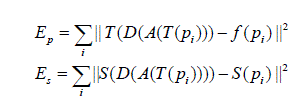
| Let T denote similarity transformation, A denote articulation that can rotate mu with respect to the joint connected to mv , D denote a deformation function that can move pi to a new position, and f denote a correspondence function from M to C. The aim is to find T , A, D and f such that the total error E is minimized: | ||||||||||
 |
||||||||||
IV.FRACTURE DETECTION |
||||||||||
| The complex problem of segmenting bone structures in x-ray images can be decomposed into several sub-problems as follows. | ||||||||||
| 1. Feature Selection In this step, stable features should be selected to obtain correct correspondence between the points in the atlas and the points in the target image. This is an important first step that will affect the result of the following steps. | ||||||||||
| 2. Global Alignment Based on the features found in the above step, global alignment is to determine a rough alignment between the atlas and the image in terms of scaling, rotation and translation. This step is performed with respect to the whole bone structure. In addition, articulations between connected bones should be taken into account. | ||||||||||
| 3. Local Refinement Local refinement is to accurately register the atlas contours to the boundaries of the bones. It needs to take into account the articulation and shapes of the bones[4]. The research plan is to solve each of these sub-problems and then integrate the algorithms into a complete solution. | ||||||||||
| The fracture detection techniques proposed can be loosely categorized into classification-based and transform-based. The method detected femur fractures by computing the angle between the neck axis and shaft axis. Since the individual classifiers tend to complement each other, the combined method improves both the accuracy and sensitivity significantly [10]. The use of Hough transformation in identifying fractures has also been proved advantages. The Hough transform (Duda and Hart, 1972) is a feature extraction technique in image analysis, computer vision, and digital image processing. It is concerned with the identification of straight lines, position of arbitrary shapes, most circles or ellipses. The important case of Hough transform is the linear transform for detecting straight lines. Compared with other algorithms that detect straight lines, Hough transform can be used to find and link segments in an image. A line in the image space is mapped to a point in the parameter space. Similarly, each pixel of the image space is transformed to a parameterized curve of the parameter space. Each transformed point in the parameter space is considered as a candidate for being a line and accumulated in the corresponding cell of an accumulator.[7,8] Finally, a cell with a local maximum of scores is selected, and its parameter coordinates are used to represent a line segment in the image space. The main advantage of the Hough transform technique is that it is tolerant of gaps in feature boundary description and is relatively unaffected by image noise. However, using Hough transform introduces computation complexity, which in turn slows the feature extraction and fracture detection process. | ||||||||||
VI. CONCLUSSION |
||||||||||
| In this proposal, a thorough review of the general medical image segmentation algorithms, particularly atlas-based medical image segmentation algorithm and x-ray image segmentation algorithms is presented. General segmentation algorithms are categorized into six classes, namely thresholding, region-based, edge-based, graph-based, classification-based and deformable models. Apart from deformable models, they usually work on either individual pixels or local image patches, and are very sensitive to noise. In comparison, deformable models perform segmentation by fitting a connected contour to the features (usually edges) in the image. Therefore, they are less sensitive to noise. | ||||||||||
| From the visual results, it could be seen that the proposed algorithm produces better segmentation results than the traditional region growing and active contour algorithms. Fact that the proposed method combining wavelets and morphological transform based segmentation algorithm has improved the existing system and can be used by other bone image analysis algorithms. In future, the effect of segmentation during fracture detection is analyzed. | ||||||||||
ACKNOWLEDGMENT |
||||||||||
| Valuable discussion and proper suggestions from Assit. Prof.P.H.ZOPE, Prof. S.R.SURALKAR Sir and all the senior faculties of SSBT, COE Jalgaon,(M.S.),India. Millions of thanks to all my friends who support me for the research. | ||||||||||
Tables at a glance |
||||||||||
|
||||||||||
Figures at a glance |
||||||||||
|
||||||||||
References |
||||||||||
|


