ISSN ONLINE(2319-8753)PRINT(2347-6710)
ISSN ONLINE(2319-8753)PRINT(2347-6710)
R K Singh1 F W Bansode2 and A. K. Meena1
|
| Related article at Pubmed, Scholar Google |
Visit for more related articles at International Journal of Innovative Research in Science, Engineering and Technology
The non-hormonal contraceptive, named RISUG (an acronym for Reversible Inhibition of Sperm under Guidance) has expected to provide a valuable addition to the current options of male contraception. The present study was conducted to determine the reproductive functional success, safety of vas occlusion by RISUG, and its reversal by dimethylsulphoxide (DMSO), followed by multigenerational (F1-F3) teratogenicity studies in rats. RISUG - a copolymer of styrene maleic anhydride (SMA) dissolved in 0.01 ml DMSO was injected into the lumen of the vas deferens bilaterally at the dose levels of 0.25, 0.50 and 1.00 mg/vas/rat. Control rats injected with 0.01 ml DMSO only. Results showed that the vas-occlusion by RISUG-injection caused sperm abnormality in ejaculated sperms and inhibited pregnancy in female rats. But, it did not affect testicular spermatogenesis, daily sperm production rate (DSP) and epididymal sperm counts (ESC) after a post-injection period of 70 days. Vas occlusion reversal of 60 days restored sperm morphology and fertility profile without causing any significant changes in the body and reproductive organ weights, DSP, ESC, and embryological/teratological aspects in F1-F3 progenies. Results indicate that the intra-vasal injection of RISUG, inhibits pregnancy in female rats may be due to affecting sperm characteristics but not testicular function.
Keywords |
| RISUG, Contraceptive, Vas occlusion, DMSO. |
INTRODUCTION |
| The non-hormonal contraceptive, named RISUG (an acronym for Reversible Inhibition of Sperm Under Guidance) has expected to provide a valuable addition to the currently limited options of male contraceptio1 (Ananthaswamy, 2002). RISUG (Mark sans Pharma, Mumbai, India) consists of a co-polymer styrene maleic anhydride (SMA) dissolved in 99.9% pure dimethylsulphoxide (DMSO) (Guha, 1996). A single injection (therapeutic dose of 60 mg SMA in 120 ml of DMSO) bilaterally into the vas deferens of the male in a minimally invasive manner has been shown to cause the disintegration of sperms and leads to necrozoospermia (Chaudhury et al., 2004). SMA is one such agent which when placed in the vas deferens lumen lowers the pH of the luminal micro-environment and inhibits sperm acrosomal reaction within 3h post-injection before the ejaculation in human (Guha, 2007; Sharma et al., 2008). The reported reversal studies of vas occlusion have been shown to cause normospermia with normal sperm motility and viability 30 days after reversal in langur monkeys (Lohiya et al., 1998). |
| Phase II clinical trials of RISUG clearly indicate that the absence of living sperm is not to be interpreted as a total obstruction of the vas deferens as obtained with vasectomy (Guha et al., 1997). Mishra et al., (2003) have also investigated the status of spermatogenesis and sperm parameters in langur monkeys following long-term vas occlusion with RISUG. The ejaculated sperms were found to be necroasthenoterato-zoospermic, suggesting instant sterility. The residues of cells which were seen could be due to disruption of the membrane system (Guha, 1996). An additional advantage of this technique is that it causes a partial blockage of the vas deferens with concomitant flow of functionally inactive cells (Sharma et al., 2001; Chaudhury et al., 2002). RISUG is retained in the folds of the inner wall of the vas deferens for a long period of time despite not being tissue adherent. Phase I (Guha et al., 1993) and Phase II (Guha et al., 1997) clinical trials have been successfully completed and currently a Phase III multicentre trial is underway (Guha, 2007). The short term studies on semen and accessory gland function in phase III clinical trials subjects confirmed azoospermia between 1-4 months post-injection period and absence of pregnancy during 6 months study period (Chaki et al., 2003). Male mediated non-toxic teratogenic potential have been demonstrated in rabbits by Sethi et al., (1990a). Teratogenic safety on reversal (Sethi et al., 1990b) and reversibility has also been confirmed in rats previously (Koul et al., 1998; Guha, 1999). |
| However, information on the reproductive function and multigenerational teratogenicity studies is lacking. Therefore, the present study was conducted on testicular function and multigenerational teratogenicity so as to examine the functional status and safety of reversal of RISUG by DMSO in vas occlusion, based on the reproductive organ weights, testis histology, estimation of daily sperm production rate, epididymal sperm counts, sperm morphology, fertility ratio, embryo-toxicity and teratological changes (if any) in F1, F2 and F3 progenies/offspring of reversal rats. |
MATERIALS AND METHODS |
Chemicals |
| Styrene maleic anhydride, a copolymer of styrene and maleic anhydride (1: 1 ratio), prepared by irradiation of these compounds at a dose of 0.2–0.24 megarad for every 40 g of copolymer, precipitated, dried and stored in a vaccum desiccator (Trade name – RISUG) dissolved in the solvent vehicle DMSO (spectroscopic grade) was kindly provided by Prof. S. K. Guha, School of Medical Science and Technology, Indian Institute of Technology (IIT), Kharagpur, India. All other chemicals used in this study were of analytical grade and purchased from Sigma Aldrich Chemical Company (India). |
Animals |
| Eighty adult male albino rats (Body weight 180-200 g) and 160 females of proven fertility (Body weight 140-150 g) of Charles Faster strain used in the study were obtained from Institute’s animal house. Animals were acclimatized for 1 week, maintained in standard laboratory conditions (24±20C) with 12: 12 h light and dark cycles in individual polypropylene cages. Animals were fed with pelleted standard rat diet (Lipton India Ltd., Bangalore) and water was provided ad libitum. Experimental protocol was approved by the ‘Institutional Animal Ethical Committee’ (IAEC) and was in compliance with the ‘Guidelines for Care and Use of Animals for Scientific Research (Indian National Science Academy, 2000). |
Experimental design |
Vas occlusion |
| Apparently healthy and disease-free rats were selected in the present study. Adult male rats were divided into four groups (Gr. I-IV) containing twenty animals each and subjected to bilateral vas occlusion under ether anesthesia. The vasa were exposed by a single median incision surgically just above the urethral opening as per method described previously (Sethi et al., 1990b). The distal ampullary portion of vas deferens was located beneath the fat bodies having greater diameter of duct where SMA was injected into the lumen of each vas deferens. Rats injected with 0.01 ml DMSO in the lumen of vas deferens, served as controls (Gr. I). Rats in Gr. II, III and IV were injected with 0.25mg, 0.50 mg and 1.00 mg SMA dissolved in 0.01 ml DMSO (SMA-DMSO complex) per vas per rat, respectively. The vasa of treated rats were placed properly in their original position and the incision was closed by stitching with catgut internally and upper skin incision with nylon thread. Post-operative care was taken by dressing with Neosporin antibiotic powder and merbromin solution (2% w/v) and anti-inflammatory drugs. The antibiotic, Terramycin (Pfizer Ltd, Bombay) was injected intramuscularly to each rat for 5 consecutive days as per method of Sethi et al, (1989). Animals were kept under watch to observe gross behavioral changes (if any) for 70 days. To ascertain the effects of vas occlusion by a single intra-vassal injection of SMA-DMSO complex on testicular spermatogenesis cycle, the testicular cross sections were examined, daily sperm production (DSP) rate was calculated, cauda epididymal sperm counting was done and a fertility test was conducted with normal females on days 71-74 of post-occlusion. The sperm morphology was checked microscopically in vaginal smears of mated rats for functional success of vas occlusion. After fertility test ten vehicle-control and ten vas-occluded rats by SMA-DMSO were necropsied for organ weight/histology/DSP/sperm counting studies. The remaining ten animals, each from control and vas-occluded groups, were subjected to vas occlusion reversal. |
Vas occlusion reversal |
| Vas occlusion reversal was done in SMA-treated male rats under ether anesthesia after 70 days of vas occlusion. The vasa were exposed and injected with 0.01 ml DMSO bilaterally into the lumen of vas deferens to flush out the SMA into the lumen of vas in a similar way to that of vas occlusion for a period 3-4 minutes so to dissolve SMA properly in DMSO and wash out of vassal duct (Sethi et al., 1992). The vasa were placed again into their original position, closed the incision with catgut and nylon thread and post-operative care was taken as described previously (Sethi et al., 1989; 1990c). Functional success of occlusion-reversal was ensured by fertility test after a post-reversal period of 60 days. Five animals from control-reversal group (Gr.I) and vas occlusion-reversal groups (Gr. II-IV) were mated with female rats and necropsy was done for organ weights/histology/DSP/sperm counting studies. |
F1-F3 progeny |
| F1 progenies were obtained by mating the vehicle control and SMA-treated (0.25, 0.5 and 1.0 mg RISUG) vas occlusion-reversal male rats with normal female rats in the ratio of 1:2. Following next day morning, the vaginal smears/plugs were observed for presence of spermatozoa and confirmation of pregnancy on day 1 post-coitum. Also the sperm morphology was checked. |
| The pregnant rats were observed throughout gestation period (21 days) for any change in appetite and gross behavioral changes such as irritation, depression and vaginal bleeding. On day 19-20 of gestation period prior to delivery, caesarian delivery was performed in 50% of pregnant rats for recording the number of resorption, implantations/implantation sites, corpora lutea, live and stillborn fetuses, size and weight, sex and gross abnormality (if any) in each fetuses as per method described previously (Sethi et al., 1989; 1992). |
| The remaining 50% pregnant rats were used for next generartion fertility test. The sister or brother pups were separated and did not use for fertility test with the same parents. The resultant offsprings of F1 progeny (n = 320) were maintained for 90 days to grow up. The next generations, F2 and F3 progenies were obtained in similar manners from the same groups and maintained separately in different groups (Gr. I-IV) in which they were initially treated with DMSO and SMA-DMSO complex as described to obtain F1 progeny. The resultant pups were kept to grow up to adulthood for fertility test. The 50% of the offsprings were again used for recording the number of resorptions, implantations, corpora lutea, live and stillborn fetuses, size and weight, sex and gross abnormality (if any). Necropsy for organ weight and sperm characteristics was done after completion of fertility test using five animals from each group. |
Parameters |
| The animals from vehicle-control, vas occlusion, reversal vehicle-control, vas occlusion reversal and F1, F2 and F3 offsprings obtained from initially reversal rats were evaluated for the following- |
Body and organ weights |
| Body weights were recorded initially (day 0), after 70 days post-injection and 60 days vas occlusion-reversal in male rats. Body weights of F1, F2 and F3 offsprings were also recorded on days 90 (assumed as day 0), 160 (as 70 days post - injection period) and 220 (as 60 days reversal period) of age. The weights of reproductive organs were also recorded at every scheduled sacrifice, i.e. 60 days post-reversal for all three F1, F2 and F3 progenies. |
Testis histology |
| Bouin’s fixed testes from vehicle control and SMA-treated (vas occluded) and reversal groups (Gr. I-IV) of rats were dehydrated in graded series of ethanol, cleared in xylene and infiltrated/embedded in paraffin wax (at 580C). Serial cross sections (5 μm) were stained with haematoxylin and eosin for histopathology. |
Daily sperm production rate in testis |
| Measurements of sperm production (DSP) in testicular homogenates were carried out as per method described previously (Amann et al., 1976). Briefly, testicular parenchyma was cut into small pieces, placed in 0·25 mol/l sucrose solution (pH 7·5) and homogenized in a fluid containing 150 mM NaCl/l, 3·8 mM NaN3/l, and 0·05% (v/v) Triton X- 100 using an Ultra Turrax® (Janke and Kunkel, Staufen, Germany). The homogenates were further diluted with the same medium, and homogenization-resistant spermatid nuclei in steps 17–19 of stages IV–VIII of the spermatogenic cycle were counted under the Olympus Trinocular Microscope (Olympus, Japan) using a Neubauer’s haemocytometer(Amann et al., 1976; Amann & Lambiase, 1969). DSP per gram testicular parenchyma was calculated by dividing the number of spermatid nuclei with the product of weight of parenchyma and time divisor of 6.3 days for rat as reported previously (Johnson et al., 1980). The values were represented as mean ± standard deviation (SD) for five animals in each group. |
Epididymal sperm counts |
| For estimation of epididymal sperm counts, cauda epididymis was excised, weighed, cut into small pieces in suspension medium containing 140 mmol NaCl, 0.3 mmol KCl, 0.8 mmol Na2HPO4, 0.2 mmol KH2PO4 and 1.5 mmol D-glucose (pH adjusted to 7.3 by adding 0.1 (N) NaOH) and sperms were collected by centrifugation at 100 × g for 2 min. The resultant precipitate was resuspended in fresh suspension medium, diluted and sperm counting was carried out using hemocytometer in duplicate in each rat under Phase contrast Olympus Trinocular microscope at x 200 magnification. The values were expressed as an average of total sperm counts per ml of suspension (Robb et al., 1978). |
Fertility test |
| Fertility testing of each male rat from control group (Gr. I) and SMA-injected groups (Gr. II-IV) was done on days 71– 73 of post-injection periods. These treated rats were cohabitated overnight with proestrous females in the ratio (1:2) and the positive mating was confirmed by presence of sperms in vaginal plugs or vaginal smears. The mated females were separated and maintained in individual cages to record the number of pups after delivery in F1 progeny. Fifty per cent of pregnant females were used for the implantation record, viz. number of corpora lutea, number of implantations and number of resorptions (if any) for the evaluation of embryo toxicity and teratogenicity. The remaining 50% of the pregnant females were allowed to complete the term for F2 progeny and like wise F3 progeny was studied. |
Skeletal evaluation |
| The selected pups/offspring from the control ⁄ reversal, vas-occluded ⁄ reversal group of animals were fixed in 70% alcohol and examined for external and visceral abnormalities following the method of Wilson and Warkany (Wilson, 1985). Some of the fetuses were cleared in 0.1 % potassium hydroxide (KOH) and stained by alizarin Red Technique (Sethi et al., 1990c; Christian et al., 2003) for skeletal evaluation. |
Statistical analysis |
| Student’s ‘t’ test and one-way ANOVA (one factor analysis of variance) was applied for statistical significance and comparisons between control and treated groups of rats. P values < 0.1 and 0.2 were considered as non-significant and p values < 0.05 considered as significant. |
RESULTS |
General behavior |
| The female rats of proven fertility were mated earlier with SMA-treated males did not show any change in appetite, water intake and in general observable behavior, depression/irritation, during gestation period. The vas occluded control and SMA- injected and reversal animals had adopted the normal behavior within a week of surgery. None of them showed hypo and hyper excitability of nervousness during implants, post-operative reversal and handling thereafter. The gestation period observed in treated rats was 20.5±0.71 days comparable to that of control (20.00±1.41 days) rats. There was no vaginal bleeding in female animals. No abnormal effect on labor and delivery of female rats was noticed. No effect on lactation was observed. No mortality was observed during gestation period. There was no any effect observed on estrous cycles (normal estrous cycle duration 4.5 ±0.5 days) and mating behavior. |
Body and reproductive organ weights |
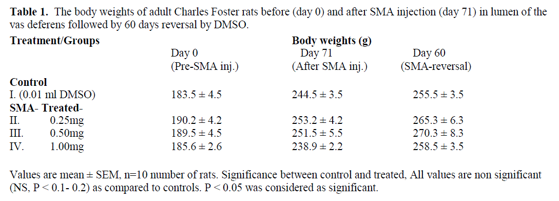
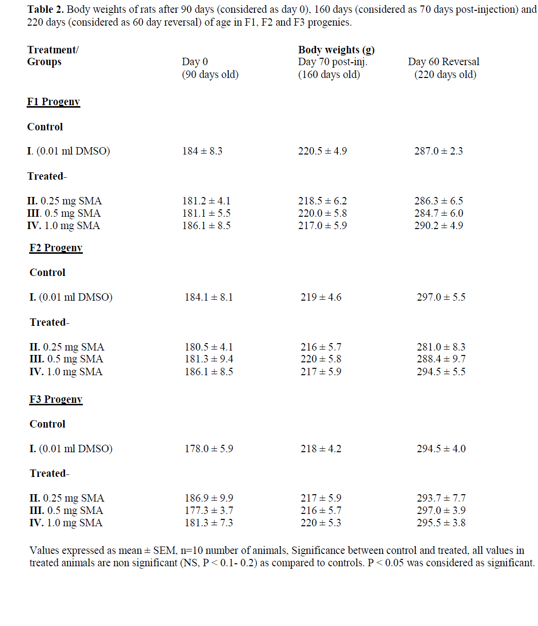
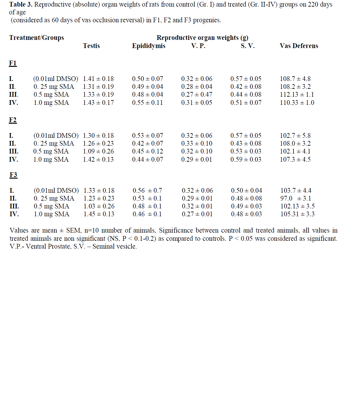
| The data on body weights and reproductive organ weights from different groups (viz. control and SMA-treated, vas occlusion-reversal) of rats and from F1, F2 and F3 progenies is summarized in Tables 1-3. The results showed no significant changes (P < 0.1-0.2) in body weights before vas occlusion by SMA-injection (day 0), after vas occlusion with single SMA-injection (on day 70) and after vas occlusion reversal (on day 60) in treated as compared to control male rats injected with 0.01ml DMSO only. The body weights in F1, F2 and F3 progenies have also not shown any significant changes (P < 0.1-0.2) in offsprings obtained from different group of treated rats as compared to control on days 90 (as day 0), 160(as 70 days post-injection) and 220 (as 60 days reversal) of age (Table 2). The Reproductive organ weights viz. testes, epididymis, ventral prostate, seminal vesicles and vas deferens did not show any significant change (P < 0.1-0.2) in treated-reversal as compared to corresponding controls in F1, F2 and F3 offsprings (Table 3). |
 |
 |
 |
 |
 |
Daily sperm production rate and epididymal sperm counts |
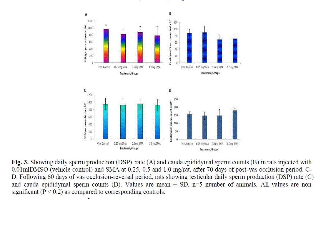
| The DSP/gm testicular parenchyma in SMA-injected (vas occluded) and occlusion-reversal rats was comparable to that in vehicle control rats. The cauda epididymal sperm counts were observed to be similar in vehicle control, SMA-injected reversal group of rats (Fig. 3 A-D). Testicular DSP rate and cauda epididymal sperm counts were also observed to similar in F1, F2 and F3 progenies as compared to corresponding control group of rats (Fig. 4 A, B). |
 |
Fertility record |
| Fertility test carried out in normal adult rats before injection of SMA and in DMSO injected control (Gr. I) rats which were mated with normal females in 1:2 ratio, showed 100% fertility as per microscopic observation of presence of spermatozoa in vaginal smears/ plugs of female rats. In rats injected with SMA at doses 0.25, 0.5 and 1.0 mg/vas (Gr. II-IV), showed 0% fertility when mated with normal adult female rats. The vas occlusion reversal rats mated with female rats showed 100% fertility. Fertility record for F1, F2 and F3 progenies showed comparable normal fertility index to that of controls (Table 4). |
| Teratological parameters observed were the number of corpora lutea, number of implantation sites/implants, live births, number of resorptions, average fetal body weight, average fetal body length and average fetal body width of offsprings. These teratological observations of SMA-reversal rats were comparable (P < 0.1- 0.2) with that of control group of rats when vas occlusion reversal rats mated with normal cyclic female rats in all three (F1, F2 and F3 progenies) generations (Table 5). |
Gross Observations |
| In foetuses, the abnormalities such as kinking of tail, everted claw and valgus deformity were seen in control as well as in treated group of animals. These are common in foetuses delivery in Institute's breeding colony. |
Visceral and Skeletal morphology |
| No visceral abnormality was seen in fetuses from F1, F2, and F3 progeny of either control or SMA-treated reversal groups of rats. Skeletal morphology of the F1, F2, and F3 progeny by alizarin red stain showed no detectable abnormalities in both control and vas occluded-reversal animals. |
DISCUSSION |
| The surgical vasectomy, a male contraceptive method blocks the passage of spermatozoa permanently and there is no vasectomy reversal in further. Such surgical contraceptive methods also have shown to cause epididymal dysfunction, abnormality in spermatozoa and unable to restore fertility (Srivastava et al., 2000). Recently, chemical occlusion of the vas deferens by RISUG (Reversible Inhibition and Sperm Under Guidance) has been considered to be an ideal male contraceptive method which is in Phase III clinical trial after successful completion of phase I and II clinical trials. The polymer SMA can be used to block the lumen of the vas deferens over an extended period of time. In our laboratory, systemic toxicity evaluation of RISUG-injection had been carried out in detail in rat, rabbit and rhesus monkeys previously (Sethi et al., 1989; 1990a, b, c, d; 1991). Findings indicated that SMA-injection did not cause any systemic toxicity, male mediated teratogenicity and carcinogenic potential in experimental animals. |
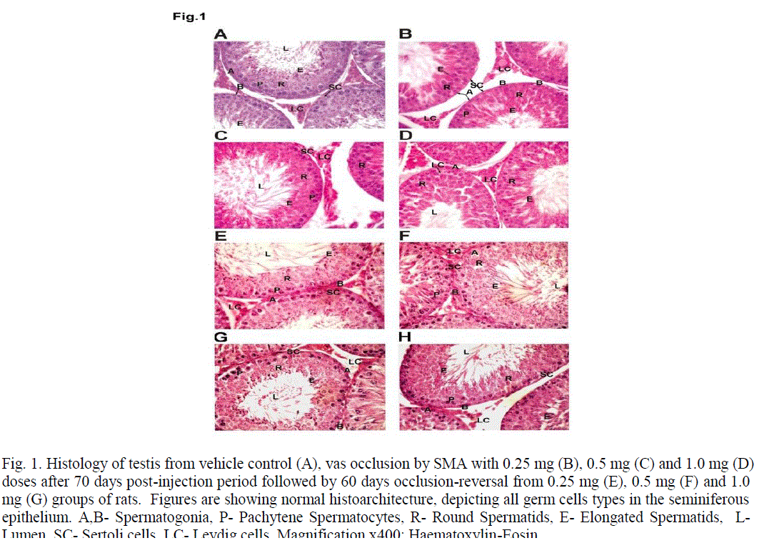
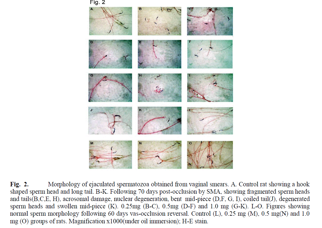
| Present study on multigenerational teratogenicity in Charles Foster rats injected with RISUG showed no observable changes in gonadal function (viz. organ weights, testicular spermatogenesis, DSP, ESC), estrus cycles, mating behavior, conception rates, early stages of gestation, organogenesis, gestation period, labor and delivery, litter size, litter weights during different days of weaning and lactation was not affected. The vas occlusion by SMA-injection after a postinjection period of 70 days in rats caused 100% inhibition of fertility when treated males were mated with normal females. The testicular spermatogenesis, daily sperm production rates and epididymal sperm counts were not affected by vas occlusion. The sperm morphology was observed to be tremendously disturbed in SMA-treated males after 70 days post-injection period. It showed breakage of head and tails, blabbing of heads, curved tails, acrosomal damages, nuclear degeneration, etc. Similar changes in sperm characteristics and morphology causing damage to head (acrosome) and midpiece have been reported to cause sterility within short period of vas occlusion by RISUG-injection in rats (Lohiya et al., 1998; 2010). The intrinsic property of such polymers by lowering pH leads to inhibition of functional ability of spermatozoa to fertilize the ovum has been reported earlier (Sethi et al., 1992; Misro et al., 1979; Guha et al., 1985). Further, previous studies have been clearly demonstrated the polyelectrolytic nature of RISUG that induces a surface charge imbalance on the human sperm membrane system and the integrity which is essential for sperm-oocyte interaction (Chaudhury et al., 2004; Sharma et al., 2008; Jha et al., 2009). In-Vitro studies with RISUG (at the concentration of 1.0 mg SMA dissolved in 100 ml of DMSO) have been shown to cause significant damage to the acrosome and its contents indicating loss of functional ability of sperms as evidenced by decreased levels of hyaluronidase, proacrosin and acrosin enzymes activity (Chaudhury et al., 2004). In the present study 0.01 ml DMSO is a very small dose and it did not cause any toxic effect even at 0.03 ml (Sethi et al., 1989; 1990b). This polymer injection reacts with cellular secretion and forms a stable precipitate within the lumen of vas and makes the microenvironment by lowering pH as well as enzymatic profile of sperms leading to sperm cell death. Moreover, magnetic field-directed Fe3O4–Cu–SMA–DMSO Cuproferrogel drug interaction with sperm cells that may open new dimensions in the field of biomagnetic technology. RISUG action on sperm cells had not altered by higher pulse magnetic field (PMF). But, PMF of 760 mT or above, causes 30% decrease in the sperm count and 70% decrease in sperm motility that induce the spermicidal enhancement potential (Jha et al., 2009). In females, effects SMA-injection in intra-fallopian tube / oviduct have been shown to cause oocyte abnormality, enlargement of zona, membrane disintegration and viability within 72 h (Jha et al., 2010). |
| The vas occlusion reversal (SMA-injected Gr. II-IV) mated with female rats, showed non significant (P < 0.1-0.2) changes in the body and reproductive organ weights, number of copra lutea /implantation sites/ resorptions and litter size when compared with that of control group (DMSO-injected control reversal males) of rats and in F1, F2 and F3 progenies as compared to appropriate controls. The sperm morphology was restored and testicular sperm production rate and epididymal sperm counts were similar to that in control rats. The visceral and skeletal morphology was same in treated- and control-reversal groups similar to recently reported study of Lohiya et al., (2010) However, in foetuses, the abnormalities such as kinking of tail, everted claw and valgus deformity were occasionally seen in control as well as in treated group of animals. These abnormalities are very common in the foetuses delivery in our Institute's breeding colony. No visceral abnormality was observed except minor findings in skeletal morphology such as nonossified skull bones, ribs bent inside or intercostal spaces, seen occasionally in all groups. It has no relevance to teratogenic action of SMA in foetuses from F1, F2, and F3 progenies of either control or SMA-treated reversal group of rats. After an analysis of the data generated in three segmental studies up to three generations, it was concluded that RISUG - injection did not cause any teratogenic effect in Charles Foster rats at the doses (0.25, 0.5 and 1.0 mg/vas/rat) used in the present study. Therefore, reversal of RISUG vas occlusion and by DMSO is safe, feasible and effective leading to 100% fertility reversal. Thus, it may indicate an important non-invasive approach for humans. Further, multi-centric Phase III clinical trials in humans are in progress. |
ACKNOWLEDGEMENTS |
| Authors are grateful to Prof. S. K. Guha, School of Medical Science and Technology, Indian Institute of Technology (IIT), Kharagpur, India for providing the RISUG. Authors thanks to Dr. (Mrs.) N. Sethi for excellent guidance and Mr. R. K. Srivastava for technical assistance. This project was funded by I.C.M.R. and C.S.I.R., New Delhi. |
CONCLUSION |
| In conclusion, our results demonstrated that saraca indica inhibits the formation of free radicals in rats showing potential antioxidant effect. It was also exhibiting significant protection on blood parameters. Saraca indica might be potent therapeutic agent in treating hematinic related disorders since they possess both antioxidant and Hematoprotective potentials. |
References |
|