Keywords
|
| Cardiac arrhythmias, Electrocardiogram (ECG) , Discrete Wavelet Transform(DWT),P-QRS-T Segment. |
INTRODUCTION
|
| An ECG machine interprets and records the electrical impulses of the heart for diagnostic purposes. It is not a form of treatment for heart conditions. An electrocardiogram (ECG) records the electrical activity of the heart. The heart produces tiny electrical impulses which spread through the heart muscle to make the heart contract. These impulses can be detected by the ECG machine. An ECG to help find the cause of symptoms such as palor chest pain. The ECG test is painless and harmless (The ECG machine records electrical impulses coming from your body) it does not put any electricity into your body [1]. The ECG consists of several electrodes which are attached to the body of the patient and are connected by wires to the device. The device itself consists of a graphing device (originally paper, although electronic recorders are becoming more common). Each one of the sensors can detect a change in electrical charge in the skin that can only be the result of the impulses that are travelling through the heart and on to the rest of the body. It starts in the upper right ventricle, which contracts in response, and quickly travels through the other three chambers of the heart, which contract all at once. This impulse travels very quickly and is also transmitted to the cells surrounding the heart as it dissipates throughout the body. This forms a very regular pattern which establishes a control for patients that do have heart problems, such as arrhythmia. ECG signals are recorded on paper chart or on a patient monitor. ECG signal tracing are described by wave components or segments [2]. |
| An electrocardiogram (EKG or ECG) is done to: |
| • Check the heart's electrical activity, how fast your heart is beating. |
| • Find the cause of unexplained chest pain, which could be caused by a heart attack, inflammation of the sac surrounding the heart (pericarditis), or angina. |
| • Find the cause of symptoms of heart disease, such as shortness of breath, dizziness, fainting, or rapid, irregular heartbeats (palpitations). |
| • Find out if the walls of the heart chambers are too thick (hypertrophied). |
| • Check how well medicines are working and whether they are causing side effects that affect the heart. |
| • Check how well mechanical devices that are implanted in the heart, such as pacemakers, are working to control a normal heartbeat. |
| • Check the health of the heart when other diseases or conditions are present, such as high blood pressure, high cholesterol, cigarette smoking, diabetes, or a family history of early heart disease. |
| An ECG also can show: |
| • Lack of blood flow to the heart muscle (coronary heart disease) |
| • A heartbeat that's too fast, too slow, or irregular (arrhythmia) |
| • A heart that doesn't pump forcefully enough (heart failure) |
| • Heart muscle that's too thick or parts of the heart that is too big (cardiomyopathy) |
| • Birth defects in the heart (congenital heart defects) |
| •Problems with the heart valves (heart valve disease) |
| • Inflammation of such that the surrounds the heart (pericarditis). |
| The test also shows how long it takes for electrical signals to travel through the heart [2] [3]. |
HISTORY
|
| EKG stands for electrocardiogram. It should probably and sometimes is abbreviated ECG, but EKG seems to have stuck as more popular. ECG or EKG is short for electrocardiogram and is called both an ECG and an EKG, as abbreviations for the word electrocardiogram (derived from the Greek electro for electric, cardio for heart, and graph for “to write”) and the German word electrocardiogram. An initial breakthrough came when Willem Einthoven, working in Leiden, the Netherlands, used the string galvanometer he invented in 1901. This device was much more sensitive than both the capillary electrometer Waller used and the string galvanometer that had been invented separately in 1897 by the French engineer Clement Ader. Rather than using today's self-adhesive electrodes Einthoven's subjects would immerse each of their limbs into containers of salt solutions from which the ECG was recorded. Einthoven assigned the letters P, Q, R, S, and T to the various deflections, and described the electrocardiographic features of a number of cardiovascular disorders. In 1924, he was awarded the Nobel Prize in Medicine for his discovery. Though the basic principles of that era are still in use today, many advances in electrocardiography have been made over the years. The instrumentation, for example, has evolved from a cumbersome laboratory apparatus to compact electronic systems that often include computerized interpretation of the electrocardiogram [1] [4]. |
THE HEART
|
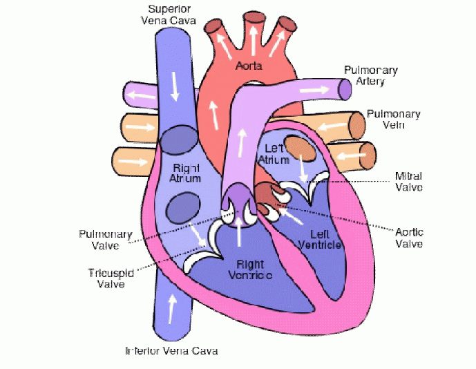
| The heart is composed of four chambers which make up two pumps. There are two upper chambers, called the right and left atria, and two lower chambers, called the right and left ventricles. The purpose of the Atria, is receive blood from the body, the right atrium receives oxygen-devoid blood from the body and the left atrium receives oxygen-rich blood from the lungs. The heart is controlled by a very precise electrical system. The right pump receives the blood returning from the body and pumps it to the lungs. The left pump gets blood from the lungs and pumps it out to the rest of the body. Each Pump is made up of two chambers, an atrium and a ventricle. The atrium collects the Incoming blood, and when it contracts, transfers the blood to the ventricle. When the ventricle contracts the blood is pumped away from the heart. The pumping action of the heart is regulated by the pacemaker region, or sinoatrial node, located in the right atrium. An electrical impulse is created in this region by the diffusion of calcium ions, sodium ions, and potassium ions across the membranes of cells [5]. The impulse created by the motion of these ions is first transferred to the atria, causing them to contract and push blood into the ventricles. After about 150 milliseconds, the impulse moves to the ventricles, causing them to contract and pump blood away from the heart. This system regulates the mechanical pumping action of the heart so that the entire cardiovascular system can function properly. The ECG device detects and amplifies the tiny electrical changes on the skin that are caused when the heart muscle depolarizes during each heartbeat. At rest, each heart muscle cell has a negative charge, called the membrane potential, across its cell membrane. Decreasing this negative charge toward zero, via the influx of the positive cat ions, Na+ and Ca++, is called depolarization, which activates the mechanisms in the cell that cause it to contract. During each heartbeat, a healthy heart will have an orderly progression of a wave of depolarization that is triggered by the cells in the sinoatrial node, spreads out through the atrium, passes through the atrioventricular node and then spreads all over the ventricles. This is detected as tiny rises and falls in the voltage between two electrodes placed either side of the heart, which is displayed as a wavy line either on a screen or on paper. This display indicates the overall rhythm of the heart and weaknesses in different parts of the heart muscle. Structure of Human Heart [6]. |
METHODOLOGY& PRINCIPLE OF ECG
|
| The ECG device detects and amplifies the tiny electrical changes on the skin that are caused when the heart muscle depolarizes during each heartbeat. At rest, each heart muscle cell has a negative charge, called the membrane potential, across its cell membrane. Decreasing this negative charge toward zero, via the influx of the positive cat ions, Na+ and Ca++, is called depolarization, which activates the mechanisms in the cell that cause it to contract [6]. During each heartbeat, a healthy heart will have an orderly progression of a wave of depolarization that is triggered by the cells in the sinoatrial node, spreads out through the atrium, passes through the atrioventricular node and then spreads all over the ventricles. This is detected as tiny rises and falls in the voltage between two electrodes placed either side of the heart, which is displayed as a wavy line either on a screen or on paper[7] [8]. This display indicates the overall rhythm of the heart and weaknesses in different parts of the heart muscle [9]. |
THE WAVES AND INTERVALS OF ECG
|
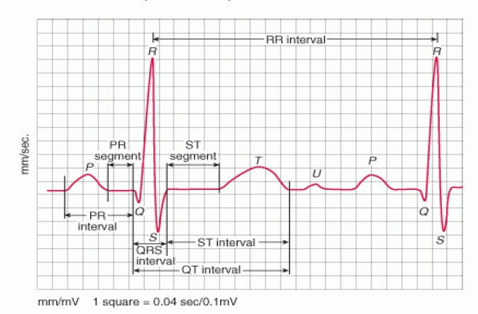
| A typical ECG tracing of the cardiac cycle (heartbeat) consists of a P wave, a QRS complex, a T wave, and a U wave, which is normally invisible in 50 to 75% of ECGs because it is hidden by the T wave and upcoming new P wave. The baseline of the electrocardiogram (the flat horizontal segments) is measured as the portion of the tracing following the T wave and preceding the next P wave and the segment between the P wave and the following QRS complex (PR segment) [10] [11]. In a normal healthy heart, the baseline is equivalent to the isoelectric line (0 mV) and represents the periods in the cardiac cycle when there are no currents towards either the positive or negative ends of the ECG leads. However, in a diseased heart, the baseline may be depressed (e.g., cardiac ischaemia) or elevated (e.g., myocardial infarction) relative to the isoelectric line due to injury currents during the TP and PR intervals when the ventricles are at rest. The ST segment typically remains close to the isoelectric line as this is the period when the ventricles are fully depolarized and thus no currents can be in the ECG leads. Since most ECG recordings do not indicate where the 0 mV line is, baseline depression often gives the appearance of an elevation of the ST segment and conversely baseline elevation gives the appearance of depression of the ST segment. The electrocardiographic deflections are termed P, QRS complex, T and U [12]. |
| •the P wave represents Atria activation (Contractions). |
| • QRS complex represents ventricular activation or depolarization. |
| •the T wave represents ventricular recovery or re-polarization |
| •S-T segment, the T wave and the U wave together represent the total duration of ventricular recovery [13]. Normal ECG wave form show in figure below [14]. |
CLINICAL IMPORTANT PARAMETERS
|
| A. RR-Interval: - The interval between an R wave and the next R wave; normal resting heart rate is between 60 and 100 bpm Duration of RR-Interval 0.6 to 1.2s. |
| B. P-Wave: - During normal atria depolarization, the main electrical vector is directed from the SA node towards the AV node and spreads from the right atrium to the left atrium. This turns into the P wave on the ECG. Duration of P-Wave is 80ms. |
| C. PR-Interval: - The PR interval is measured from the beginning of the P wave to the beginning of the QRS complex. The PR interval reflects the time the electrical impulse takes to travel from the sinus node through the AV node and entering the ventricles. The PR interval is, therefore, a good estimate of AV node function. Duration of PR-Interval is 120 to 200ms. |
| D. PR-Segment: - The PR segment connects the P wave and the QRS complex. The impulse vector is from the AV node to the Bundle of His to the bundle branches and then to the Purkinje fibers. This electrical activity does not produce a contraction directly and is merely traveling down towards the ventricles, and this shows up flat on the ECG. The PR interval is more clinically relevant. Duration of PR-Segment is 50to120ms. |
| E. QRS-Complex: - The QRS complex reflects the rapid depolarization of the right and left ventricles. The ventricles have a large muscle mass compared to the atria, so the QRS complex usually has much larger amplitude than the P-wave. Duration of QRS-Complex is 80 to 120ms. |
| F. ST- Segment: - The ST segment connects the QRS complex and the T wave. The ST segment represents the period when the ventricles are depolarized. It is isoelectric. Duration of ST-Segment is 80to120ms. |
| G. ST- Interval: - The ST interval is measured from the J point to the end of the T wave. Duration of ST-Interval is 320ms. |
| H. QT-Interval: - The QT interval is measured from the beginning of the QRS complex to the end of the T wave. A prolonged QT interval is a risk factor for ventricular tachyarrhythmia and sudden death. It varies with heart rate and, for clinical relevance, requires a correction for this, giving the QTc. Duration of QT-Interval is up to 420 ms in heart rate of 60 bpm. |
ARRHYTHMIA AND CORRESPONDING ABNORMALITIES
|
| An arrhythmia (ah-RITH-me-ah) is a problem with the rate or rhythm of the heartbeat. During an arrhythmia, the heart can beat too fast, too slow, or with an irregular rhythm. A heartbeat that is too fast is called tachycardia (TAK-ih-KAR-de-ah). A heartbeat that is too slow is called bradycardia (bray-de-KAR-de-ah). Most arrhythmias are harmless, but some can be serious or even life threatening. During an arrhythmia, the heart may not be able to pump enough blood to the body. Lack of blood flow can damage the brain, heart, and other organs. Many things can lead to, or cause, an arrhythmia, including: |
| • A heart attack that's occurring right now. |
| • Scarring of heart tissue from a prior heart attack. |
| • Changes to your heart's structure, such as from cardiomyopathy. |
| • Blocked arteries in your heart (coronary artery disease) |
| • High blood pressure. |
| • Diabetes. |
| The four main types of arrhythmia are |
| A. Premature (extra) beats: - Premature beats are the most common type of arrhythmia. They're harmless most of the time and often don't cause any symptoms. Premature beats that occur in the atria (the heart's upper chambers) are called premature atrial contractions, or PACs. Premature beats that occur in the ventricles (the heart's lower chambers) are called premature ventricular contractions or PVCs. Some heart diseases can cause premature beats. They also can happen because of stress, too much exercise, or too much caffeine or nicotine. |
| B. Supraventricular Arrhythmias: - Supraventricular arrhythmias are tachycardia (fast heart rates) that start in the atria or atrioventricular (AV) node. The AV node is a group of cells located between the atria and the ventricles. Types of supraventricular arrhythmias include atria fibrillation (AF), atria flutter, paroxysmal supraventricular tachycardia (PSVT), and Wolff-Parkinson-White (WPW) syndrome. |
| C. Ventricular Arrhythmias: - These arrhythmias start in the heart's lower chambers, the ventricles. They can be very dangerous and usually require medical care right away. Ventricular arrhythmias include ventricular tachycardia and ventricular fibrillation (v-fib). Coronary heart disease, heart attack, a weakened heart muscle, and other problems can cause ventricular arrhythmias. |
| D. Brady arrhythmias: - Brady arrhythmias occur if the heart rate is slower than normal. If the heart rate is too slow, not enough blood reaches the brain. This can cause you to pass out. In adults, a heart rate slower than 60 beats per minute is considered a brad arrhythmia. Some people normally have slow heart rates, especially people who are very physically fit. For them, a heartbeat slower than 60 beats per minute isn't dangerous and doesn't cause symptoms. But in other people, serious diseases or other conditions may cause brad arrhythmias [15]. |
CARDIAC DISEASES DETECTED BY ECG:-
|
| A. Tachycardia: Tachycardia is an abnormally rapid beating of the heart, defined as a resting heart rate of 100 or more beats per minute in an average adult. |
| B. Bradycardia: Bradycardia (from Greek Brady=slow and cardiac=heart), as applied in adult medicine, is defined as a resting heart rate of under 60 beats per minute. |
| C. Long QT syndrome (LQTS): Long QT syndrome (LQTS) is a heart disease in which there is an abnormally long delay between the electrical excitation (and depolarization) and relaxation (depolarization) of the ventricles of the heart. |
| D. Short QT syndrome: Short QT syndrome is a genetic disease of the electrical system of the heart. It consists of a constellation of signs and symptoms, consisting of a short QT interval on EKG (≤ 300 ms). |
| E. First-degree heart block: First-degree heart block (PR interval >200 msec) is a disease of the electrical conduction system of the heart. It may be due to conduction delay in the AV node. |
| F. Second-degree block: Occasional absence of QRS and T after a P wave of sinus origin. |
| G. Third-degree block: Absence of any relationship between P waves of sinus origin and QRS complexes (AV dissociation) [16] [17] [18]. |
LEAD POSITIONING OF ECG
|
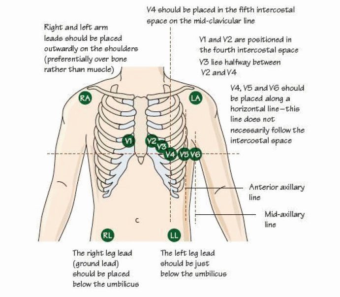
| The ECG is one of the most useful investigations in medicine. Electrodes attached to the chest and/or limbs record small voltage changes as potential difference, which is transposed into a visual tracing. Many heart problems are present all the time, and a resting 12-lead ECG will detect them [19] [20]. |
FEATURE EXTRACTION OF ECG SIGNAL
|
| It has now gone beyond the capacity of the expert cardiologist to take care of large numbers of cardiac patients efficiently & effectively. Therefore, computer- aided feature extraction and analysis of ECG signal for disease diagnosis has become the necessity. The first step in computer aided diagnosis is the identification & extraction of the features of the ECG signal. The QRS complex is the most prominent feature and its accurate detection forms the basis of extraction of other features and parameters from the ECG signal. There are a number of methods, some of which deal with detection of ECG wave segments, namely P, QRS and T, while others deals with detection of QRS complexes[21]. A good amount of research work has been carried out during the last five decades for the accurate and reliable detection of QRS segment in the ECG signal. The QRS detection algorithms developed so far can be broadly placed into four categories: (i) syntactic approach (ii) non-syntactic approach (iii) hybrid approach and (iv) transformative approach. |
| A. Syntactic Approach |
| The syntactic approach is basically pattern recognition based QRS detection techniques. The ECG signal is first reduced into a set of elementary patterns like peaks, durations, slopes, inter wave segments and thereafter use rule based grammar. The signal is represented as a composite entity of peaks, duration, slopes and inters wave segments. These patterns are then used to detect the QRS complexes in the ECG signal. These methods are time consuming and require inference grammar in each step of execution for QRS detection. Even then the motivation for using a syntactic approach resides in the fact that human inspection of ECG waveform is firstly an extraction of structural and qualitative information. Once this information is obtained and some typical forms (like a QRS complex) are recognized then the numerical values of the durations and amplitudes are measured for use in diagnosis [22]. |
| B. Non-syntactic Approach |
| Non-syntactic type is the most widely used class of ECG feature extraction techniques. In this class, we find the use of amplitude, slope and threshold limit as well as the use of different filters, mathematical functions and models. Okada reported a five step digital filter, which removes components other than those of QRS complex from the recorded ECG [23]. The final step of the filter produces a square wave and its on-intervals correspond to the segments with QRS complexes in the original signal. Thacker et al carried out power spectral analysis of ECG waveform, as well as of isolated QRS complexes and episodes of noise and artifacts [24]. A band pass filter was used to maximize the signal (QRS complex) to noise (T-waves, 60 Hz, EMG etc.) ratio to detect the QRS complex. Due to the inherent variability of ECG from different persons, as well as variability due to noise and artifacts, the filter design was suboptimal in specific situations [25]. Recently, some new techniques have also been developed base on artificial neural network, fuzzy logic and genetic algorithms for accurate QRS detection .In these approaches, the basic methodology is to learn and later on to generalize the knowledge gained through the learning process to identify the known QRS complexes out of an exhausted set of the ECG segments. |
| C. Hybrid Approach |
| In hybrid approach, the syntactic and non-syntactic approaches are combined to detect the QRS complex. These are not in common use, as in syntactic approach, the trace is being made on actual morphology of the ECG signal and in nonsyntactic approach; there is no consideration to maintain the morphology of the ECG signal. |
| D. Transformative Approach |
| Transformative Techniques, namely Fourier Transform, Cosine Transform, Pole –zero Transform, Differentiator Transform, Hilbert Transform and Wavelet Transform are being used for the QRS detection. The use of these transforms on ECG signal helps to characterize the signal into energy, slope, or spike spectra, and thereafter, the temporal locations are detected with the help of decision rules like thresholds of amplitude, slope or duration. Murthy and Prasad proposed a solution to the fundamental problem of ECG analysis, viz., delineation of the signal into its component waves [26]. In recent times, the use of Wavelet Transform (WT) in QRS detection has shown upper edge in terms of accuracy of detection, simplicity in calculations and no need of pre-processing [26] [27]. |
CONCLUSION
|
| This research aims to design an ECG analysis system that will measure the rate and regularity of heartbeats. This system need a good quality and accurate of analysis output to make sure the result of the heart problem are correct. Basically the goals of this research as follows. |
| • To determine the viability of ECG signal and characterization of ECG waveform in classifying the heart disease problem. |
| • To implement an analysis system for ECG signals using Discrete Wavelet Transform (DWT) and Adaptive Neural Inference System (ANIS) as a Neural classifier. |
| • To evaluate the performance of ECG analysis using DWT and ANIS that can allow for more accurate diagnoses in classifying the heart disease. |
| • To analyze the ECG signal waveform in classifying Normal, Arrhythmia, of ECG signals. |
Figures at a glance
|
 |
 |
 |
 |
| Figure 1 |
Figure 2 |
Figure 3 |
Figure 4 |
|
| |
References
|
- http://www.patient.co.uk/health/electrocardiogram-ecg.
- http://house.wikia.com/wiki/Electrocardiogram.
- http://www.akwmedical.com/blog/what-ekg-machine-and-how-does-it-work
- Abbreviated from the German word Elektro-Kardiographie
- http://en.ecgpedia.org/wiki/Basics
- http://upload.wikimedia.orgwikipediacommonsthumbee5Diagram_of_the_human_heart_%28cropped%29.svg400px-Diagram_of_the_human_heart_%28cropped%29.svg.png
- Deepak Kumar Garg, Diksha Thakur, Seema Sharma, ShwetaBhardwaj, “ECG Paper Records Digitization through Image Processing Techniques”International Journal of Computer Applications (0975-888) Vol. 48- No. 13, June 2012.
- Gaurav Kumar Jaiswal, Ranbir Paul, “ECG Classification with the help of Neural Network” International Journal of Electrical and ElectronicsResearch” Vol.2, Issue2, pp (42-46), April-June 2014
- Leif Sornmo, Pablo Laguna, “Electrocardiogram (ECG) Signal Processing”WileyEncyclopedia of Biomedical Engineering, Copyright 2006 JohnWiley & Sons, Inc.
- YongjinWang,FoteiniAgrafioti, DimitriosHatzinakos, and KonstantinosN.Plataniotis “Analysis of Human Electrocardiogram for BiometricRecognition” Hindawi Publishing Corporation EURASIP Journal on Advances in Signal Processing Volume 2008, Article ID148658,dol:10.1155/2008/148658 2007
- S.Karpagachelvi, Dr.M.Arthanari, M.Sivakumar “ECG Feature Extraction Techniques- A Survey Approach”, International Journal of ComputerScience and Information Security, Vol. 8, No. 1, April 2010.
- YasmineBenchaib, Mohamed Amine Chikh, “Artificial Metaplasiticity MLP Results on MIT-BIH Cardiac Arrhythmias Data Base” InternationalJournal of Advanced Research in Computer Engineering & Technology Vol.2, Issue 10, October 2013.
- Kiran Kumar Jembula, Prof. G. Srinivasulu, Dr. Prasad K.S, “Design of Electrocardiogram System on FPGA” International Journal Of EngineeringAnd Science Vol., Issue 2 (May 2013), PP21-27.
- http://www.merckmanuals.commediaprofessionalfiguresCVS_ECG_waves.gif
- Prasad, G.K. and Sahambi, J.S., Classification of ECG arrhythmias using multi-resolution analysis and neural networks. Proceedings of IEEEConference on Convergent Technologies, 1, 227-231,2003.
- http://house.wikia.com/wiki/Electrocardiogram.
- http://www.meditech.com.cn/Meditech-Library/What-is-ECG-machine-and-how-it-works
- "ECG- simplified. Aswini Kumar M.D.". LifeHugger.Retrieved 11 February 2010.
- John, M.M., Mithilesh, K.D., Anil, V.Y.D.B., Girish, N. and Cesar, A. (2006) Value of the 12-lead ECG in wide QRS tachycardia. CardiologyClinics, 24, 439-451. doi:10.1016/j.ccl.2006.03.003
- http://download.e-bookshelf.dedownload0003894768L-X-0003894768-0002290752.XHTMLimagesfig1.2.jpg
- S. C. Saxena, A. Sharma, and S. C. Chaudhary, “Data compression and feature extraction of ECG signals,” International Journal of Systems Science,vol. 28, no. 5, pp. 483-498, 1997
- Rajiv Ranjan, V.K. Giri, “A Unified Approach of ECG Signal Analysis” International Journal of Soft Computing and Engineering ISSN: 2231-2307,Vol.-2, Issue-3, July 2012.
- Okada M, A digital filter for the QRS complex detection, IEEE Trans on BME, vol. 26, no 12, pp 700-703, December 1979.
- Thakor N V, Webster J G & Tompkins W J, Estimation of QRS complex power spectra for design of a QRS filter, IEEE Trans on BME, vol.31, no11, pp 702-706, November 1984.
- 15. N.V. Thakor, Z. Yi-Sheng, Applications of adaptive Filtering to ECG analysis: noise cancellation And arrhythmia detection, IEEE Trans.Biomed.Eng. 38 (1991) 785–794.
- Murthy I S N & Prasad G S D, Analysis of ECG from Pole-zero models, IEEE Trans on BME, vol. 39, no. 7, pp 741-751, 1992.
- Chetan S. Patil1 &Shailaja Gaikwad2 Arrhythmias detection using ECG feature extraction and wavelet Transform International Journal ofElectronics,Communication& Instrumentation Engineering Research and Development (IJECIERD) ISSN(P): 2249-684X; ISSN(E): 2249-7951 Vol. 4,Issue 1, Feb 2014, 109-114.
|