Keywords
|
| Automated External Defibrillator (AED), Sudden Cardiac attack (SCA), Insulator Gate Bipolar Transistor (IGBT), Biphasic multipulse and monophasic multipulse. |
| Note: One complete heart beat in the ECG waveform is referred as PQRST waveform which shown in fig.1. Heart rate for adults ranges from 60-100 beats per minute. |
| • PQR refers to atrial contraction |
| • QRS refers to contraction of ventricles |
| • ST refers to relaxation of ventricles |
INTRODUCTION
|
| VENTRICULAR fibrillation is a condition in which there is non-coordinated contraction of the cardiac muscle of the ventricles in the heart, making them expand rather than contract. Ventricular Fibrillation is most commonly identified arrhythmia in cardiac arrest patients. Most cases of VF occur in patients with pre-existing known heart disease with a apex of incidence of VF occurring in people aged 45-75 years. VF is a form of heart rhythm disturbance that causes cardiac arrest. During VF, the heart ceases beating customarily and simply expanding. No blood is pumping. Sudden cardiac apprehend is the term that describes when a heart ceases pumping blood around the body. When a victims heart ceases pumping blood and he or she ceases breathing (Which transpires within a few seconds of the heart ceasing) the victim is clinically dead. If the victim’s heart doesn’t start again (or) Cardiac pulmonary resuscitation (CPR) isn’t commenced within 4 minutes of cardiac arrest, brain damage will occur. |
| In order to overcome the situation, AED kit is utilized to distribute an electrical shock to the victim’s heart to restore a normal rhythm of the patient. AED is a portable device. It is a common treatment for sudden cardiac tachycardia. AED is classified into two types, one is internal defibrillator, were electrodes are placed directly to the victim’s heart. For example Pacemaker. It is a diminutive contrivance that’s placed in the chest (or) abdomen to avail control abnormal heart rhythm. Whereas, External defibrillator electrodes are placed directly on the victim’s heart. For an example AED: It is a portable device utilized within the first 3-5 minutes of a person suffering from a sudden cardiac arrest (SCA) can increase a victim’s chances of survival from currently less than 5% to as much as 70% and higher with the defibrillator on the scene. The shock distributed to the patient’s heart in terms of energy. The waveforms (or) pulses are mainly two types. They are namely Biphasic multipulse and Monophasic multipulse. Monophasic is a unidirectional pulse, energy is distributed in one direction through the victim’s heart. Higher energy up to 200 to 300 joules is applied. Whereas Biphasic multi pulse is bidirectional pulse, energy distributed in both direction through the victim’s heart. Lower energy up to 120 to 200 joules is applied. So, the tissues of the victim’s body will not be affected compared to monophasic pulse[5]. |
LITERATURE SURVEY
|
| 1. Yong qin Li, Member, IEEE, Joe Bisera, Max Harry, Weil Senior Member, “An Algorithm used for ventricular fibrillation detection without interrupting chest compression”,IEEE, and Wanchun Tang IEEE Transactions on biomedical engineering, vol.59, no.1, January 2012. In this paper it describes the detection of ventricular fibrillation and how the automated external defibrillator has been used are explained. |
| 2. Qiao Li, Cadathur Rajagopalan, senior Member, “ Ventricular fibrillation and tachycardia classification using a machine learning approach”. IEEE, and Gart D.Clifford, Senior Member, IEEE Transactions biomedical engineering, vol. 61. No, 6, june 2014. In this paper explaining the classification of ventricular fibrillation, tachycardia and bradychardia and their approaches. |
| The fig.2 shows the restoration of normal rhythm in fibrillating heart by direct current shock. |
| The fig.3 shows the waveforms of both monophasic and biphasic pulses |
| This fig.4 shows the direction of an electrical shock is given to the patient’s heart with monophasic and biphasic pulses. |
AED ALGORITHM
|
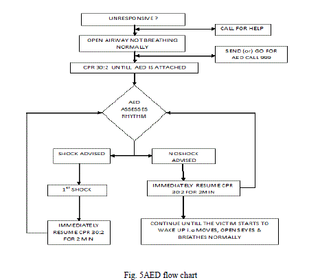
| The following sequences are applied to utilize an automatic AED’s in a victim who is found to be unconscious and not breathing normally[2][5]. The AED flow chart is shown in fig.5. |
| 1. Don’t delay, starting CPR, unless the AED is available immediately. |
| 2. As soon as the AED arrives. |
| • If more than one rescuer present, continue CPR while the AED is switched on. If you are alone, stop CPR and switch on the AED. |
| • Follow the voice /visual prompts. |
| • Annex the electrode pads to the patient’s bare chest. |
| • Make sure that nobody physically contacts the victim while the AED is analyzing the rhythm. |
| 3a. If a shock is needed |
| 1. Ensure that nobody physically contacts the victim. |
| 2. Push the shock button as directed (AED’s will distribute the shock automatically) |
| 3. Continue as directed by the voice/visual prompts. |
| 4. Minimize, as for as possible, interruption in chest compressions. |
| 3b If no shock is needed |
| 1. Resume CPR immediately utilizing a ratio of 30 compressions to 2 rescue breaths. |
| 2. Continue as directed by the voice/visual prompts. |
4. Continue to follow the AED prompts until
|
| The victim commences to show designations of consciousness such as coughing, opening his eyes, speaking (or) moving and commences to breathe normally. |
DEFIBRILLATOR ELECTRODES PLACEMENT
|
A. Placement of Defibrillator pads
|
| Place one of the AED pad to the right of sternum (breast bone), below the clavicle (collar bone). Place the other pad in the left anterior mid-axillaries line, over the position of the V6 ECG electrode. It is important that this pad is placed sufficiently laterally and that it is pellucid of any breast tissue. The victim’s impedance value should be measured. It lies up to 25Ω to 225Ω[3][4]. The normal impedance of the victim is 50Ω and the chest pad will prevent the pads adhering to the skin and will interfere with electrical contact. Shave the chest hair if it is excessive. Do not delay defibrillation if a razor is not immediately available. |
B. Electrode Pads for Adults
|
| These Adult AED Defibr illation Pads are non -polarized so that ei ther pad can be placed in ei ther locat ion, simpl ifying the rescue. The electrodes need to be replaced every 2 years or after they are used in a cardiac emergency which is shown in fig.7 |
C. Electrode Pads for Adult/Child
|
| Cardiac attacks in infants and children mainly results from respiratory failure, not cardiac failure. The priority is to establish and maintain a clear airway and provide breathing and circulation. AED pads are suitable for use in children below 8 years. Special pediatrics pads should be utilized in children aged between 1 and 8 years, if they are available, if not standard adult sized pads should be utilized. The utilization of an AED is not recommended in children aged less than 1 year which is shown in fig.6. |
AED SPECIAL CONSIDERATIONS
|
A. Defibrillator if the Victim is wet
|
| Dry the victim’s chest so that the adhesive AED pads will stick and take particular care to ensure that no one is physically contacting the victim when a stock is distributed. |
B. Voice Prompts
|
| The sequence of action and voice prompts provided by an AED are usually programmable and it is recommended that they can be set as follows. |
| 1. Distribute a single shock when a suitable rhythm is detected. |
| 2. No rhythm analysis immediately after the shock. |
| 3. A voice prompts for resumption of CPR immediately after the shock. |
| 4. A period of 2 min of CPR afore further rhythm analysis. |
C. Storage and Utilization of AED
|
| AED should be stored in locations that are immediately accessible to rescuers they should not be stored in locked cabinets. People with no antecedent training have utilized AED’s safety and efficiently. |
METHODS
|
A. Monophasic Pulse/Waveform Method
|
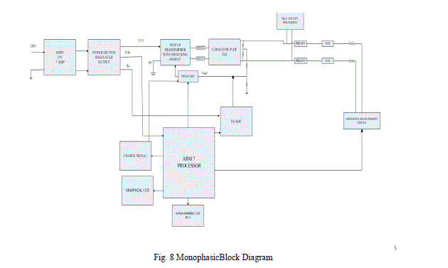
| Monophasic pulse is one of the methods of distributing an electrical shock to the victim’s heart with the energy up to 200J to 300J. This method is utilized in-hospitals nowadays. The working principle of a monophasic method is explicated in below section and the corresponding block diagram is shown in fig.8. |
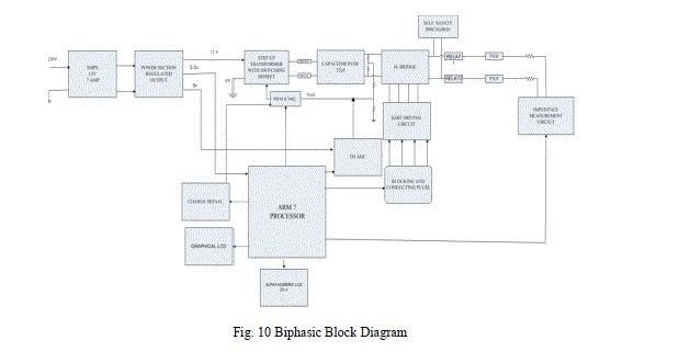
| The main aim of this method is to generate an electric shock with monophasic pulse. This method has 3 sections namely power supply, charging section and discharging section. In power supply section, an alternating current of 230v is applied into Switch Mode Power Supply (SMPS) circuit it splits up into 15v and 7amps of required current and voltage to the next power regulated output section. In power regulated output section, pulsating output is converted into pure amount of direct current with constant output and equally splits its voltage into +12v,+3.3v,and +5v which is applied to the respective blocks. In charging section step up transformer with switching MOSFET (metal oxide semiconductor field effect transistor), PWM IC3482, capacitor PVDF 32μf, 2500vdc and ADC (analog to digital) circuit are involved. Output of power supply section is fed into corresponding blocks, +12v is applied for step up transformer, +5v for ADC circuit and +3.3.v for ARM 7 processor. Working principle of step up transformer is to increase the voltage with subsequent decrease in current this voltage is charged with the capacitor in terms of energy with the avail of switching MOSFET. |
| E= ½ CV2 |
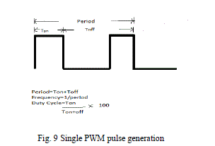
| Pulse width modulation (PWM) is a method of transmitting the duration of a pulse with respect to the analog input. The duty cycle of a square wave is modulated to encode a specific analog signal level. The PWM signal is digital because at any given instant of time, the full DC supply is either ON or OFF completely. The duty Cycle is a measure of the time of modulated signal in high state. PWM pulse is shown in fig.9. |
| The generated voltage of PWM pulse is to be stored in the capacitor (32μf, 2500vdc) in terms of energy (joules) with the avail of ADC and charging signal from the ARM7 processor. The discharging section consists of self and safety relay, high speed patient relay, electrode pads, and impedance quantification circuits. The charge stored in the capacitor is discharged through high speed patient relay with the avail of electrode pads to the victim’s heart. Before discharging the electric shock to the patient we have to measure the impedance of the patient body, if it is 50Ω denotes then only the shock is distributed to the victim up to 320ns. After 320ns completed remaining energy is discharged through self/safety relay switch. PQRST waveforms, ventricular fibrillation, ventricular tachycardia waveforms are exhibited utilizing graphical LCD (128×64). Alphanumeric LCD is utilized to exhibit the commands which are used in AED kit. |
A. Biphasic Pulse/Waveform Method
|
| This is one of the methods of distributing electrical shock to the victim’s heart with biphasic multipulse. As we discussed in above section, the working principle is similar to monophasic method. The main aim is to distribute the shock with biphasic multi pulse with less amount of energy requirement up to 120J to 200J. Similar to monophasic block diagram, this method consists of power supply section, charging section, discharging section, and displaying section with graphical LCD (128×64), in integration to that an IGBT (Insulator Gate Bipolar Transistor) with H-bridge blocking and conducting pulses are built. IGBT with H-Bridge circuit is a three terminal semiconductor contrivance utilized as an electronic switch with ON and OFF conditions. It has high input impedance, low output impedance and medium switching speed. It has four switches, if any two opposite switches are short circuited remaining two switches are open circuited and the output is taken from the mid-point of the IGBT driving circuit. After taking output from the mid-point of IGBT, the output is charged to the capacitor in terms of energy and discharged through patient relay up to 320ns the same procedure is followed for this method. Graphical LCD (128×64) is to exhibit the waveforms and alphanumeric LCD is to exhibit the commands which are acceptable in AED kit |
RESULT
|
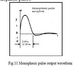
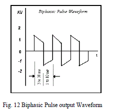
| In this work various methods are discussed and it is implemented using Keil μ-vision and proteus simulation software we get an output waveform is shown in fig. 11 and fig.12. Biphasic pulse will generate raised defibrillation efficacy with respect to Monophasic pulses. |
CONCLUSION AND FUTURE WORK
|
| In this paper various methods for generating and delivering an electric pulses and Comparative study of monophasic and biphasic output waveforms are discussed. As we mentioned earlier monophasic pulse is unidirectional pulse, electric shock is applied only one side (200J to 300J) of the victim’s heart so tissues of the body will affect more. Whereas, biphasic multi pulse is bidirectional pulse, electric shock is applied to both the direction (120J to 200J) of the victim’s heart so the tissues of the body will not be affected more. AED is easy to use and more appropriate for layrescuers. In future work this paper is helpful to implement AED in hardware kit using either Monophasic or biphasic pulse waveform. |
ACKNOWLEDGMENT
|
| We are very thankful to the department of Electronics and Communication Engineering, Dr.SJS Paul Memorial College of Engineering and Technology,Puducherry,India for the support rendered to carrying this work. |
Figures at a glance
|
 |
 |
 |
 |
 |
| Figure 1 |
Figure 2 |
Figure 3 |
Figure 4 |
Figure 5 |
| |
 |
 |
 |
 |
 |
| Figure 6 |
Figure 7 |
Figure 8 |
Figure 9 |
Figure 10 |
|
| |
References
|
- Yongqin Li, Member, IEEE, Joe Bisera, Max Harry, Weil Senior Member, “An Algorithm used for ventricular fibrillation detection withoutinterrupting chest compression”,IEEE, and Wanchun Tang IEEE Transactions on biomedical engineering, vol.59, no.1, January 2012.
- Qiao Li, CadathurRajagopalan, senior Member, “ Ventricular fibrillation and tachycardia classification using a machine learning approach”.IEEE, and GartD.Clifford, Senior Member, IEEE Transactions biomedical engineering, vol. 61. No, 6, june 2014.
- Willis A tacker, “External Defibrillator”, in the biomedical engineering handbool, j.bronzino (ed) CRC press, 1995.
- T.D.Valenzuela, K.B.kern, L.L.clark, and R.A.Berg, “interruptions if chest compression during emergency medical systemsresuscitation,”circulation, vol.112, pp. 1259-1265.
- N.v.Thakor, Z.Y. Sheng, and P.K. Yan, “Ventricular tachycardia and fibrillation detection by a sequential hypothesis testin g algorithm”, IEEE.Trans. Biomed.Eng., vol.37, no.9, pp.837-843, sep. 1990.
- S.chen, N.V.Thakor, and M.M.Mower, “Ventricular Fibrillation detection by a regression test on the autocorrelation function, “Med.Biol. comput,vol.25, no.3, pp.241-249, 1987.
- R. Clayton, A. Murray, and R. Campell, “Changes in the surface ECGfrequency spectrum during the onset of ventricular fibrillation,” inProc.Comput. Cardiol. Los Alamos, CA: IEEE Computer Society Press,1991, pp. 515–518.
- L. Khadra, A. S. Al-Fahoum, and S. Binajjaj, “A quantitative analysis approach for cardiac arrhythmia classification using higher order spectraltechniques,” IEEE Trans. Biomed. Eng., vol. 52, pp. 1840–1845, 2005.
- X. S. Zhang, Y. S. Zhu, N. V. Thakor, and Z. Z.Wang, “Detecting ventricular tachycardia and fibrillation by complexity measure,” IEEE. Trans.Biomed. Eng., vol. 46, no. 5, pp. 548–555, May 1999.
- F. M. Roberts, R. J. Povinelli, and K. M. Ropella, “Identification of ECG arrhythmias using phase space reconstruction,” in Proc. Eur. Conf.PrinciplesPract. Knowl. Discovery Databases, 2001, vol. 2168, pp. 411–423.
|