Keywords
|
| Image processing, MRI, segmentation, watershed segmentation, CNN, k-Means clustering, |
RELATED WORK
|
| Rajesh C. Patil and Dr. A. S. Bhalchandra have analysed that processing of MRI images is the most challenging and emerging field. Magnetic Resonance Imaging (MRI) is an advanced medical imaging technique used to produce high quality images of the parts contained in the human body. The paper brings out the methodology, which includes preprocessing of the given MRI image, its segmentation and lastly morphological operations on it, for the detection and extraction of brain tumour of real time patient’s MRI scan images. |
| Azian Azamimi Abdullah, Bu Sze Chize and Yoshifumi Nishio developed an automated detection method for brain tumor using Cellular Neural Network (CNN). For this purpose they combined many templates to obtain the accurate results. Simulation results using the new algorithm prove that brain tumor can be detected in shorter time. |
| Ishita Maiti and Dr. Monisha Chakraborty have used the technique of Watershed algorithm in combination with Edge Detection operation for the Brain Tumor segmentation of an MRI image. The paper includes Marker based Watershed algorithm. And the Edge detection is carried out by the Canny Edge Detector. The two methods are performed on the HSV Color model of an image. The results have been shown to be very accurate through the paper. |
| J.Vijay and J.Subhashini have concluded that k-Means Clustering is a recognized as a powerful tool for the detection and extraction of Brain tumor from MRI images. The paper uses pixel-based k-Means clustering technique to achieve the aim. The paper has deduced that unsupervised segmentation methods are better than the supervised methods. This technique is proved to be lesser time consuming and achieves maximum lossless compression. The paper proves this technique to be very efficient. |
| Anam Mustaqeem, Ali Javed, Tehseen Fatima have concluded that Watershed based segmentation and thresholding based segmentation is a powerful tool for the detection of Brain tumour in MRI images. The paper further contains the analysis of persons suffering and dying from this fatal problem of brain tumour. The main emphasis of the paper lies on the various segmentation techniques. The methodology mentioned in the paper consists of Image Acquisition, Preprocessing, Processing and Post Processing of the input MRI image. |
| Dibyendu Goshal1, Pinaki Pratim Acharjya have introduced a new technique of Marker Controlled Watershed Algorithm to carry out the segmentation of MRI images. The paper shows how this technique overcomes the problem of over segmentation with watershed algorithm. The detailed algorithm with images at each step has been given the paper. |
INTRODUCTION
|
| Medical Image processing has emerged as one of the major area of research. In medical science, MRI (Magnetic Resonance Imaging) is a very popular technique which is used in Radiology to analyze internal structures of the body such as brain, kidney etc. Other techniques in comparison to MRI are Computed Tomography (CT) and X-Ray. X-Rays are a type of radiation, and when they pass through the body, dense objects such as bone block the radiation and appear white on the x-ray film. CT uses ionization but MRI combines a powerful magnet with radio waves (instead of x-rays) and a computer to manipulate the magnetic elements and create highly detailed images of structures in the body. An important and more specialized imaging technique which uses short-lived radioactive substances to produce images with three-dimension of the objects functioning within the body. The output of this scanning machine is called as PET scan.PET scan also gives details about the complete chemistry or metabolic activity of the human body. SPECT stands for Single Photon Emission Computed Tomography, which is a scanning procedure based on nuclear medicine. |
| Brain tumor can be easily detected and extracted from an MRI image. The word tumor is a synonym for a word neoplasm which is formed by an abnormal growth of cells Tumor is something totally different from cancer. Tumors can damage the normal brain cells by producing inflammation, exerting pressure on parts of brain and increasing pressure within the skull. Figure1 shows that the presence of tumor in the brain. Figure 2 shows the MRI result of the brain tumor image. |
| Figure 2 shows the MRI result of the brain tumor image. |
| For some applications, such as image recognition or compression, we cannot process the whole image directly for the reason that it is inefficient and unpractical. Therefore, several image segmentation algorithms were proposed to segment an image before recognition or compression. Before segmenting an image following steps are taken for improving the image quality: |
| 1) Noise Removal: Median filter acts as noise removal non linear tool. In this filtering technique each image pixel is replaced by the neighborhood median pixel. |
| 2) Morphological Opening: Morphological opening is another important preprocessing (skull removing) step. Two gray scale morphological operations, Erosion and Dilation is used for this purpose. Here, 3 x 3 square Structuring Element (SE) is considered for tumor detection. |
VARIOUS SEGMENTAION TECHNIQUES
|
(I) CELLULAR NEURAL NETWORK:
|
| In 2012, Azian Azamimi Abdullah, Bu Sze Chize and Yoshifumi Nishio proposed a brain tumor detection method based on cellular neural networks (CNNs). CNN can be defined as any spatial arrangement of locally-coupled cells, where each cell is a dynamical system which has an input, an output, and a state evolving according to some prescribed dynamical laws. The proposed algorithm is as follows: |
| Step 1: Start |
| Step 2: Original |
| Step 3: Improved SOE |
| CNN |
| [ 1] |
| Step 4: An improved logicNOT template |
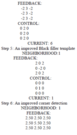 |
 |
| As preliminary result, sample brain images (tumor and non tumor) with new algorithm simulation are shown in Figure 11 and 12. From these results, it can be shown that the new proposed algorithm can detect the location of brain tumor successfully. It seems that the proposed algorithm gives clearer result in detecting brain tumor symptoms. |
| The output of the images is the image segmentation based on black and white area. Black represented as normal area and the white area represented as tumor area. Simulation results using the new algorithm prove that brain tumor can be detected in shorter time. |
| The advantage of using CNN is increasing of throughput due to the massive parallelism of the structure, together with the analog way of signal processing. However, this algorithm still has its weakness where the skull (white ring) unable to filter with CNN template. |
(II) WATERSHED and EDGE DETECTION ALGORITHM IN HSV COLOR MODEL:
|
| In 2012, Ishita Maiti and Dr. Monisha Chakraborty developed another method for brain tumor detection. They used Watershed method in combination with Edge Detection operation. It is a color based brain tumor detection algorithm using color brain MRI images in HSV color space. For the detection of edges Canny edge detector is applied to the output image. The step-by-step algorithm is as follows: |
| Step 1: Read the original image in |
| RGB format |
| Step 2: Convert the image in HSV |
| Step 2: Convert the image in HSV format (divide the image into three regions) |
| Step 3: Contrast enhancement for each region |
| Step 4: Watershed Transform applied in each region |
| Step 5: Edge detection (canny operator) applied to each region |
| Step 6: Combination of three segmented regions |
| Step 7: Final segmented image |
| The paper describes the Marker based Watershed algorithm and HSV Color Model. Gradient magnitude is used here for preprocessing of the image to overcome the over segmentation problem. Markers are used for modifying gradient images. Here watershed transform of the distance transform of the internal marker is computed. Finally the gradient image is modified by imposing regional minima at a location of both internal and external markers using the MATLAB toolbox. |
| The total algorithm is based on HSV color model. The brain tumor image is converted into HSV color model which separate the total image into three regions hue, saturation and intensity. Then the total process as described in the flowchart is executed using each of the three regions. First histogram equalization is done for contrast enhancement of hue region. Histogram equalization is a method for modifying the dynamic range and contrast. Then marker based watershed algorithm described in section A is applied to the contrasted enhanced image. Then edge of the image is obtained by applying canny operator to the output of the watershed algorithm. The whole process is repeated for saturation and intensity region of the image. Finally, the three output images obtained from canny edge detection is combined. |
| The combined image is then converted to RGB color model, the model in which the image was taken. |
| In this work, it is found that when this developed algorithm is applied on gray image to segment the tumor region the performance is not so well as obtained in the color model as HSV color space carries more information than the gray image. This can be said by comparing the Fig. 6 with Fig. 4(j)q |
| Using this algorithm image can be segmented more precisely but there are some challenges regarding this algorithm, Over Segmentation is a common problem for this algorithm. To avoid this problem marker based segmentation process is used in this paper. |
(III) K-MEANS CLUSTERING:
|
| In 2013 J.Vijay,J.Subhashini proposed an efficient method for automatic brain tumor segmentation for the extraction of tumor tissues from MR images. In this method segmentation is carried out using K-means clustering algorithm. This enhances the tumor boundaries more. |
| The limitations of K-means clustering are many iterative rounds may be required. The main argument of the proposed modifications is on the reduction of intensive distance computation that takes place at each run (iteration) of K-means algorithm between each data point and all cluster centers. To reduce the intensive distance computation, a simple mechanism by which, at each iteration, the distance between each data point and the cluster nearest to it is computed and recorded in a data structure is suggested. Thus, on the following iterations the distance between each data point and its previous nearest cluster is recomputed. |
| In the proposed method, segmentation and the K-means clustering have been combined. A brain Image consists of four regions i.e. gray matter (GM), white matter (WM), cerebra spinal fluid (CSF) and background. Therefore, an input image needs to be divided into these four classes. In order to avoid the chances of misclassification, the outer elliptical |
| shaped object should be removed. After the enhancement of image morphological process is carried out to extract the required region. The Next step is by implementing K-means with clusters exact result is produced. |
| The execution time for K-means clustering was less compared to the other clustering methods. The proposed work also reduces the computational complexity and also provides an accurate method of extracting the Region of Interest (ROI).More importantly, the supervised segmentation method requires considerable amount of training and testing data which comparatively complicates the process. This study can be applied to the minimal amount of data with reliable results. |
RESULT AND DISCUSSION
|
| Studying the various segmentation techniques, it is seen that each of the technique has given the best results. Each has its own advantage and disadvantage which has been tried to overcome using its advanced version. Let’s have a look at the result of each technique discussed above. |
| The Cellular Neural Network (CNN) Algorithm has been worked on the gray scale MRI images. This technique has been shown to take lesser time to detect the brain tumor. Also the results of this technique have been compared with the previous results. |
| The Watershed and Edge Detection Algorithm is being applied on the colored MRI images of the brain. The advanced watershed technique, which is Marker Based Watershed Technique has been used. This algorithm has proved that brain tumor is better detected from colored MRI image than the gray scale image. Figure 4(j) shows the detected brain tumor from colored MRI image. |
| Figure 6 shows the detected brain tumor from gray scale MRI image. |
| The K- Means Clustering Algorithm has combined the segmentation of MR image with the k-means clustering. The basic emphasis is given on removing the outer scull before the morphological processes. The algorithm is proved to take lesser execution time. |
CONCLUSION AND FUTURE SCOPE
|
| To examine the location of tumor in the brain, Magnetic Resonance Imaging (MRI) is used. Radiologists will evaluate the grey scale MRI images. This procedure is really time and energy consuming. This paper presents a review of various techniques proposed for segmenting an MRI image which comparatively take lesser time than manual operations to detect and extract the brain tumor. In future work, the techniques will be compared on the basis of other parameters along with the execution time parameter. |
Figures at a glance
|
 |
 |
 |
| Figure 1 |
Figure 1a |
Figure 2 |
 |
 |
 |
| Figure 3 |
Figure 4 |
Figure 4a |
 |
 |
 |
| Figure 4j |
Figure 5 |
Figure 6 |
 |
 |
| Figure 6a |
Figure 11 |
|
| |
References
|
- AzianAzamimi Abdullah, Bu SzeChize and Yoshifumi Nishio, “Implementation of An Improved Cellular Neural Network Algorithm For Brain Tumor Detection,” International Conference on Biomedical Engineering (ICoBE) , Penang, pp. 27-28, February 2012.
- Yu-Hsiang Wang, Tutorial: Image Segmentation.
- IshitaMaiti, Dr. MonishaChakraborty, “A New Method for Brain Tumor Segmentation Based on Watershed and Edge Detection Algorithms in HSV Color Model, ” National Conference on Computing and Communication Systems (NCCCS), Vol. 73, No. 3, pp. 329–345, March 2012.
- J.Vijay, J.Subhashini, “An Efficient Brain Tumor Detection Methodology Using K-Means Clustering Algorithm,” IEEE International conference on Communication and Signal Processing, pp. 653-657, April 3-5, 2013.
- Bilotta.E.,Cerasa.A., Pietro.P., Quattrone.A., Staino.A., Stramandinoli.F., “A CNN Based Algorithm for the Automated Segmentation of Multiple Sclerosis Lesions,” EvoApplications, Part I, pp. 211-220, 2010.
- K. S. Angel Viji, J. Jayakumari, “Performance evaluation of standard image segmentation methods and clustering algorithms for segmentation of MRI brain tumour images,” European Journal of Scientific Research, Vol.79, No.2, pp.166-179, 2012.
- Laxman Singh, R.B.Dubey, Z.A.Jaffery ,Zaheeruddin,” Segmentation and characterization of brain tumor from MR images,” IEEE International Conference on Advances in Recent Technologies in Communication and Computing, 2009.
- ArashAzimZadehIrani and BahariBelaton "A K-means Based Generic Segmentation System" Sixth International Conference on Computer Graphics, Imaging and Visualization, 2009.
- AmitavaHalder, ChandanGiri and AmiyaHalder, Brain Tumor Detection using Segmentation based Object Labeling Algorithm.
- K.S.Tamilselvan, Dr.G.Murugesan and B.Gnanasekaran, “Brain Tumor Detection from Clinical CT and MRI Images using WT-FCM Algorithm,” IEEE International Conference on Green Computing, Communication and Conservation of Energy (ICGCE), pp. 260-263, 2013.
- AnamMustaqeem, Ali Javed, Tehseen Fatima, “An Efficient Brain Tumor Detection Algorithm Using Watershed & Thresh Holding Based Segmentation,” International Journal of Image, Graphics and Signal Processing, Vol. 10, pp. 34-39, 2012.
|