Keywords
|
| Simultaneous identification of vessels, Analysis of blood vessel, Bifurcations. |
INTRODUCTION
|
| The Digital image processing methods stems from two principal application areas; improvement of pictorial information for human interpretation and processing of scene data for autonomous machine perception [5]. The medical imaging is used in several technologies for viewing the internal structure and for medical diagnosis. Image techniques are used for identification of many diseases especially cardiac diseases. Several diseases in retinal vasculature like diabetes, hypertension and arteriosclerosis can be diagnosed by observing some retinal vessel characteristics like caliber, colour, and tortuosity [4]. For diagnosis and treatment many algorithms were developed and proposed for extracting the blood vessels from the retinal image. |
| There are four techniques that are used for the automatic segmentation of blood vessels. First technique is based on the supervised learning in which each and every pixel of the respective retinal image is classified into vessel or non-vessel. The second category is to apply the matched filtering technique to a retinal image [6]. The third category technique is based on the mathematical morphology are proposed [7]. And the last category is based on the tracking of vessels from a collective set of seed points [1]. Basically we know that the normal snapshot of the retinal image describes what is actually happening in the human body. To quantify the retinal vascular structure, respective measurements are done and their properties are also determined. These results are highly useful in measuring the heart diseases and stroke. But then for this we need an accurate extraction of the true blood vessels from the retinal images. For this here use a tracing algorithm for acquiring the true blood vessels. |
RELATIVE WORK
|
| The extraction of the retinal vessel involves in the segmentation of vasculature and identifying the distinct vessels. Only the proper segmentation and identification the blood vessels will lead to the accurate identification of the retinal diseases like diabetic retinopathy (DR), Central Retinal vein occlusion (CRVO), Central Retinal artery Occlusion (CRAO). Among our branch of works one is named as Vessel tracking which here performs the blood vessel segmentation and identifying the blood vessels simultaneously. But this approach is not that much suitable because it does not provide the sufficient information that we require. |
| And another work is termed as the identification of vessels as the post processing step to the segmentation of the blood vessels. In paper [1], the graph formulation is acquired by using the Dijktra’s shortest path algorithm. Similarly in [2], also used the same algorithm for identifying the vessels at a time and they have evaluated the results on 15 number of images. But then it results in identification of incorrect blood vessels because, for acquiring the correct blood vessel, the bifurcations and cross over points in the vessel requires some information from the other nearby vessels present. |
| The work that we have focused here is the identification of the true blood vessels as a post processing step to the segmentation process. Our work is entirely different from the other existing work. Here we identify the multiple numbers of true blood vessels simultaneously by neglecting the false blood vessels from the retinal image. |
BLOOD VESSEL SEGMENTATION
|
| The segmentation of blood vessels in the retinal image is used for the early diagnosis of the diseases. Segmenting automatically provides several bents including the subjectivity. The examination of the blood vessels in the retina is used for identification of the eye diseases like diabetic retinopathy and glaucoma. Blood vessel segmentation is nothing but dividing the blood vessels into many number of parts. |
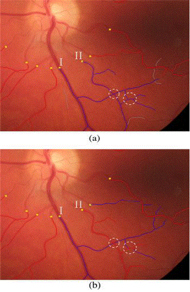
| Blood vessel segmentation can be obtained using several approaches like pattern recognition technique, tracking based approaches, artificial based and model based approaches. The wrong and correct identification of the blood vessels in the retinal image is shown in Fig.1 [1]. |
| Pattern recognition technique deals with the classification, automatic detection of the blood vessels and their features which includes region growing methods, skeleton based, ridge based, mathematical morphology based methods. Tracking based approach needs an initial point to start the approach and to track the vessel center lines, boundaries by finding the pixels that orthogonal to the tracking direction. |
| But it has a disadvantage that it cannot be detected automatically as it needs an intervention for the selection of initial and end point. Artificial intelligence based approaches is used to provide some important information to guide the segmentation process and also to delineate the boundaries of the vessel structure. This system need some prior information and also employs some low level processing algorithms like thinning and thresholding. |
| In model based approaches it provides some explicit vessel models for the vessel extraction. Basically it is classified into deformable models and template matching. The accuracy obtained in this approach is crucial and it is must to obtain a simple, precised and repeatable system for automatic detection of blood vessels. |
PRELIMINARY WORK
|
| As a preliminary work we must first define the area of interest or zone of interest in the image. The measurements that are obtained from this area are used in number of useful clinical studies and experiments. Then we do the existing methods for vessel segmentation process and any outline procedure to acquire the line image with respect to the area of interest. |
| The segmentation process is done by extracting a seed point, then acquiring the connection of pixels and then vessel verification. |
| The lines that are present in the acquired line image in Fig.2. (b) Show the connectivity of the structure of the blood vessel in topological level [1]. From the line image let the set of all white pixels present be considered as P. We have to first determine the connected pixels, that is if pi, pj are connected that belongs to the set P if the adj (pi,pj) belongs to P- {pi,pj}. |
| Next we have to identify the pixel crossing number. Let us consider 8 neighborhood pixels P1 to P8 that surround the main pixel “P”. And the term xnum(p) represents the black to white transitions in order to the neighborhood pixels of P shown in Fig.3.[1]. |
| Even after acquiring the connected set of pixels it still contains some non-vessel points. The final process of our proposed work is to neglect the false blood vessels and acquire the true blood vessels from the respective retinal image. |
| To acquire the vessel point first the perpendicular profile of the point and two profiles of the consecutive points (preceding and succeeding points) are averagely combined into a single intensity profile. The intensity of the background level in the averaged profile is approximated with a straight line equation using a robust M estimator [3]. If the intensity of the hypothetical point is below the value of approximate straight line, we consider the point as vessel. Otherwise the point is considered as non-vessel and is rejected [4]. |
GRAPH TRACING TECHNIQUE
|
| Our method is proposed for segmenting the retinal image by acquiring the true blood vessels in the form of binary tree. Here by using this process we must overcome the problems in the set of vessel trees that occur due to the crossover points and bifurcations. As a preliminary step first we have to identify the cross over points because the blood vessels in retinal image cross each other very frequently. So it is necessary to identify the crossovers. First we define it as cross over points then later as crossover segments. We must first know the crossover location then we can identify the problems and challenges that are caused by the crossover points. |
| The short crossover segments need not be a true crossover segments and so we plan to use the directional change between the adjacent segments and their pixel intensity values to differentiate the crossover segments [1]. The directional change between the segments can be calculated by considering two crossover segments sa and sb which are adjacent to a common junction. Let us consider Pa and Pb as end points of sa and sb and the vector Va, starts on sa and it ends at Pa whereas vector Vb starts from Pb and ends on sb. Then the directional change between sa and sb can be calculated as [1] |
 |
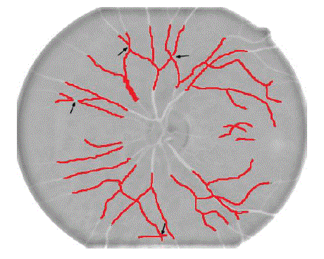
| Whereas ΔD(sa,sb) lies between 0° to 180°. The identified cross over segments for fig 2(a) is shown in fig 4, which is highlighted in red colour indicated by black arrows. As our next step we must find the optimal forest (set of vessels), we here model the segments as graph segment and we use some optimization technique to select the best set of vessels from the graph. Segment graph is defined as connection which is set to 2pt segment automatically. Blood vessels are the tubular structure carrying blood through the tissues and organs; a vein, artery or capillary. |
| After acquiring the segmented graph connection set we must do vessel verification procedure. Because even after connected set of points are obtained it may still contain non vessel points. We do vessel verification and we obtain the true blood vessels from the retinal image using the graph tracing algorithm. |
RESULTS AND DISCUSSION
|
| Quantitative results are acquired from our proposed method. We have used retinal images from publicly available retinal image database called DRIVE (Digital retinal images for vessel extraction) for our experimental approach. Each image is a colour retinal fundus images acquired by a Canon CR5 non-mydriatic 3CCD camera with 45 degree Field of view (FOV)[4]. The flow of our experiment is explained using the pictorial representation in Fig.5. |
| Our experimental results are used to identify major parts of the blood vessels efficiently as we compare the results we obtained with the same results of those images provided by the database creators. As we check our image results and also results provided in the database for comparison, we can observe clearly that we have obtained all the major vessels and most of the branch vessels too. We obtain these results by overcoming the challenges and problems caused due to crossovers and bifurcations. Our method provides the vessel skeleton and also major blood vessels that are present in the respective retinal image. But then fewer branch vessels are not obtained in our method. In near future our experiment can be proceeded in further to obtain better results. |
CONCLUSION
|
| In our work, we propose a method to segment the retinal vessels in the image by neglecting the non-vessel points and acquiring the true blood vessels from the retinal image. After the basic processing steps, the work starts with the extraction of the seed points followed with the graph tracing algorithm and vessel verification. The proposed work overcomes the challenges and problems due to the crossover points and bifurcations. Promising results are obtained by using retinal images from the “DRIVE” database. The work still left is to handle some other better ways to acquire the left branch vessels too from the respective retinal images. |
Figures at a glance
|
 |
 |
 |
 |
 |
| Figure 1 |
Figure 2 |
Figure 3 |
Figure 4 |
Figure 5 |
|
| |
References
|
- Qiangfeng Peter Lau*, Mong Li Lee, Wynne Hsu, and Tine Yin Wong: “Simultaneously Identifying All True Vessels from Segmented Retinal Images” IEEE TRANSACTIONS ON BIOMEDICAL ENGINEERING, VOL.60, NO. 7, JULY 2013.
- Thomas H, et.al: “Introduction to algorithms” 3rd edition, The MIT press, pp-658-664, 2009.
- Haralic, et.al: “Computer and Robot vision” VOL.1 Addition- Wesley ISBN: 0201108771, 1991.
- SuhitRattanpad, BunyaritUyyanovara “Vessel segmentation in retinal images using Graph theoretical vessel tracking” MVA 2011 APR conference on machine vision application June 13-15, 2011, Nara, JAPAN.
- M. Sangeetha, S. Nirmala Devi, Dr. N. Kumaravel: “Wavelet transform based coronary blood vessel segmentation using entropy thresholding” Department of Electronics and communication, CEG , Anna university, Chennai.
- Al-Rawi M, et.al: “An improved matched filter for blood vessel detection of digital retinal images” Computer in Bio and medicine pp262-267, 2007.
- F.Zana: “Segmentation of vessel like pattern using mathematical morphology and curvature evaluation” IEEE Transactions on Image processing, pp. 1010-1019, 2001.
- V. S. Joshi, M. K. Garvin, J. M. Reinhardt, and M. D. Abramoff, “Automated method for the identification and analysis of vascular tree structures in retinal vessel network,” in Proc. SPIEConf.Med. Image.,vol. 7963, no. 1, pp. 1–11,2011.
- W. E. Hart, M. Goldbaum, P. Kube, and M. R. Nelson, “Automated measurement of retinal vascular tortuosity,” in Proc. AMIA Fall Conf., 1997,pp. 459–463.
- E. Grisan, A. Pesce, A. Giani, M. Foracchia, and A. Ruggeri, “A new tracking system for the robust extraction of retinal vessel structure,” in Proc. IEEE Eng.Med.Biol.Soc., Sep. 2004, vol. 1, pp. 1620–1623.
|