International Journal of Advanced Research in Electrical, Electronics and Instrumentation Engineering
ISSN ONLINE(2278-8875) PRINT (2320-3765)
ISSN ONLINE(2278-8875) PRINT (2320-3765)
P.Ganesh1, J. Jai Jaganath Babu2, S.SuganthKannan3
|
| Related article at Pubmed, Scholar Google |
Visit for more related articles at International Journal of Advanced Research in Electrical, Electronics and Instrumentation Engineering
This paper presents a novel automated detection of thyroid nodular boundaries of various shapes and resolutions in ultrasound images. Detection of boundaries is required in diagnostic and treatment procedures for thyroid diseases. The boundary detection algorithm is comprised of three stages. In first stage, uniform sticks are applied for contrast enhancement and also for reducing speckle noise in ultrasound thyroid images. The resultant image is then smoothed using fourth order partial differential equation. In the third stage, an edge based active contour model is applied for detection of boundaries. The algorithm was tested on different ultrasound images from patients with thyroid nodules. The results show that the performance of the proposed system provides an efficient delineation of thyroid nodules
INTRODUCTION |
||||||||
| In the world population 12 percent of adults suffer from some form of thyroid disease. In this disease, thyroid cancer is a rare cancer, constituting only about 2 percent of all cancers but now it is considered to be a fastest rising cancer among women [1], [2]. Most thyroid cancers are first found as nodules in thyroid gland. Thyroid nodules are solid or cystic lumps and can either be benign or malignant. There are two different techniques for identification of thyroid nodules, invasive and non invasive. Non invasive techniques are methods by which diagnosis is carried out without harming the patient. Medical imaging techniquesuch as MRI, CT, and Ultrasonography belong to this type of these CT and MRI use ionising radiation and also expensive techniques. | ||||||||
| Ultrasonography is recognised as a cost effective imaging technique, which does not involve ionising radiation and acquisition time also less compared to the above technique. However, the main disadvantage of medical ultrasonography is the poor image quality due to backscattered echo signals called speckle [3]. It is difficult task for the sonologist to analyse and diagnose the image in the presence of speckle noise and impossible to do feature extraction, analysis and recognition [4]. As a result, speckle filtering is a preprocessing step for segmentation, feature extraction and classification [5]. A number of methods have been proposed for speckle reduction. Some of the local statistics filters are Lee [6], the Frost et al [7] and the Kuan et al [8] are based on a speckle noise as multiplicative. Armand lopes et al [9], uses local statistics of the image to estimate the areas of the image to be processed and other [10] methods have been proposed. | ||||||||
| Some segmentation methods have been introduced based on region and edge. In this paper an algorithm that automatically identifying the thyroid nodule within the ultrasound image is proposed. It consists of three sequential stages including contrast enhancement, smoothing and segmentation. The preprocessing stages are stick filtering and fourth order partial differential equation for speckle noise reduction and contrast enhancement. The last stage is thyroid nodule segmentation based on the edge based active contour model. | ||||||||
| The remainder of this paper is organised as follows. In section II, algorithm design of the proposed model is provided. In section III, results of the proposed model in thyroid nodules segmentation is presented. Finally, section IV summarises the conclusion of the study. | ||||||||
ALGORITHM DESIGN |
||||||||
| The segmentation system consists of two stages. The first part is preprocessing. The objective of this stage is to enhance the contrast of image. The enhanced image is then used to segment the thyroid nodule in ultrasound images. The preprocessing step includes two steps A) Contrast enhancement and B) Image smoothing. In the next stage,for accurate delineation of thyroid nodular boundaries, distance regularised level set based active contour model [15] was implemented. | ||||||||
A) Contrast Enhancement |
||||||||
| Medical ultrasound imaging suffer from a unique type of noise called speckle, which has granular appearance [11].A stick technique proposed by Czerwinski et al. [12]to reduce speckle as well as improve edge information which is visible in the original images has been used.In stick filter of size N×N there are 2N-2 discrete lines passing through the center pixel.Typical sticks of length five pixels are shown in Fig. 1. | ||||||||
| In this figure the white and black pixels have a value of 0 and 1/5 respectively. Each mask in a filter banks represents orientation of line segmentation. A 2N-2 mask is superimposed on a current pixel in the original image. Speckle reduction is obtained by determining the mean intensity of all pixels along each stick orientation and current gray value is replaced by maximum of 2N-2 averages. The process is repeated for all the pixels in the image. | ||||||||
 |
||||||||
| Where f(x,y) ,s(x,y),g(x,y) and i represents the original image, stick filter, stick filtered output and number of stick orientation respectively. Fig. 2shows the result of applying sticks to thyroid nodule ultrasound image. It is seen that, in the output image speckle is suppressed and edge information is enhanced. | ||||||||
B. Image smoothing |
||||||||
| The image obtained by high frequency ultrasound scanner doesnot have the uniform speckle pattern. Using a stick filter with uniform size will not achieve satisfactorily results [11]. To improve the image smoothness and edge enhancement we introduce a nonlinear fourth order partial differential equation after the stick filtering. Using of second order partial differential equation [10] cause blocky effects and multiple false edges in image, so we go for fourth order partial differential equation.We used L2- curvature gradient model proposed by Yu-Li You et al [13] as given below. | ||||||||
 |
||||||||
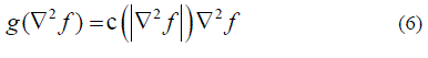
| Where 2 f is the Laplacian of the image f and c(.) is the diffusion coefficient proposed by Perona and Malik [14] and it is defined by | ||||||||
 |
||||||||
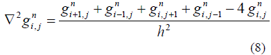
| Rate of diffusion depends on the diffusion coefficient. The discrete form of non linear fourth order partial differential equation (2) is defined as follows. | ||||||||
| The above equation can be solved in three steps. First step , finding Laplacian of the original image. | ||||||||
 |
||||||||
| Second step, calculate the following function as | ||||||||
 |
||||||||
| Third step, calculate Laplacian of gradient as | ||||||||
 |
||||||||
| Where ?t the time step size and h is is space grid size. | ||||||||
C. Segmentation |
||||||||
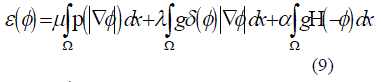
| In image segmentation, we have used edge based geometric active contour model proposed by Chunming Li et al [15]. Active contours are dynamic curves that move towards the object of the boundaries based on the energy function. The energy function is defined as | ||||||||
 |
||||||||
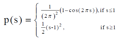
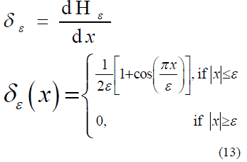
| Where Φ is a level set function, p is is a potential function, g is a edge indicator function, ? is a dirac delta function and H is a Heaviside function.In this active contour model, applieda first derivative of double well potential function and it is defined as | ||||||||
 |
||||||||
 |
||||||||
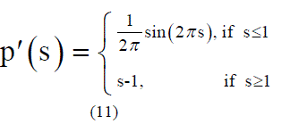
| Used Heaviside function as proposed in [16], is | ||||||||
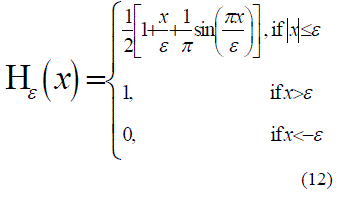 |
||||||||
| Dirac delta function is derived from, | ||||||||
 |
||||||||
 |
||||||||
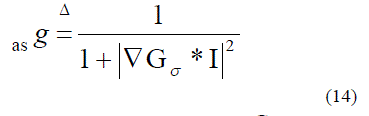
| Where I be the original image, Gσ is a Gaussian kernel with standard deviation σ . The equation (9) can be deduced to | ||||||||
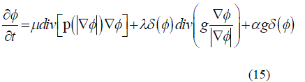 |
||||||||
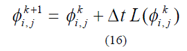
| First term in the right hand side of the above equation (15) represents the distance regularisation energy term while second and third term represents the gradient flow of energy term. The linear form of above equation can be written as, | ||||||||
 |
||||||||
| Where denotes the right hand side equation in (15), (i,j) denotes the spatial index and k represents temporal index. | ||||||||
EXPERIMENTAL RESULTS |
||||||||
| In this section, we present the experimental results of our proposed method. The proposed method was implemented in Matlab 7.6 on Intel core2 Duo PC operating at 1.6GHz, 1GB RAM and windows7 ultimate operating system. For active contour model time step Δt =1, grid spacing h=1,? =1.5 and λ =5 was used for all the experiments in this section. The only one controlling parameter α is adjusted for different characteristics of thyroid nodular ultrasound images with different resolutions.The proposed model was compared with other methods.In Fig. 4 first column shows the original ultrasound thyroid nodular image with initial contour. The second column shows the results of segmentation with frost filter [7] as a preprocessing stage, in this the image has poor resolution the boundaries are not perfectly delineated. The third column shows the result of implementing fourth order partial differential equation [13] as a preprocessing stage. The fourth column shows the result of non local means based filter [17]. The last column shows the result of proposed preprocessing scheme.In this all the images with different resolutions are exactly delineated. | ||||||||
CONCLUSION AND FUTURE WORK |
||||||||
| In this paper, an image processing method for perfect delineation of ultrasound thyroid nodules which are with blurred and irregular boundaries is presented. In the presence of speckle noise, it is difficult to trace the boundaries of the nodular ultrasound image. So to improve the diagnostic examination a speckle reduction filter is needed.Three different speckle reduction filters and compared with proposed model. The comparison of filters is based on effective boundary detection. The model was tested with 14 different thyroid ultrasound images with nodule.From the results theproposed model performs well than the others. In future this work is continued for classification of different nodules and identifying whether the nodule is benign or malignant. | ||||||||
Figures at a glance |
||||||||
|
||||||||
References |
||||||||
|


