ISSN ONLINE(2278-8875) PRINT (2320-3765)
ISSN ONLINE(2278-8875) PRINT (2320-3765)
Mr.M.Arun1, Mr. Ashok Kumar1, N.P.Ria2, H.Sandhya Bhargavi2
|
| Related article at Pubmed, Scholar Google |
Visit for more related articles at International Journal of Advanced Research in Electrical, Electronics and Instrumentation Engineering
The conventional method of detection and classification of brain tumor is by human inspection with the use of medical resonant brain images. But it is impractical when large amounts of data is to be diagnosed and to be reproducible. And also the operatorassisted classification leads to false predictions and may also lead to false diagnose. Medical Resonance images contain a noise caused by operator performance which can lead to serious inaccuracies classification. The use of artificial intelligent techniques for instant, neural networks, and fuzzy logic shown great potential in this field. Probabilistic Neural Network gives fast and accurate classification and is a best tool for classification of the tumors. Probabilistic Neural Network with image and data processing techniques is implemented for automated brain tumor classification. Decision making was performed in two stages: feature extraction using the principal component analysis and the Probabilistic Neural Network (PNN). The performance of the PNN classifier was evaluated in terms of training performance and classification accuracies. Probabilistic Neural Network gives fast and accurate classification and is a promising tool for classification of the tumors
Keywords |
||||||||||||||
| Principal Component Analysis, Probabilistic Neural Network, Magnetic Resonance Image | ||||||||||||||
INTRODUCTION |
||||||||||||||
| The mere thought of someone close harboring a brain tumor is devastating. Besides most brain tumor victims are children or adults in their prime of life. This not only makes it more poignant but also creates a need to look after such victims who can often, with the medical resources available, be restored to near normalcy. This, however, needs a multi-disciplinary approach, which is expensive and rarely available under one roof. Even after the completion of the hospital treatment (surgery, radiotherapy or chemotherapy), many patients require intensive rehabilitation at their homes and sometimes in special institutions. | ||||||||||||||
| Automated classification and detection of tumors in different medical images is motivated by the necessity of high accuracy when dealing with a human life. Also, the computer assistance is demanded in medical institutions due to the fact that it could improve the results of humans in such a domain where the false negative cases must be at a very low rate. It has been proven that double reading of medical images could lead to better tumor detection. But the cost implied in double reading is very high, that’s why good software to assist humans in medical institutions is of great interest nowadays. Conventional methods of monitoring and diagnosing the diseases rely on detecting the presence of particular features by a human observer. Due to large number of patients in intensive care units and the need for continuous observation of such conditions, several techniques for automated diagnostic systems have been developed in recent years to attempt to solve this problem. Such techniques work by transforming the mostly qualitative diagnostic criteria into a more objective quantitative feature classification problem. | ||||||||||||||
| In this paper the automated classification of brain magnetic resonance images by using some prior knowledge like pixel intensity and some anatomical features is proposed. Currently there are no methods widely accepted therefore automatic and reliable methods for tumor detection are of great need and interest. The application of PNN in the classification of data for MR images problems are not fully utilized yet. These included the clustering and classification techniques especially for MR images problems with huge scale of data and consuming times and energy if done manually. Thus, fully understanding the recognition, classification or clustering techniques is essential to the developments of Neural Network systems particularly in medicine problems. | ||||||||||||||
1.1 BRAIN TUMOR AND ITS TYPES: |
||||||||||||||
| A brain tumor is a mass of abnormal tissue growing in any part of the brain. For some unknown reason, some brain cells multiply in an uncontrolled manner and form these tumors'. These tumors can arise from any part of the brain, spinal cord or the nerves. Broadly these tumors can be divided into benign and malignant tumours. | ||||||||||||||
`1.2 METHODS OF DETECTION: |
||||||||||||||
| CT or MRI Scan produce special X-ray pictures that show the detailed structure of the brain and spine and pick up any abnormality. To get a clearer picture, Iodine or Gadolinium contrast dyes are given intravenously. Some people can develop an allergic reaction to the iodine contrast agent and you should always tell the doctor if you have any allergies. The more expensive non-ionic contrast agents reduce the risk of allergic reaction. There is a strong magnetic field during the MRI scan and you should inform the doctor if you have any Pacemaker or metallic clip or prostheses inside your body. For these scans which take about half an hour, the patient lies down on the couch of these CT or MRI machines. The couch moves the patient through the large aperture or tunnel of these machines. The whole procedure is painless but the noise created by the MRI machines can be disturbing for some patients. During the scan the patient should not move and for small children who may move a lot, sometimes a minor anesthesia is given. | ||||||||||||||
| ïÃâ÷ Angiogram is an X-ray taken after injecting an iodine dye through catheters placed into the arteries. This shows the details of the blood supply to the tumor. For vascular malformation like AVM it is essential to plan embolisation, surgery or stereotactic radiation. | ||||||||||||||
| ïÃâ÷ Cerebro Spinal Fluid (CSF) Study is done after removing the CSF from the spine by a long needle (lumbar puncture). This is done in certain tumours which have a high chance of spreading to the spine or to rule out infections or bleeding. | ||||||||||||||
| ïÃâ÷ Hormonal Blood Tests are done for tumours like pituitary adenoma, craniopharyngioma, optic chiasmal or hypothalamic glioma. | ||||||||||||||
| ïÃâ÷ Electroencephalogram (EEG) is occasionally done to study the pattern of seizures. | ||||||||||||||
NEED FOR THE PROJECT |
||||||||||||||
| Automated classification and detection of tumors in different medical images is motivated by the necessity of high accuracy when dealing with a human life. Also, the computer assistance is demanded in medical institutions due to the fact that it could improve the results of humans in such a domain where the false negative cases must be at a very low rate. It has been proven that double reading of medical images could lead to better tumor detection. But the cost implied in double reading is very high, that’s why good software to assist humans in medical institutions is of great interest nowadays. Conventional methods of monitoring and diagnosing the diseases rely on detecting the presence of particular features by a human observer. Due to large number of patients in intensive care units and the need for continuous observation of such conditions, several techniques for automated diagnostic systems have been developed in recent years to attempt to solve this problem. Such techniques work by transforming the mostly qualitative diagnostic criteria into a more objective quantitative feature classification problem. | ||||||||||||||
| Here the automated classification of brain magnetic resonance images by using some prior knowledge like pixel intensity is proposed. Currently there are no methods widely accepted therefore automatic and reliable methods for tumor detection are of great need and interest. The application of PNN in the classification of data for MR images problems are not fully utilized yet. These included the clustering and classification techniques especially for MR images problems with huge scale of data and consuming times and energy if done manually. Thus, fully understanding the recognition, classification or clustering techniques is essential to the developments of Neural Network systems particularly in medicine problems. | ||||||||||||||
EXISTING AND PROPOSED SYSTEM |
||||||||||||||
2.1 SYSTEM ANALYSIS EXISTING SYSTEM |
||||||||||||||
| The conventional method for medical resonance brain images classification and tumors detection is by human inspection. Any doctor or analysist who had specialized on the brain structures, brain diseases, symptoms & remedies, brain features, MRI images can examine the MRI image by vision through it and classify the tumor. Medical Resonance images contain a noise caused by operator performance which can lead to serious inaccuracies classification. A CT scan is a single analysis image, where as the MRI image is visually 2D, but wholly 3D. Because as the tumor is not 2D, the 3D tumor should be scanned in layer by layer. So, there will be a number of scanned images in terms of MRI consideration. The Operator-assisted classification methods are impractical for large amounts of data and are also non-reproducible. Thus there should be an automatic classifier. | ||||||||||||||
2.2 DRAWBACKS OF THE EXISTING SYSTEM |
||||||||||||||
| ïÃâ÷ Medical Resonance images contain a noise caused by operator performance which can lead to serious inaccuracies classification. | ||||||||||||||
| ïÃâ÷ Operator-assisted classification methods are impractical for large amounts of data and are also non-reproducible. | ||||||||||||||
| ïÃâ÷ Large data is to be stored in memory of the analysist for the immediate remedy suggestion or for continuous monitoring of the tumor at the required last stages. | ||||||||||||||
2.3 PROPOSED SYSTEM |
||||||||||||||
| There is a proposed method as mentioned in literature survey that artificial intelligence like probabilistic neural network can be used for the classification of brain tumor whether the image has a tumor or it is a normal image, but is not implemented. So, in this project, the concept of Probabilistic Neural Network is used for the effective and accurate classification eliminating the drawbacks of the existing system. | ||||||||||||||
| The use of artificial intelligent techniques for instant, neural networks, and fuzzy logic shown great potential in this field. Hence, the Probabilistic Neural Network was applied for the purposes. Decision making was performed in two stages: feature extraction using the principal component analysis and the Probabilistic Neural Network (PNN). This project classifies a brain tumor if it is a benign tumor or a malignant one. Probabilistic Neural Network gives fast and accurate classification and is a promising tool for classification of the tumors. | ||||||||||||||
2.5 REQUIREMENT SPECIFICATION |
||||||||||||||
| The requirements specification is a technical specification of requirements for the hardware and software products. It is the first step in the requirement analysis process. The purpose is of the hardware and software requirements specification is to provide a detailed overview of the project, its parameter and goals. The only hardware requirement is a PC(personal computer). | ||||||||||||||
PROPOSED SYSTEM MODULES |
||||||||||||||
3.1 SYSTEM DESIGN & DEVELOPMENT SYSTEM BLOCK DIAGRAM |
||||||||||||||
| ïÃâ÷ Pre-processing for noise removal | ||||||||||||||
| ïÃâ÷ Principle component analysis for feature extraction | ||||||||||||||
| ïÃâ÷ Probabilistic neural network for the classification of brain tumor | ||||||||||||||
| In the block diagram, The training images are the MRI images given as the database for each neuron in the PNN. The database has the features of the image such as pixel intensity.But the information such as pixel intensity is larger, so the dimensionality is reduced by extracting principal components by using PCA which is principal component analysis.The test image which is also an MRI image, is to be converted to principal components by the use of PCA. The test image principal components are compared with the PNN’s trained image’s principle components & the best with less covariance is selected as resembled image & is output as classified image. | ||||||||||||||
3.2 MODULE I - PRE-PROCESSING |
||||||||||||||
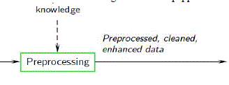
| Noise presented in the image can reduce the capacity of region growing filter to grow large regions or may result as a fault edges. When faced with noisy images, it is usually convenient to preprocess the image by using weighted median filter. Weighted Median (WM) filter have the robustness and edge preserving capability of the classical median filter. WM filters belong to the broad class of nonlinear filters called stack filters. This enables the use of the tools developed for the latter class in characterizing and analyzing the behavior and properties of WM filters, e.g. noise attenuation capability. The fact that WM filters are threshold functions allows the use of neural network training methods to obtain adaptive WM filters (Scherf and Roberts, 1990). A weighted median filter is implemented as follows: | ||||||||||||||
| W(x, y) =median {w1 x x1…wn x wn} | ||||||||||||||
| x1…..xn are the intensity values inside a window centered at (x,y) and w x n denotes replication of x, w times. | ||||||||||||||
| There are several types of noise that influences images like salt and pepper noise, Gaussian noise, shot noise, quantization noise, anisotropic noise etc,. but the noise that influence the MRI images is salt and pepper noise. | ||||||||||||||
 |
||||||||||||||
MODULE II - PRINCIPLE COMPONENT ANALYSIS (PCA) |
||||||||||||||
| PCA is a mathematical procedure that uses an orthogonal transformation to convert a set of observations of possibly correlated variables into a set of values of linearly uncorrelated variables called principal components. The number of principal components is less than or equal to the number of original variables. This transformation is defined in such a way that the first principal component has the largest possible variance (that is, accounts for as much of the variability in the data as possible), and each succeeding component in turn has the highest variance possible under the constraint that it be orthogonal to (i.e., uncorrelated with) the preceding components. Principal components are guaranteed to be independent only if the data set is jointly normally distributed. PCA is sensitive to the relative scaling of the original variables. Depending on the field of application, it is also named the discrete Karhunen–Loève transform (KLT), the Hotelling transform or proper orthogonal decomposition (POD). | ||||||||||||||
| Steps: | ||||||||||||||
| ïÃâ÷ Convert the 2D images into one dimensional image using reshape function-for both test image and database images. | ||||||||||||||
| ïÃâ÷ Find the mean value for each one dimensional image by dividing sum of pixel values and number of pixel values. | ||||||||||||||
| ïÃâ÷ Find the difference matrix for each images by [A]=(Original pixel intensity of 1D image) – (mean value) | ||||||||||||||
| ïÃâ÷ Find the covariance matrix L | ||||||||||||||
| ïÃâ÷ Find the Eigen vector for 1D image V, By using this, get the Vector and diagonal matrix of the 1D images. | ||||||||||||||
| ïÃâ÷ Find the Eigen face of 1D image, | ||||||||||||||
| By using these principal components, we can identify the image from database which is similar to the features of test image. | ||||||||||||||
MODULE III - PROBABILISTIC NEURAL NETWORK |
||||||||||||||
NEURAL NETWORKS |
||||||||||||||
| Neural networks are predictive models loosely based on the action of biological neurons. | ||||||||||||||
| The selection of the name “neural network” was one of the great PR successes of the Twentieth Century. It certainly sounds more exciting than a technical description such as “A network of weighted, additive values with nonlinear transfer functions”. However, despite the name, neural networks are far from “thinking machines” or “artificial brains”. A typical artificial neural network might have a hundred neurons. In comparison, the human nervous system is believed to have about 3x10¹⁰ neurons. We are still light years from “Data”. The original “Perceptron” model was developed by Frank Rosenblatt in 1958. Rosenblatt’s model consisted of three layers, (1) a “retina” that distributed inputs to the second layer, (2) “association units” that combine the inputs with weights and trigger a threshold step function which feeds to the output layer, (3) the output layer which combines the values. Unfortunately, the use of a step function in the neurons made the perceptions difficult or impossible to train. A critical analysis of perceptrons published in 1969 by Marvin Minsky and Seymore Papert pointed out a number of critical weaknesses of perceptrons, and, for a period of time, interest in perceptrons waned. | ||||||||||||||
GENERAL REGRESSION NEURAL NETWORK: |
||||||||||||||
| This neural network like other probabilistic neural networks needs only a fraction of the training samples a back propagation neural network would need. The data available from measurements of an operating system is generally never enough for a back propagation neural network. Therefore the use of a probabilistic neural network is especially advantageous due to its ability to converge to the underlying function of the data with only few training samples available. The additional knowledge needed to get the fit in a satisfying way is relatively small and can be done without additional input by the user. This makes GRNN a very useful tool to perform predictions and comparisons of system performance in practice. The probability density function used in GRNN is the Normal Distribution. | ||||||||||||||
SIMULATION RESULTS: |
||||||||||||||
CONCLUSION |
||||||||||||||
| PNN has been implemented for classification of MR brain image. PNN is adopted for it has fast speed on training and simple structure. Fifteen images of MR brain were used to train the PNN classifier.Here we created a neural network that could classify whether it has a brain tumor or a malignant one. The future work can be the classification that gives the number, diagnose and may be remedy of and for the stage of the tumor in the MRI images taken. The further improvement can be locating the exact location of the tumor in the brain by observing the pixel intensity variation. | ||||||||||||||
Figures at a glance |
||||||||||||||
|
||||||||||||||
References |
||||||||||||||
|