Medical image compression applications are quality-driven applications which demand high quality for certain regions that have diagnostic importance in an image, where even small quality reduction introduced by lossy coding might alter subsequent diagnosis, which might cause severe legal consequences. Due to this, lossless techniques have been extensively used. As an alternative, owing to the observation that only some part of the image actually is of interest to the practitioners, ROI-based techniques are becoming popular. This paper proposes four techniques for this purpose. The four techniques are based on Mixed Raster Content layering, block-based thresholding, region growing and active contour algorithms. All the four techniques are enhanced and have the common objective of determining a ROI that can improve the compression process. Experiments results prove that all the four algorithms are efficient in determining the ROI and are efficient in terms of segmentation, compression and speed.
Keywords |
| ROI, MRC Layering, Block-based
Segmentation, Region Growing, Active Contour. |
INTRODUCTION |
| Medical imaging is an evolving and growing area of
research and development both in academia as well as in
industry. It involves interdisciplinary research and
development encompassing diverse domains. New
techniques and directions are being proposed in the
literature every day. The medical equipments of today‟s
modern era are creating huge number of high resolution
images that are used by medical practitioners during
analysis and diagnosis. These images while are
revolutionizing the healthcare industry creates the
problem of storage and transmission. For example, an
image of size 512 x 512 pixels created by CT (Computed
Tomography) requires about 1/4 MB of storage space,
thus stressing the need for image compression
algorithms. |
| Image compression is the process of eliminating
redundant data in an image in a fashion that minimizes
the storage space requirement while maintaining the
quality of the image. The algorithms used for this
purpose are categorized as lossy and lossless, out of
which lossless techniques are more popular in the
medical domain. The reason behind this popularity is the
need for recovering the decompressed image which is
exactly the same as the original image. As healthcare
professionals require accurate and clear picture, lossless
techniques are not frequently used. Owing to the great
demand for high compression ratio while maintaining
high image quality, recently, Region of Interest (ROI)
techniques have become acknowledged in medical
compression. The main advantage of using ROI-based
compression techniques is that it combines the usage of
both lossy and lossless technique to compress images.
Here, an image is initially segmented into two regions,
interested and not-interested regions. It is assumed that
the Interested Region (IR) consist of the most important
part that has diagnostic/medicinal important, while the
Not-Interested Region
(NIR) has data that are not considered vital for diagnosis
purposes. During compression, a lossless technique is
used for IR while a lossy technique is used on NIR. |
| The method used for determining the ROI in medical
images is still an active research area. The method used
can be either manual or automatic, both with the same
aim of achieving optimal compression balance between
lossy and lossless regions. In this paper, different
techniques are used to separate the IR and NIR regions
of the medical images. All the techniques have the
common aim of determining a ROI that can improve the
compression process. Four techniques are proposed for
IR and NIR segmentation. Lossy JPEG algorithm and
lossless JPEG algorithm are used to compression the IR
and NIR regions respectively. In this paper, the IR
region represents the human organ and the NIR region
represents the background of the acquired image. |
PROPOSED ROI TECHNIQUES AND
COMPRESSION MODEL |
| This section presents the four techniques that are used
during the initial stage of ROI-based medical image
compression. The four techniques are |
| (i) Layer-based segmentation |
| (ii) Block-based segmentation |
| (iii) Enhanced Region Growing Segmentation |
| (iv)Enhanced Active Contour-based Segmentation |
Layer-based Segmentation |
| In layer-based approach, the original image is divided
into rectangular and mask layers (planes). The
rectangular layers represent the IR and NIR and the
mask layer is used to specify which pixels of a particular
layer should be included in the final composite image.
After successful division, each layer is compressed with
a specific compression method. As the compression
model proposed needs to separate the medical image into
two regions, a two layer-based approach is used in this
paper. Most layered coding algorithms use the standard
three layers Mixed Raster Content (MRC) representation
([6], [4]) and has wide usage in compound image
compression domain. The main appeal of this approach
is that it is the shortest path to supporting multiple region
compression with existing standards. One main
drawback of the traditional algorithm is the creation of
halo effect on decompressed image, which affect the
image quality. In this paper, this problem is solved using
an average data filling method. The 3-layer MRC model
represents a color image as two multi-level layers
(Foreground or IR, and Background or NIR) and one
binary layer image (Mask or M). The mask layer
describes how to reconstruct the final image from the
IR/NIR layers (Equation 1) |
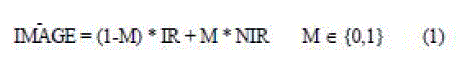 |
| Depending on the mask value on a certain position, a
pixel from the IR or NIR on the corresponding position
is selected (e.g. 0 for IR selection and 1 for NIR). Thus,
the IR layer is poured through the mask onto the NIR
layer. The imaging model, however, is composed of
basic elementary plane (layer) pairs, IR and mask. Given
a NIR, a IR plane is imaged onto it through the mask
plane composing a new NIR image. The compressed
layers are packaged in a format like TIFF-FX (file
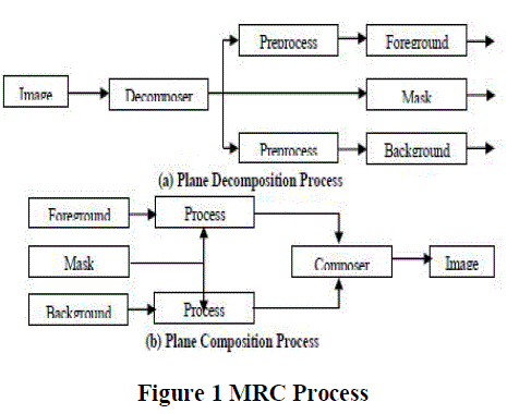
format for Internet Fax). The MRC approach has two
processes. The first process is plane decomposition and
the second stage is the plane composition process. The
plane decomposition is used to segment the image and the plan composition is used during decompression. The
steps involved during decomposition and composition is
shown in Figure 1. One consequence of MRC
segmentation is that the layers produced after
composition are actually sparse matrices and hence have
missing parts which create a „halo‟ effect. These are
often termed as “don‟t care” regions. Each layer (IR or
NIR) may contain unused pixels, since final pixels in
some positions will be selected from the other layer.
Thus, these pixels can be replaced by any color in order
to enhance compression. Redundant regions are marked
in each IR/NIR layer and the main goal here is to replace
the redundant data with other data that will enhance
compression. |
| The technique discussed for this purpose can be used to
process both IR or NIR layers and the steps are given
below. |
| Step 1: Divide the image into blocks of size 8 x 8. |
| Step 2: Blocks lying entirely in the NIR are left intact. |
| Step 3: Blocks lying entirely in the IR are filled with the
overall image mean. |
| Step 4: Partially empty blocks are then filled by using an
iterative approach that propagates values from the
existing pixels, using overall image mean value. |
| The proposed compression model that uses the enhanced
MRC-based ROI segmentation has the following steps. |
| Step 1: Segment the image into IR and NIR layers |
| Step 2: Create the binary mask layer |
| Step 3: Apply data filling to remove the halo effect |
| Step 4: Compress IR using lossless JPEG, NIR using
lossy JPEG and mask layer using JBIG compressors. |
| Step 5: Combine results into one meaningful stream for
transmission or storage. |
| After identifying the IR and NIR regions along with the
binary mask layer, the IR and NIR regions are
compressed using lossless and lossy JPEG coders
respectively. The JBIG (Joint Bi-level Image Experts
Group) algorithm is used to losslessly compress the
mask layer. The mask layer contains only two values „0‟
or „1‟ to indicate whether a pixel is taken from IR block
or background block in MRC layering. |
| As JBIG is a method for compression bi-level image
data, it is considered as the right
the mask layer. JBIG encodes redundant image data by
comparing a pixel in a scan line with a set of pixels
already scanned by the encoder. |
 |
| These additional pixels are called a template and they
form a simple map of the pattern of pixels that surround
the pixel that is being encoded. The values of these
pixels are used to identify redundant patterns in the
image data. These patterns are then compressed using an
adaptive arithmetic compression coder. JBIG is capable
of compressing color or gray scale images up to 255 bits
per pixel. |
| Block-based Segmentation |
| The proposed block-based segmentation technique uses
a block classification algorithm to segment the input
image into IR and NIR. Initially, the block-based
segmentation technique divides the image into 16 x 16
sized blocks. The classification algorithm is based on
two features, namely, histogram and gradient of the
block. The pixels of each block are grouped into three
classes, namely, low-gradient pixels, mid-gradient pixels
and high-gradient pixels, according to pixel‟s gradient
value. Then the histogram distribution for each pixel
group is computed. This method is based on the fact that
the gradient-histogram distribution of IR and NIR is
different. |
| The NIR blocks typically contain only low gradient
pixels and show one peak at the low-gradient histogram.
On the other hand the IR region shows several peaks at
the mid-gradient and high-gradient histograms. Each
block is identified using a block map which is
compressed using an Arithmetic coder. The block-based
segmentation is given in Figure 2, where B denotes a
block and N denotes the number of 16 x 16 blocks in an
image and after many tests, the thresholds T1, T2, T3
and T4 were set as 50, 45, 10 and 2 respectively.
After segmentation, the IR region is compressed using
lossless JPEG and NIR region is compressed using lossy JPEG coder. The block Map is compressed using an
Arithmetic coder. |
| Enhanced Region-Growing Segmentation |
| The goal of region growing segmentation algorithm is to
group regions having common properties between a
pixel and its neighbour. The properties can be intensity
values of the original image or unique texture patterns of
each region or spectral profiles that provide
multidimensional image data. The algorithm provides
multiple merits during segmentation. The borders of
regions found by region growing are perfectly thin and
well connected. |
| The algorithm is very stable with respect to noise. Most
importantly, membership in a region can be based on
multiple criteria. It is possible to take advantage of
several image properties, such as low gradient or gray
level intensity value, at once, while using region
growing algorithm. The traditional region growing
algorithm has two major issues. The first is the selection
of initial seed points. Incorrect selection leads to
inaccurate segmentation and therefore an automatic
process is always preferred. The second is that even with
automated process, the selected seed point may lie on an
edge. In this paper, the steps in Figure 2 is used to solve
both the issues. The last step in the algorithm consist of
two conditions that examine the candidate pixels and
makes sure that the selected seed point is highly similar
to its neighbour and is not a boundary region. For this
purpose, the relative Euclidean distance between the
seed point and its neighbours is calculated using
Equation 2. |
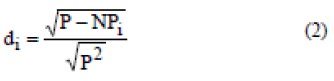 |
| Where P denotes the seed point and i denotes its 8
neighbouring pixels. After integrating the automated
process of initial seed selection, the enhanced region
growing algorithm consists of the following steps. |
| 1. Apply automatic seed selection algorithm to obtain
initial seeds for region growing |
| 2. Calculate distance between seed point and its
neighbours |
| 3. Check the neighbouring pixels and add them to the
region if they are similar to the seed |
| 4. Repeat steps 2 and 3 until no more pixels can be
added. |
| Enhanced Active Contour based Segmentation |
| In active contour based segmentation algorithm, the user
specifies an initial guess for the contour, which is then
moved by image driven forces to the boundaries of the
desired objects. The idea behind active contours, or
deformable models, for image segmentation is quite
simple. Using the user specified initial guess, the contour
is moved by image driven forces to the boundaries of the
desired objects. In such models, two types of forces are
considered - the internal forces, defined within the curve,
are designed to keep the model smooth during the
deformation process, while the external forces, which are
computed from the underlying image data, are defined to
move the model toward an object boundary or other
desired features within the image. |
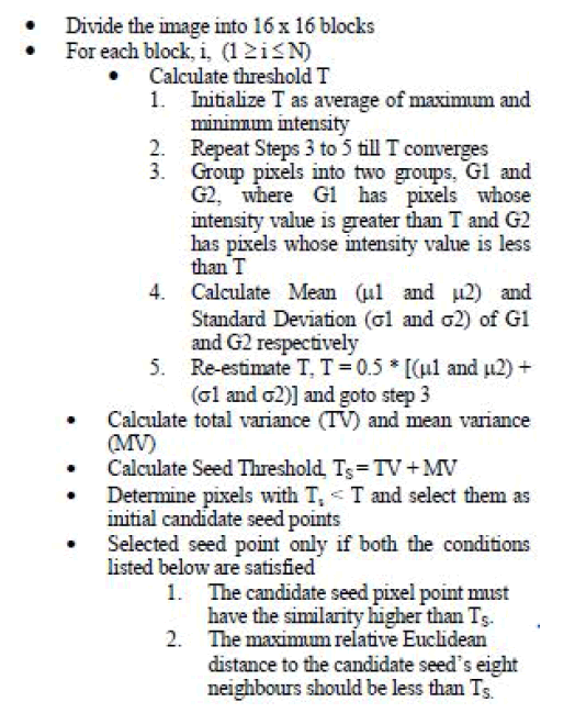 |
| Figure 2 Automatic Selections of Initial Seeds |
| The main challenge while using active contour models
for both segmentation is the initial seed selection.
Different initial seed values leads to different
segmentation result and often incorrect selection
produces inaccurate segmentation. To solve this
problem, this paper proposes the use of region growing
algorithm first to estimate the initial seeds which are
then used by the active contour model. |
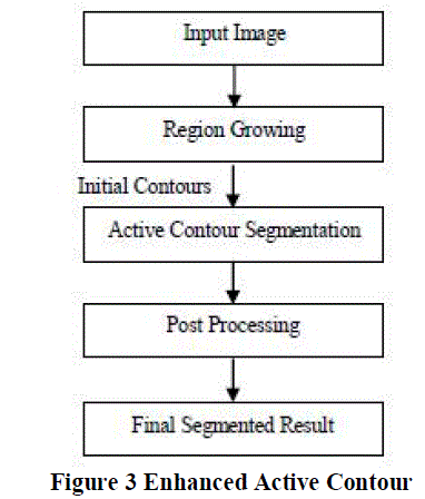 |
| The growing parameter adopted is the average between
the maximal and minimal intensities of the input image.
A post processing step that performs region merging to
merge small regions is included to improve the
segmentation result. |
| This method merges several small segments and isolates
image background by considering the distance between
regions intensity. All groups with similar intensities are
grouped together. |
RESULTS |
| The ROI segmentation was evaluated in two stages. The
first stage evaluated the performance of the proposed
ROI algorithms and the second stage analysed the effect
of ROI algorithms on compression. The quality metrics
used during performance evaluation are compression
efficiency in terms of bits per pixel, Peak Signal to Noise
Ratio (PSNR), time complexity of both ROI and
compression process and visual comparison of the
results. All the experiments were conducted using a
Pentium IV machine with 512 MB RAM and the
implementation was done in MATLAB 2009a. The
compression results are compared with the traditional
JPEG lossless algorithm. |
| In the projected results, MRC-T, BLK-T, REG-T and
ACT-T represent the enhanced MRC-based algorithm,
block-based thresholding algorithm, enhanced region
growing algorithm and enhanced active contour based
algorithm respectively. |
ROI-SEGMENTATION RESULTS |
| Table 1 shows the time taken by each of the proposed
algorithms to segment the IR from the original image. |
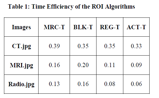 |
| From stage I experiments, it is evident that the enhanced
active contour ROI algorithm is efficient in separating
the IR and NIR regions of an image. This is followed by
the enhanced region growing algorithm and MRClayering
based segmentation algorithm. Out of the four
proposed technique, the block based algorithm showed
significant decrease in performance, which might be due
to its sensitivity to manual initialization of the threshold
values. |
| While considering the speed of the proposed ROI
segmentation algorithms (Table 1), again the active
contour-based algorithm enhanced with region growing
initial seed estimation and post processing proved to be
the fastest among the four proposed algorithm. Again the
trend proved that the block-based segmentation
technique is the slowest. |
COMPRESSION RESULTS |
| The four ROI segmentation algorithms were evaluated in
terms of compression efficiency and is shown is Table 2.
The experiment used lossy JPEG and lossless JPEG to
compress IR and NIR region. An JBIG algorithm was
used to compress the mask layer of MRC segmentation
and arithmetic coder was used to compress the block
map of the block-based segmentation algorithm. |
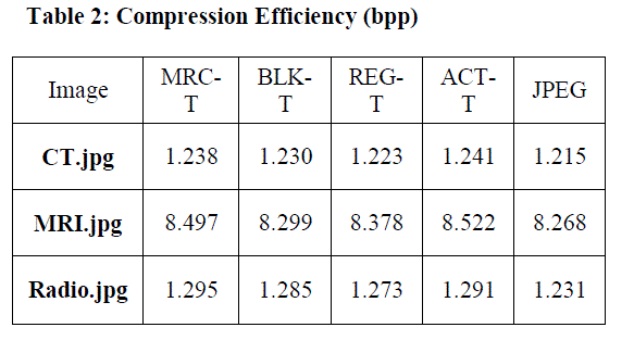 |
| From the table, it is clear that all the four algorithms
show significant compression improvement when
compared to JPEG. While comparing the proposed four
algorithms, the active contour model ranks first,
followed by MRC algorithm and block based
segmentation algorithm. The region growing algorithm
shows decreased compression efficiency in terms of
compression gained in terms of bits per pixels. Table 3
shows the Peak Signal to Noise Ratio obtained for the
original and decompressed images. |
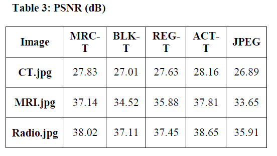 |
| The results in Table 2 again conform that the usage of
ROI segmentation to compress medical images is
positive as evident from the higher PSNR values
obtained when compared to JPEG algorithm. The
enhanced active contour model again outperforms the
rest of the three algorithms. |
| Table 4 shows the time taken for compressing the
images. The compression and decompression time is the
execution time taken by the system to perform the
compression and decompression processes. The total
compression time is calculated as the sum of
compression time and decompression time. From the
above data, it is clear that the trend of performance with
respect to compression speed has changed and JPEG
algorithm is considerably faster than the four proposed compression models. This result is obvious as the
proposed algorithm includes a ROI segmentation
process. However, the efficiency gain obtained with
respect to compression ratio and visual quality
encourages to the use of the proposed compression
models. |
| From the results obtained it is clear that the inclusion of
ROI segmentation yields efficiency compression and
produces good image quality after decompression and
therefore can be considered suitable by many medical
imaging systems. |
CONCLUSION |
| Medical image compression applications are qualitydriven
applications which demand high quality for
certain regions that have diagnostic importance in an
image, where even small quality reduction introduced by
lossy coding might alter subsequent diagnosis, which
might cause severe legal consequences. Due to this,
lossless techniques have been extensively used. As an
alternative, owing to the observation that only some part
of the image actually is of interest to the practitioners,
ROI-based techniques are becoming popular. This paper
proposes four techniques for this purpose. The first
method uses the Master-Raster Content layering based
segmentation approach enhanced to use an average block
averaging algorithm to avoid the halo effect. The second
method is an enhanced block-based thresholding
algorithm, while the third technique is an enhanced
version of the traditional region growing algorithm. The
fourth method enhanced the active contour model to
separate the input image into IR and NIR. Various
experiments on the performance of segmentation and
compression revealed that the enhanced active contour
model followed by MRC based segmentation and region
growing algorithm is efficient and can be considered as a
promising candidate by medical imaging systems. In
future, tailor-made methods for lossless and lossy
compression of the IR and NIR images are to be
designed and tested with the proposed ROI algorithms. |
References |
- Aggarwal, P. and Rani, B. (2010) Performance Comparison of Image Compression Using Wavelets, International Journal ofComputer Science and Communication, Vol. 1, No. 2, Pp. 97-100.
- Assche, S.V., Rycke, D.D., Philips, W. and Lemahieu, I. (2000)Exploiting interframe redundancies in the lossless compression of 3Dmedical images, Data Compression Conference, P. 575.
- Duraisamy, R., Valarmathi, L. and Ayyappan, J. (2008) IterationFree Hybrid Fractal Wavelet Image Coder, International Journal ofComputational Cognition, Vol. 6, No. 4, Pp. 34-40.
- Eri, H., YI, J. and Charles A,B. (2007) Segmentation for MRCcompression, Proceedings of SPIE, The International Society forOptical Engineering, Color imaging. Conference, Vol.6493, San Jose,California, USA)
- Hu, J., Wang, Y. and Cahill, P.T. (1997) Multispectral code excitedlinear prediction coding and its application in magnetic resonanceimages, IEEE Transactions on Image Processing, Vol. 6, No. 11, Pp.1555 -1566.
- ITU-T Recommendation T.44 (1998) Mixed Raster Content(MRC), Study Group-8 Contribution.
- Palanisamy, G. and Samukutti, A. (2008) Medical imagecompresssion using a novel embedded set partitioning significant andzero block coding, The International Arab Journal of InformationTechnology, Vol. 5, No. 2, Pp. 132-139.
- Pennebaker, W.B. and Mitchell, J.L. (1993) JPEG Still Image DataCompression Standard. New York: Van Nostrand Reinhold.
- Rahul, S., Vignesh, J., Santhosh Kumar, S., Bharadwaj, M. andVenkateswaran, N. (2007) Comparison of Pyramidal and PacketWavelet Coder for Image Compression Using Cellular Neural Network(CNN) with Thresholding and Quantization, International Conferenceon Information Technology (ITNG'07), Pp.183-184.
- Riazifar, N. and Yazdi, M. (2009) Effectiveness of ContourletvsWavelet Transform on Medical Image Compression: a ComparativeStudy, World Academy of Science, Engineering and Technology, Vol.49, Pp.837-842.
- Saffor, A., Ng, K.H., Ramli, A.R. and Dowsett, D. (2002)AComparison of JPEG and Wavelet Compression Applied to ComputedTomography Brain, Chest, and Abdomen Images, The Internet Journalof Medical Simulation and Technology, Vol. 1, No. 1.
- Shih, F.Y. and Cheng, S. (2005) Automatic seeded region growingfor color image segmentation, Image and Vision Computing, ScienceDirect, Vol. 23, Pp. 877âÃâ¬Ãâ886.
- Takaya, K. and Tannous, C.G. (1995) Information preservedguided scan pixel difference coding for medical images, WESCANEX95, Communications, Power, and Computing, IEEE ConferenceProceedings., Vol. 1 , Pp. 238 -243.
|