The processing of ECG signal plays a significant role in diagnosing most of the cardiac diseases. One cardiac cycle in an ECG signal consists of the P-QRS-T waves. The feature extraction scheme determines the amplitudes and intervals in the ECG signal for subsequent analysis. The shape of ECG waveform can reveal the current state of functionality of the heart. For all these observation of anomalies and selection of feature vector is important. Based on the selected feature vector classification is done. The classification is done by using RBF neural network for the analysis and evaluation of Feature Vector Selection of ECG Arrhythmias.
Keywords |
| ECG, baseline wander, zero phase filter, Atrial Fibrillation (AF), Cardiac Ischemia (CI), Sudden Cariac
Arrest (SCA), RBFNN. |
INTRODUCTION |
| Cardio-vascular disease is one of leading causes of death. Any disturbance in normal rhythm of heart is called cardiac
arrhythmia. Electrocardiogram (ECG) is one of the most important noninvasive tools for the diagnosis of cardiac
arrhythmia. The electrocardiogram (ECG) signal represents the changes in electrical potential during the cardiac cycle
recorded using surface electrodes on the body [1]. |
| The characteristics of an ECG signal helps to distinguish various arrhythmias. Manual approach of ECG analysis is
time taking process and also requires expert. ECG analysis is difficult in a small setup like clinics, health centers etc.
Hence, method for automatic classification with high accuracy is needed. Also, automatic classification will be helpful
to paramedical and emergency medical staff in cases where immediate action is to be taken. |
| In this paper, automatic classification of 4 different types of ECG beats- normal sinus rhythm, atrial fibrillation, cardiac
ischemia, sudden cardiac arrest is implemented. The R- peak detection is done using Pan Tompkins’ algorithm. The
other characteristic waves are detected by taking the R-peak as reference. The anomalies in the shape of arrhythmic
ECG are observed. The mean and standard deviation of the anomalies are taken as the features. |
| Database: MIT-BIH arrhythmia database [2] is used for testing the proposed algorithm. |
LITERATURE SURVEY |
| Pan, Tompkins [3] developed a real-time algorithm for detection of the QRS complexes of ECG signals. It reliably
recognizes QRS complexes based upon digital analyses of slope, amplitude, and width. The algorithm automatically
adjusts thresholds and parameters periodically to adapt to such ECG changes as QRS morphology and heart rate. |
| George B. Moody et al [4] studied on the factors that are involved in the automatic detection of Atrial Fibrillation. They
stated that R-R interval difference can be taken as the measure for the automatic detection of Atrial Fibrillation. |
| Recent studies suggest that Tpeak-end interval /QT interval (Tp-e/QT) may be more meaningful to predict sudden
cardiac arrest. SU Xianming et al [5] found that Tp-e/QT ratio can be as an index in predicting sudden cardiac death . |
| Shameer et al [6] implemented an automatic classification of 3 beat types – normal sinus rhythm, premature ventricular
contraction and left bundle branch block. Beat classification is implemented by using Support Vector Machine (SVM).
The main drawback of SVM is that they are binary classifiers. i.e., they can classify only two classes. So Shameer et al
designed 3 SVMs and the multiclass SVM is implemented using One Against One (OAO) approach.. |
CARDIAC DISORDERS |
| Atrial fibrillation: Atrial fibrillation occurs when the electric current in the heart is generated from all over the atria at a
very high speed, between 300 and 500 impulses a minute. This does not allow the atria to contract in a synchronized
fashion. Because of the high number of impulses generated by the heart, and their location, the atria begin to quiver.
This is known in medical terms as fibrillation [7]. |
| Cardiac Ischemia: If a coronary artery is occluded, the transport of oxygen to the cardiac muscle is decreased, causing
an oxygen debt in the muscle, which is called ischemia. Ischemia causes changes in the resting potential and in the
repolarisation of the muscle cells, which is seen as changes in the T-wave [8]. If the oxygen transport is terminated in a
certain area, the heart muscle dies in that region. This is called an infarction. An infarct area is electrically silent since it
has lost its excitability. |
| Sudden Cardiac Arrest: The immediate cause of sudden cardiac arrest is usually an abnormality in the heart rhythm
(arrhythmia), the result of a problem with your heart's electrical system. Unlike other muscles in the body, which rely
on nerve connections to receive the electrical stimulation they need to function, the heart has its own electrical
stimulator - a specialized group of cells called the sinus node located in the upper right chamber (right atrium) of the
heart. The sinus node generates electrical impulses that flow in an orderly manner through heart to synchronize the
heart rate and coordinate the pumping of blood from heart to the rest of the body. If something goes wrong with the
sinus node or the flow of electric impulses through the heart, an arrhythmia can result, causing heart to beat too fast, too
slow or in an irregular fashion. Often these interruptions in rhythm are momentary and harmless. But some types of
arrhythmia can be serious and lead to a sudden stop in heart function (sudden cardiac arrest). |
PROPOSED METHOD |
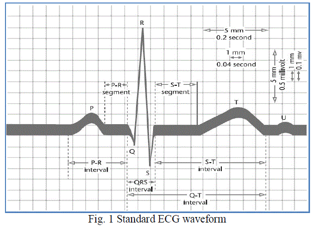
| The standard ECG waveform [9] is taken as a reference for the algorithm. The wave intervals are estimated based on
this standard waveform. The standard waveform that is considered is shown in fig 1. |
 |
| The ECG signals from the database are applied to the system developed. The block diagram of the system is shown in
fig 2. Each block in the diagram are explained below. |
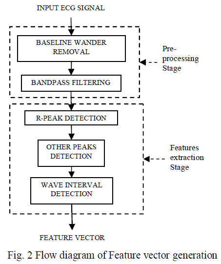 |
| Pre-processing: In this pre-processing stage first baseline wander is removed and then filtering is done to remove the
noise. First baseline wander is estimated by using first order zero phase low pass filter with cut-off frequency 0.5 Hz.
Then the baseline wander is removed from the applied ECG signal. The output signal is the applied to the band pass
filter with lower cutoff frequency 5Hz and upper cutoff frequency 40Hz. Since power line interference has the
frequency at 50Hz, this can also be removed using the above band pass filter. |
| Feature Extraction: In the feature extraction stage, all the ECG characteristic waves are identified and their intervals
are calculated. Identification of the R-peak is most important in the ECG diagnosis. First the R-peak is identified using
the PanTompkins’ algorithm [3]. By taking the R-peak as reference, the other characteristic waves are identified. The
onset and offset points of each wave are also pointed simultaneously in the above process. So that wave intervals can
be calculated. |
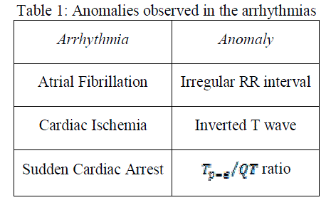
| Features are selected based on the anomalies observed in the arrhythmias. The anomaly characteristics observed in each
arrhythmia are discussed below. |
| a) For the AF case, the atria quiver irregularly due to their irregular excitation. But the ventricles quiver
regularly. So the synchronism between atria and ventricles will be disturbed. This result the irregular RR
intervals. |
| b) For the CI case, some area of the heart behaves electrically silent. Instead of propagating the electrical pulses
this part reflects them. Generally this effect is in ventricle myocardium. As an effect the T wave will be
inverted. |
| c) For the SCA case, recent studies suggest that Tpeak-end interval /QT interval (Tp-e/QT) may be more
meaningful to predict sudden cardiac arrest. SU Xianming et al [5] found that Tp-e/QT ratio can be as an
index in predicting sudden cardiac death. |
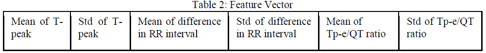
| Based on the above anomaly characteristics observed the feature vector is selected. From the above analysis, the
features selected for the classification are: mean and standard deviation of RR interval difference, mean and standard
deviation of T-peak, mean and standard deviation of ratio. The arrhythmias and their anomalies are listed in
table 1. |
 |
| The selected feature vector is based on the anomalies observed is shown in table 2. |
 |
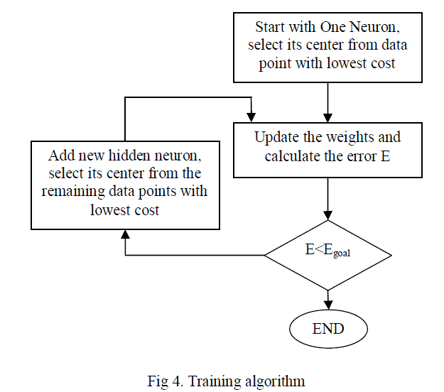
| Each ECG signal is applied to the algorithm shown in fig 2 for the feature vector generation. Based on the features
extracted the classification is done using RBF (Radial Basis Function) neural network. |
| RBF neural network: |
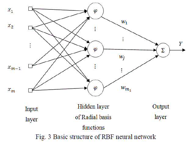 |
| The output equation can be written as |
 |
| For radial basis functions, the basis functions are |
 |
 |
RESULT AND DISCUSSION |
| The step by step result of NSR person ECG of record number ‘nsrdb 16272m’ is shown below. |
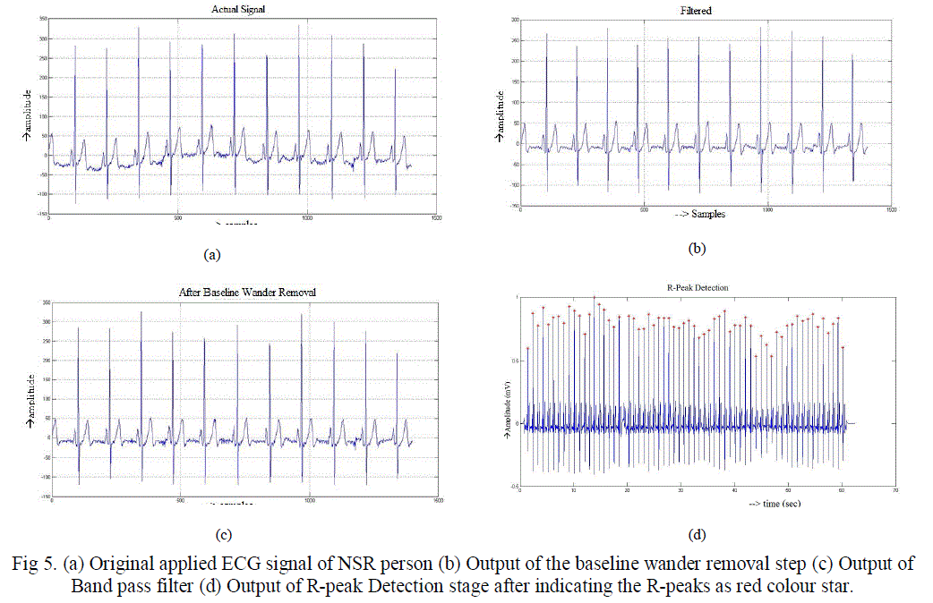 |
| In the fig 5, first original applied ECG signal is shown and then outputs of baseline wander removal step and output of
band pass filter step are shown. Finally the identified R-peaks are indicated as red colour star mark. After the detection
of R-peak the other characteristic waves i.e. P, Q, S and T peaks and their intervals are identified by taking R-peak as
reference. This process is carried out for all the ECG records. The output of one for each class of ECG signals is shown
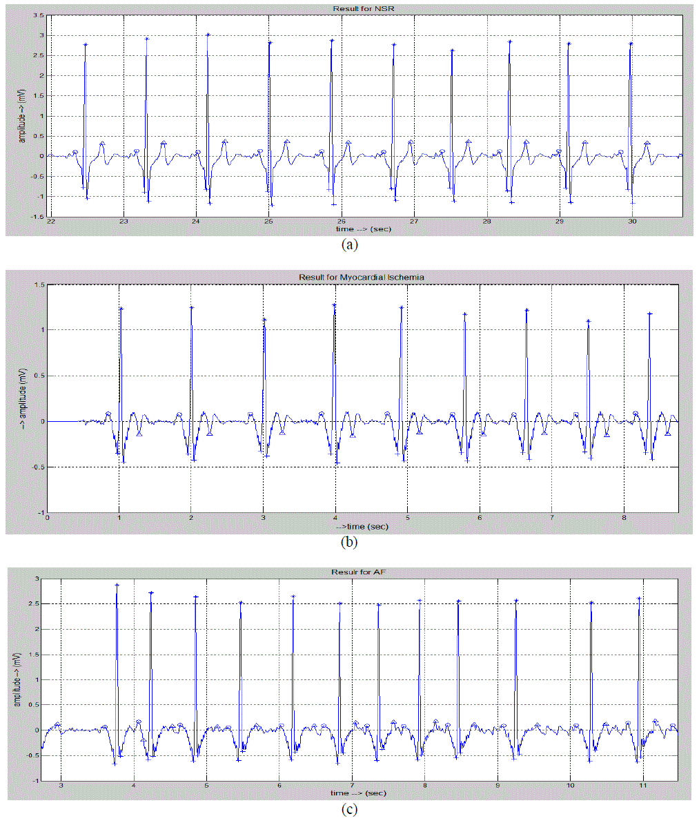
in fig 6. |
 |
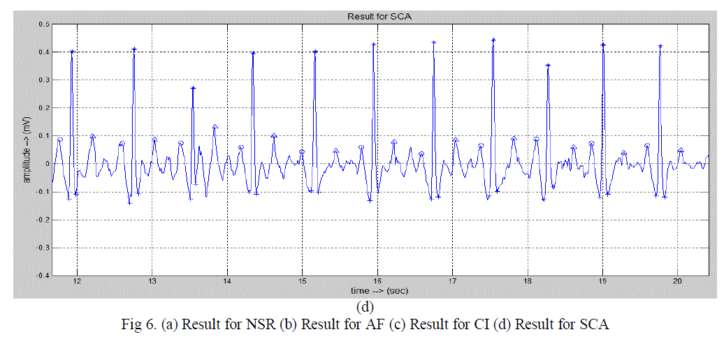 |
| In the fig 6, different peaks are indicated with different symbols. |
| * indicates R-peak |
| o indicates P-peak |
| ^ indicates T-peak |
| + indicates Q and S –peaks |
| The figure 6(a) shows the result for a typical signal of NSR person. The figure 6(b) shows the result for a typical signal
of AF person. The figure 6(c) shows the result for a typical signal of CI person. The figure 6(d) shows the result for a
typical signal of SCA person. |
| After identifying all the characteristic waves and their intervals, feature vector is calculated for each record. Here some
records are used for training the neural network and some are used for testing. |
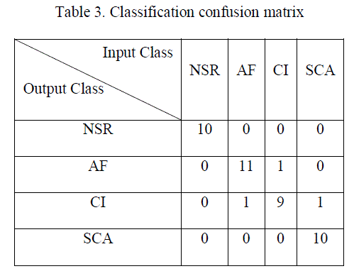 |
| Test database consists of 10 NSR ECG signals, 12 AF ECG signals, 10 CI ECG signals, and 11 SCA ECG signals. It is
applied to the trained RBF neural network. In case of NSR, AF, and CI classification results are promised. In the SCA
case, the prediction seems to be failed but it is classified correctly because before the SCA the person is already having
the CI abnormality. |
CONCLUSION |
| The anomalies of arrhythmias are observed. The feature vector is selected based on the anomalies. The neural network
is trained with the some of the collected ECG arrhythmia database and is tested with the remaining records. The results
are observed experimentally in accurate. The feature vector selected assures the requirement for the classification. With
this observation of anomalies of arrhythmias, feature vector size can be reduced compared to previous methods, so that
the time required for the feature vector extraction is reduced. |
References |
- D. 1. Rowlands, "Understanding the electrocardiogram" (Section 2: Morphological abnormalities). Imperial Chemical Industries PLC, England, 1982.
- MIT/BIH arrhythmia database: http://www.physionet.org/cgi-bin/atm/ATM
- Jiapu Pan, Willis J. Tompkins âÃâ¬ÃÅA Real-Time QRS Detection AlgorithmâÃâ¬ÃÂ, IEEE Transactions On Biomedical Engineering, VOL. BME-32, NO. 3, MARCH 1985.
- George B. Moody, Roger G. Mark âÃâ¬ÃÅ A new method for detection Atrial Fibrillation using R-R intervals.âÃâ¬Ã IEEE Transactions on Computers in Cardiology, pp. 227-230, 1983.
- SU Xianming PhD MD, ZhiXiaowen âÃâ¬ÃÅTpeak-end interval /QT interval ratio of ECG predicts sudden cardiac death and malignant Ventricular arrhythmiasâÃâ¬ÃÂ, IEEE, pp. 57-60, 2011.
- ShameerFaziludeen, Sabiq P.V âÃâ¬ÃÅECG Beat Classification Using Wavelets and SVMâÃâ¬ÃÂ, Proceedings of IEEE Conference on Information and Communication Technologies, pp. 815-818, 2013.
- Atrial fibrillation description, http://www.nhlbi.nih.gov/health/health-topics/topics/af/
- Cardiac Ischemia Theory, http://www.cardiacischemia.com/
- Gari D. Clifford Francisco Azuaje Patrick E. McSharry, Advanced Methods and Tools for ECG Data Analysis, Artech House, 2006
|