X-ray Bone image segmentation is an integral component of orthopaedic X-ray image analysis that aims at extracting the bone structure from the muscles and tissues. Automatic segmentation of the bone part in a digital Xray image is a challenging problem because of its low contrast with the surrounding flesh, which itself needs to be discriminated against the background. X ray images are found to have random noise. The random distortion makes it difficult to perform perfect image processing. The non-linear total variation based on partially differential equation algorithm proposed by Leonid et at. [7],is chosen because it effectively removes random noise present and also smoothens the image. The presence of noise and spurious edges further complicates the segmentation. In this paper, we propose an efficient Total-Variation based segmentation technique that integrates several algorithms like non-linear total variation algorithm to remove random noise present, edge detection, Otsu’s global thresholding and application of morphological operators. Experiments on several X-ray images taken from IRMA Database for different parts of the body like hand, abdomen and spine images provide encouraging results.
Keywords |
| Global thresholding, IRMA Database-X-ray Segmentation, Non linear Total Variation algorithm |
INTRODUCTION |
| Diagnostic radiology requires accurate interpretation of each and every complex component in medical images.
Especially for X-ray Images, bone segmentation is a vital step in X-Ray image analysis techniques. The main aim of
segmentation is to help medical practitioners to the study of bone structure, identify and measure bone fracture and to
plan the treatment prior to surgery. It is considered as a challenging task since the bone x-ray images are complex in
nature and the output of segmentation algorithm is affected due to various factors like partial volume effect, intensity
variation, presence of noise and artifacts and closeness in gray level of different soft tissues.[1,2] Every day large
volumes of different types of medical images such as dental, endoscopy, skull, MRI, ultrasound, radiology are
produced in various hospitals as well as in various medical centres [4]. Segmentation is a key pre-processing step that
further helps in separating the image into various regions of interest. Mohmmad et al. [16] proposed a morphological
based segmentation method for segmenting medical images. The results of the proposed method used morphological
transformations to get efficient segmentation result. A gradient analysis was initially conducted to determine the edges,
which were then used as input to subsequent dilation of eroded image. The results proved that segmentation was
efficient and was able to preserve important edges of the bone X-Ray images. Several methods have employed manual
segmentation to rule out retrieval errors due to wrong segmentation. However, despite its advantages, manual
segmentation is a tedious task. This has lead to the development of semiautomatic and automatic segmentation
algorithms to extract the regions of interest. [9]Segmentation of bone structures in x-ray images is both intrinsically and
extrinsically difficult. Intrinsic difficulties refer to those caused by the intrinsic properties of x-ray imaging systems:
Following are some of the intrinsic difficulties. |
| 1:Noise. Noise in x-ray images has a number of sources, but the most fundamental is from the x-ray source itself. This
type of noise is called quantum noise, in reference to the discrete nature of the x-ray photons. |
| 2:Overlapping. Overlapping of two bones or joint bones in some cases create exact location problem of fracture. |
| Extrinsic difficulties are usually related to the patients, and are as follows: |
| 1: Ambiguity- Neighboring tissues inside human body may have similar x-ray absorption rates. As a result, the
boundaries of the organs may be ambiguous. |
| 2: Bone density variability- Different patients may have different bone densities, which may result in significantly
different intensities in the bone regions between their x-ray images. A normal patient usually has dense bones and the x-ray images of bones are bright, whereas a patient who suffers from osteoporosis will have low-density bones, which
may results in much darker bone images. Moreover, other body tissues may also affect the intensities of bone images. |
| 3: Inter-patient shape variability-The shapes of the bones of different patients can differ quite significantly. In
particular, the shape of the pelvis bone of a female patient is quite different from that of a male patient. This is due to
the fact that females usually have much wider pelvis bones. |
| 4: Imaging pose variability- Same bones imaged at different times may be located at different locations due to different
imaging posture. [15] |
| The rest of paper is organised as follows, section II describes the related work done in segmentation of Xray images
section III describes the proposed framework of segmentation for X-ray Images followed by experimental results,
section IV put forward the concluding remarks for paper. |
RELATED WORK |
| Automatic image classification is mapping images into pre-defined classes and it involves some basic principles such
as representation where visual feature of the image are extracted and generalization which is training and evaluating the
classifier. The first and most vital component of any classification system is image representation. It is categorized into
two main approaches, (i) low-level image representation and (ii) patch based image representation. Low-level image
representation has been used by several researchers in various domains [2-9][28]. |
| Seong-Hoon et al. [2] proposed classification of medical x-ray images where texture information of medical x-ray
images are extracted using LBP; the classification accuracy obtained outperforms others who use edge histogram
descriptor. Combination of block based local binary pattern with edge histogram was used as medical image
representation for the task of automatic medical image annotation in ImageCLEF 2007 [3]. Zhy et al. [4] applied
segmentation method on ultrasound medical images based on texture features which are obtained according to GLCM.
GLCM were combined with histogram moments as feature extraction to classify ultrasound medical images in [5].
Another widely used strategy is combining different local and global descriptors into a unique feature representation.
Pixel value as global image descriptor was combined with other image representation techniques to construct feature
vector of the image [6-9]. |
| Recently, more promising studies have been focused on local patch based image representation. The bag of words
(BoW) represents images using histograms of quantized appearances of local patches. In recent years, many studies
exploited this feature in various image classification domains including medical domain [10-20]. With increasing size
of medical X-ray archives, it is important to have simplistic, discrete representations and simple matching measures to
preserve computational efficiency. It is argued that BoW paradigm provides efficient means to address the challenge of
CBIR system in large size databases such as the one in ImageCLEF [10]. They proposed X-ray image categorization
and retrieval based on local patch representations using a “bag of visual words” approach. They analyzed the effects of
various parameters on system performance. The best result was presented using dense sampling of simple features and
a nonlinear kernel-based Support Vector Machine (SVM). This was an extension of another work where visual words
dictionary were generated to represent X-Ray chest Images [11]. Deselaers et al. in [12] extracted features from local
patches of different sizes which were taken at every position and were scaled down to a common size. In that work,
rather than using a dictionary, the feature space was quantized uniformly in every dimension and the image was
represented as a sparse histogram in the quantized space. |
PROPOSED FRAMEWORK FOR X-RAY IMAGE SEGMENTATION |
| Image segmentation facilitates delineation of anatomical structures and other regions of interest. Segmentation is one of
the most difficult tasks in image processing and determines the outcome of analysis and evaluation of pathological
regions. |
| The proposed system uses image processing techniques like nonlinear total variation based noise removal, applying
Sobel Edge detection to locate region of interest (ROI), global image thresholding using Otsu method and finally post
processing through morphological operators .Figure1shows the flow diagram for the proposed work. |
Image Denoising |
| X-Ray images are found to have random noise. The random distortion makes it difficult to perform perfect image
processing. The non-linear total variation based on partially differential equation algorithm proposed by Leonid et at.
[7], is considered in this work since it effectively removes random noise present and also smoothens the image. For an
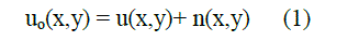
Input X-ray image(Figure 2 from IRMA database) if the observed intensity function is uo(x, y) denote the pixel values
of a noisy image. Let u(x, y) denote the desired clean image, and n be the additive noise, then their relation can be
represented as in (1). |
 |
| The total variation of the image is minimized subject to constraints involving the statistics of the noise. The constraints
are imposed using Lagrange multipliers as in (2). |
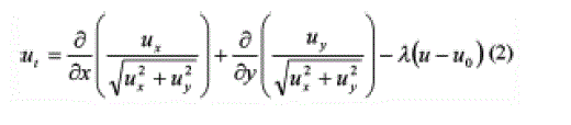 |
| The solution is obtained using the gradient-projection method with time as a parameter. As t extends to infinity the
solution converges to a steady state, the required denoised image(Figure 3) is obtained. |
Region of Interest Detection |
| Edge detection is the part of segmentation in image processing. The goal of segmentation is to simplify and change the
representation of an image into something that is more meaningful and easier to analyze. The edge function is used to
detect edges, which are those places in an image that correspond to object boundaries. To find edges this function looks
for places in the image where the intensity changes rapidly. Edge returns a binary image containing 1's where edges are
found and 0's elsewhere. Different edge detection methods Sobel, Prewitt, Roberts, Laplacian of a Gaussian (LOG),
Zero Cross, & Canny are applied on X-Ray medical images [17, 18]. Here Sobel Edge detector was applied since the
Sobel operator performs a 2-D spatial gradient measurement on an image and so it emphasizes regions of high spatial frequency that correspond to edges. Typically it is used to find the approximate absolute gradient magnitude at each
point in an input gray scale image. |
| These kernels are designed to respond maximally to edges running vertically and horizontally relative to the pixel grid,
one kernel for each of the two perpendicular orientations. The kernels can be applied separately to the input image, to
produce separate measurements of the gradient component in each orientation (call these Gx and Gy). These can then
be combined together to find the absolute magnitude of the gradient at each point and the orientation of that gradient.
The gradient magnitude is given by: |
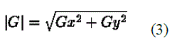 |
| Where Gx and Gy are gradients in X and Y axis orientation |
| Typically, an approximate magnitude is computed using: |
 |
| which is much faster to compute. |
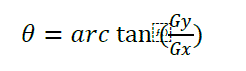
| The angle of orientation(θ) of the edge (relative to the pixel grid) giving rise to the spatial gradient is given by: |
 |
| Algorithm: |
| 1. Create Sobel horizontal edge-emphasizing filter |
| 2. Create Sobel vertical edge-emphasizing filter |
| 3. Apply Sobel horizontal edge-emphasizing filter on the noise removed image |
| 4. Apply Sobel vertical edge-emphasizing filter on the noise removed image |
| 5. Compute the gradient magnitude of horizontal and vertical component of filtered image. |
3. Image Thresholding |
| Thresholding is used to extract an object from its background by assigning an intensity value T (threshold) for each
pixel such that each pixel is either classified as an object point or a background point |
| In general T = T[x, y, p(x, y), f (x, y)] |
| • If T is a function of f(x,y) only then it is referred as Global thresholding |
| • If T is a function of both f(x,y) and local properties p(x, y) then as Local thresholding |
| • If T depends on the coordinates (x, y) Dynamic/adaptive thresholding. |
| Here global thresholding using Otsu’s method is used. It selects the threshold by minimizing the within-class variance
of the two groups of pixels separated by the thresholding operator. It does not depend on modeling the probability
density functions; however, it assumes a bimodal distribution of gray-level values.[8] |
| Consider that we have an image with L gray levels and its normalized histogram (i.e., for each gray-level value i, P(i) is
the normalized frequency of i). |
| Assuming that we have set the threshold at T, the-normalized- fraction of pixels that will be classified as background
and object will be: |
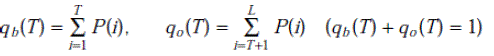 |
| Where qb and qo are fraction of pixels belonging to background and object of given image . |
| The mean gray-level(μ) value over the whole image (grand mean) is: |
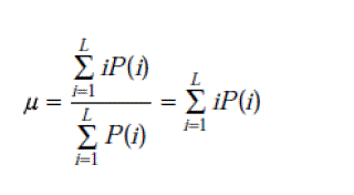 |
| The variance(σ) of the background and the object pixels will be: |
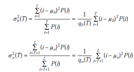 |
| where σb(T) and σo(T) are background and object variance respectively.[15] |
| The variance of the whole image is: |
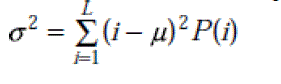 |
| The resulted threshold image is shown in figure 4. |
| Following Images have been tested with the proposed method and satisfactory results are found. |
4. Post Processing Using Morphological Operations |
| After having the thresholded image in order to remove small objects which are less than predefined threshold.
Morphologically open binary image and remove small objects.[13] |
| Algorithm: |
| 1. Find connected components in image. |
| 2. Define a threshold based on practical observations and study of images used for segmentation. |
| 3. Apply the threshold to remove small areas of connected component. |
| Finally the result of segmentation is shown through image in figure no.5. |
CONCLUSION |
| In this work different medical images of X-Ray have been segmented successfully using nonlinear toatal variation
based algorithm which is simpe and relatively fast. The results appear to be state-of the art for very noisy images as the
method is noninvasive yielding sharp edges of the image. The following experiment is tested on near about 300 images
of spine and hand X-rays and had reported satisfactory results with required fine edge detection. The results for hand
and spine image from IRMA database are tested for segmentation using total variation based denoising followed by
global thresholding using Otsu method and some morphological transformations to have original segmented image
retrieval possible. |
Figures at a glance |
 |
 |
 |
 |
 |
| Figure 1 |
Figure 2 |
Figure 3 |
Figure 4 |
Figure 5 |
|
References |
- Rafael C.Gonzalez& Richard E.Woods, “Digital Image Processing”, Second Edition, 2005.
- Rafael C.Gonzalez, Richard E.Woods&Steven L. Eddins, “Digital Image Processing UsingMATLAB”, Pearson Education 2007.
- Kenneth R.Castleman, “Digital Image Processing”, Pearson Education., 2006
- Adrian Low, “ComputerVision&Image Processing”, McGraw Hill (1991)
- B.Chanda&DuttaMajumdar, “Digital Image Processing and Analysis”, PHI (2001)www.mathworks.com
- Leonid I Rudin, “Nonlinear Total Variation Based algorithm for Noise Removal”Physica D (1990)259-268.
- MitraBasu, “Gaussian-Based Edge-Detection Methods-A Survey”IEEE Transactions on Systems Man, And Cybernetics Part: C ApplicationAnd Reviews, Vol., 32, No.3, August 2002.
- J. Canny, “A computational approach to edge detection”, IEEE Trans Pattern Anal. Machine. Intell., vol. PAMI-8, pp. 679–698, Nov. 1986.
- V. Torre and T. Poggio, “On edge detection”, IEEE Trans. Pattern Anal. Machine. Intel., vol. PAMI-8, pp. 147–163, Mar. 1986.
- T. Pavlidis and Y. Liow, “Integrating region growing and edge detection”, IEEE Trans. Pattern Anal. Machine Intell. vol. 12, pp.225–233, Mar.1990.
- Gautam A. Kudale, Shivanand .S. Gornale “Performance Analysis of Edge Detection Methods for Medical Images”, National Conference on Advancements in Information Technology and Internet Security (AITIS 2008), SIBAR, Pune. (MS). India.
- Mahesh Pawar, GautamKudale, S.S. Gornale “Deblurring of Blurred Image: for Medical Image Processing Applications”.National Conference on Emerging Trends in Information Technologies and its Application for Technical And management Education.Alwar, Rajasthan, India.
- Mahesh Pawar, GautamKudale, S.S. Gornale “Deblurring of Medical Image for Medical Daignosis”, Eighth National Conference on Advanced Computing and Applications (NCACA'09), PSG College of Technology, Coimbatore, International Journal of Computer Science and Application Issue 2010 ISSN 0974-0767 Tamilnadu.
- Kristin Norell' ”An Automatic Method for Counting Annual Rings in Noisy Sawmill Images”, Springer Berlin / Heidelberg.
- Mohamed Roushdy, “Comparative Study of Edge Detection Algorithms Applying on the Gray scale Noisy Image Using Morphological Filter”, GVIPJournal, Volume 6, Issue 4, December,2006.
- “Edge detection”, (Trucco, Chapt 4ANDJain et al., Chapt 5).
- Wang Luo, “Comparison for Edge Detection of Colony Images”, IJCSNS International Journal of Computer Science and Network Security, VOL.6 No.9A, September 2006.
|