ISSN ONLINE(2278-8875) PRINT (2320-3765)
ISSN ONLINE(2278-8875) PRINT (2320-3765)
Chandan Singh Rawat1, Vishal S. Gaikwad2
|
| Related article at Pubmed, Scholar Google |
Visit for more related articles at International Journal of Advanced Research in Electrical, Electronics and Instrumentation Engineering
Speckle noise is the main source of noise in biomedical imaging, due to which noise quality of an image degrades. During image acquisition due to constructive and destructive phenomena speckle noise arises. To reduce the noise from an image one can take multiple images or frames of the same object and can average them for noise reduction. Wavelet transform can be used for image de-noising. This paper introduces various methods of multi-frame image de-noising for biomedical as well as other multi-frame images.
Keywords |
| Biomedical image de-noising, Image de-noising using Wavelet transform, Speckle noise reduction of biomedical images. |
INTRODUCTION |
| In high speed image acquisition techniques like OCT it’s possible to take multiple frames/images of the same object. In OCT biomedical imaging for the analysis of disease or tissue structure it is very important for doctors to get clear biomedical image. In OCT biomedical imaging technique which uses Laser Sources, images are corrupted by unwanted speckle noise which arises due to constructive and destructive phenomena of light. One common component of any biomedical image is speckle noise suppression. This noise is generated due to the interference of many random scattered photons which losses in the biomedical samples. Noise reduction from biomedical images enhances the quality of image which enhances the visual structure of tissue for better visualization & study. The developments of these methods are demanding, as speckle noise differs in its properties from the Gaussian noise which is typically understood to be present in normal images taken with Charge Coupled Devices (CCD-sensors). Speckle noise in addition contains a few useful information about the structure of cell or object as its not pure noise [1]. |
| As speckle noise contains a few useful information about the image it is pattern does not change if no physical parameter of the imaging system is changed. Biomedical images are usually intensity images because of this image quality also depends on scattering property of the imaged object/tissue, hence speckle suppression can be done by frame averaging and digital de-noising algorithm. Frame averaging is further depends on change in speckle pattern due to movement of the object such as eye when imaging the retina [1]. |
| In OCT imaging techniques an image is often degraded by speckle noise during OCT signal acquisition or image acquirement. The aim of de-noising is to take away the noise as collecting the imaging object information as much as possible to retain the important signal features of an image. This can be done by various filters such as median filter, Wiener filtering. There are great literatures are available on signal de-noising using these types of techniques/filters for removing speckle noise [4]. The image de-noising process can be separated into four different steps which are data acquisition, pre-processing, data reduction, and features analysis. Speckle noise reduction from OCT images is the one of the key tasks in OCT image de-noising. Depending on quantity of the speckle noise in image various filters can be implemented to remove noise. OCT images are usually corrupted by speckle noise during its acquisition because of optical components and laser source and constructive and destructive interference of signal. As with OCT imaging it is possible to take multiple images of the same object with very fast speed, hence the main objective of OCT multiple frame de-noising technique is to take out speckle noise which degrades image quality. In OCT imaging technique the main drawback is the low quality of images, which are due to addition speckle noise in an image. Due to the presence of speckle noise it degrades image quality and it affects visual inspection to the doctors. Accordingly, speckle filtering is the central pre-processing step for image quality enhancement in medical imaging techniques [2]. |
SPECKLE NOISE AND ITS REDUCTION |
| The speckle noise present in an OCT images are multiplicative noise which is nothing but an unwanted randon signal which gets multiplied with some significant signal in the image capturing, transmission or processing. Mathematically speckle noise can be represented as follow [9], |
| Dm, n = S m, n * Um, n + V m, n (1) |
| where Dm, n is the noisy pixel, Sm, n is the noise free pixel, Um,n and Vm,n are the multiplicative and additive noise respectively and m,n are indices of the spatial locations. As the effect of additive noise is very much smaller when compared to that of multiplicative noise [9], which may be written as |
| Dm, n ≈ S m, n (2) |
| Also logarithmic compression is applied to the noisy signal which affects the speckle noise statistics and it becomes very close to white Gaussian noise. The logarithmic compression transforms multiplicative form in [9] to additive noise form as |
| log (Dm,n) = log (Sm,n) + log (Um,n) (3) |
| O(m,n) = C(m,n) + N(m,n) (4) |
| The first log term in the equation ( log (Dm,n)), represent the noisy image, after log compression it can be represented as O(m,n), and the second and third log terms log (Sm,n) and log (Um,n) are nothing but the noise free pixel and noisy components which can be further denoted as C(m,n) and N(m,n) respectively after logarithmic compression. This mathematical model proves that the speckle noise is a multiplicative noise of the image, hence the speckle noise is commonly known in the high intensity area than in a low intensity area [9]. |
| The main aim of the speckle noise suppression is to eliminate or to take out such noise and to preserve as much as achievable essential signal features of that image. As speckle noise degrades image quality and influences the analysis of that image in OCT image analysis as it is biomedical images. Now a day’s there are many filters available for the removing of speckle noise. Out of which now a day’s wavelet transform is used to remove noise from signal/image as it is a multi-resolution image-processing tool. Speckle noise in an image/signal is nothing but a high-frequency component of the image/signal, which is having added advantage in wavelet transform based de-noising method as it appear in wavelet coefficients during wavelet transform processing[5]. |
WAVELET BASED SPECKLE NOISE REDUCTION |
| Now a day’s in research domain wavelet transformed is tremendously used to remove noise from signal/image for increasing image quality of an image. As wavelet transform based de-noising try to eliminate the noise which is present in the signal and it also keep the original signal characteristics [4]. |
| Wavelet transforms normally uses wavelet thresholding for signal de-noising, thresholding can be soft-thresholding or hard-thresholding. Speckle noise normally a high-frequency component in any signal/image. We can use this property in wavelet based de-noising method, as speckle noise is high frequency component it can appeared in wavelet coefficients, hence for further processing it is very much useful in wavelet based transform. Hence one extensive technique is used for speckle noise reduction which is nothing but a wavelet thresholding procedure. |
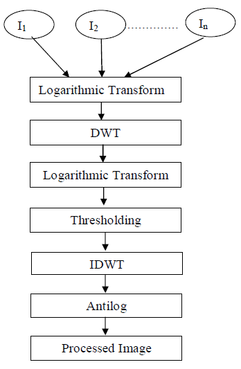
| In this de-noising method we take multiple frames/images of the same object with small modification in the position of the object. After that we apply logarithmic transform to convert speckle noise from multiplicative to additive form. After that wavelet thresholding can be applied followed by thresholding. These above steps (up to thresholding) can be applied to multiple frames and then averaging of these frames can be done to improve image quality. After that inverse discrete wavelet transform can be applied followed by antilog. After executing these entire steps one can get final noise free image. An algorithm has been designed for OCT image de-noising, as shown in Fig.1, where I1, I2…..In are image frames/images.DWT is Discrete Wavelet Transform and IDWT is Inverse Discrete Wavelet Transform. |
 Fig 1: Image Denoising Algorithm Fig 1: Image Denoising Algorithm |
RESULT AND DISCUSSION |
| The performances for de-noising of images were tested on set of grayscale images with the resolution of 512×512. The quality of an image can be examined by objective evaluation and subjective evaluation. In subjective evaluation, the image is observed by a human expert. But as the human visual system is too much difficult and this cannot predict the precise quality of an image, hence one can perform an objective evaluation for image analysis and to check image quality [3]. |
| The objective performance metrics used are (i) Peak Signal to Noise Ratio (PSNR) and (ii) SSIM. PSNR is a quality measurement between the original and a de-noised image. The higher the PSNR, the better is the quality of the compressed or reconstructed image. To compute PSNR, the block first calculates the Mean- Squared Error (MSE) and then the PSNR[6],which is explained in equation 5 and 6 . |
 |
| Where m equals to number of rows and n equals to number of columns in the input image and output image respectively The despeckling models were tested with the test image (grayscale) of 512 x 512 size. The structural similarity index measure is another method for objective performance evaluation between images x and y which is given by (7). |
 |
| where c1 and c2 are constants. |
| SSIM can be applied locally using different sliding window size as per the user which can help to move over the image by pixel by pixel horizontally and vertically covering all the rows and columns of the image, starting from top-left corner of the image [6]. |
| The SSIM is improved method for measuring the similarity between two images. SSIM gives improved result as compared to traditional methods like peak signal to noise ratio (PSNR) [7]. |
| The proposed models were implemented using MATLAB R2010a and were tested on Pentium IV machine with 1GB RAM. The amount of noise reduction is measured by the SSIM & PSNR. |
| A dataset for multi-frame image de-noising is publicly available on “Pattern Recognition Lab, Martensstrasse, Erlangen, Germany” [1].All the frames/image sets information is also available on that website which is a key requirement for this type of de-noising methods. There are total 35 image datasets of dead pig’s eye which were acquired by scanning a pig’s eye with a OCT machine named Spectralis HRA & Optical Coherence Tomography (OCT) system (Heidelberg Engineering) in high speed mode with 768 A-scans. The spacing between the pixels of the acquired images is 3.87 μm in the axial direction and 14 μ m in the transversal direction [1]. The axial resolution of the system is 7 μm. To take image of the eye it was placed in front of the OCT device. All image acquisition was performed manually. Total 35 sets were recorded each contain 13 frames with 35 different eye positions. All these 35 sets were recorded with a complete 0.384mm shift in the transversal direction. In this process speckle noise can be considered to be an uncorrelated in between the scans from the varying positions. To keep eye moisturized during imaging the opacity of the eye was increased as the eye lost humidity during moving. Hence the image quality can be decreased by a lesser signal-to-noise ratio [1]. |
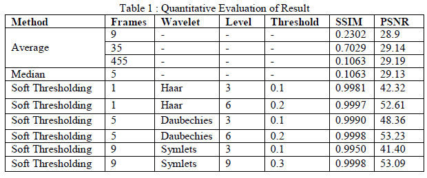
| Averaging of OCT frames are performed on 9 frames, 35 frames and 455 frames for this method SSIM are 0.2302, 0.7029, 0.8680 and PSNR are 28.9dB, 29.14 dB, and 42.16dB respectively. Soft thresholding is performed using Haar,Daubechies & Symlets wavelet using different decomposition levels(3,6,9) with 0.1,0.2,0.3 threshold values. For all these methods SSIM & PSNR calculated out of which Symlet wavelet gives best results. The results were performed by averaging and wavelet methods. For averaging method PSNR is in the range of 29.1dB and for SSIM it’s in the range of 0.1 to 0.7. |
| The result shows great changes when wavelet method is used. For Haar wavelet with 1 frame, 3 decomposition levels and with 0.1 threshold value SSIM is 0.9981 and PSNR is 42.32dB which indicate great result as compared with averaging method. For Haar wavelet with 1 frame, 6 decomposition levels and with 0.2 threshold value SSIM is 0.9997 and PSNR is 52.61dB. For Daubechies wavelet with 5 frames, 3 decomposition levels and with 0.1 threshold value SSIM is 0.9990 and PSNR is 48.36dB. For Daubechies wavelet with 5 frames ,6 decomposition levels and with 0.2 threshold value SSIM is 0.9998 and PSNR is 53.23dB. For Symlets wavelet with 9 frame ,3 decomposition levels and with 0.1 threshold value SSIM is 0.9950 and PSNR is 41.40dB For Symlets wavelet with 9 frame ,9 decomposition levels and with 0.3 threshold value SSIM is 0.9998 and PSNR is 53.09dB |
| From the above discussion it is clear that in wavelets as we increase decomposition level the SSIM and PSNR increases. SSIM=0.9998 and PSNR=53.23dB are the best case results obtained with Daubechies wavelet family with 6 and 9 decomposition levels with soft thresholding method. Symlets wavelet family also give very good results with 9 decomposition levels and 9 frames. |
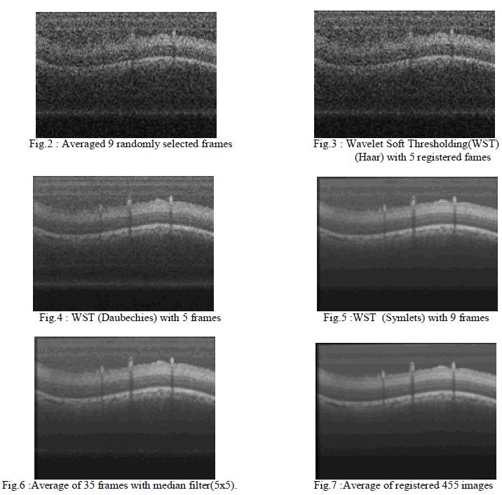
| All the subjective results are depicted in figures (Fig.2 to Fig.7).Though subjectively some processed images look like same but objectively their PSNR and SSIM are different. The result table with all details are given in Table 1. |
 |
 |
CONCLUSION |
| In this paper, OCT multi-frame image de-noising techniques are mainly focused with different frames of OCT images. All results are compared using image quality parameters SSIM & PSNR. All the results of the image de-noising are performed using multi-frame image averaging & wavelet transform out of which soft thresholding with Symlet wavelet with 7 frames and 0.3 threshold values gives best result. Gold standard image created by averaging all registered 455 frames. |
ACKNOWLEDGMENT |
| The authors would like to thanks to “Pattern Recognition Lab, Martensstrasse, Erlangen, Germany” for providing open access to OCT image dataset. We would also like to thanks Optoelectronics Division, SAMEER, IIT Campus Mumbai for providing information. |
References |
|