Keywords
|
| Specific Absorption Rate (SAR), Microwave detection and breast cancer |
INTRODUCTION
|
| In 2013, an estimated 232,340 new cases of invasive breast cancer were expected to be diagnosed in women in the U.S along with 64,640 new cases of non-invasive (in situ) breast cancer. From these statistics it is clear that for women, the mortality of breast cancer is higher than for any other cancer. |
| The current standard breast cancer screening methods includes X-ray mammography, Ultrasound and Magnetic Resonance Imaging (MRI).X-ray mammography is the most common method. Although it has shown good breast cancer detection capabilities, X-ray mammography has a number of well-known limitations, particularly when imaging dense breast, the breast is required to undergo heavy compression. In addition it employs ionizing radiation and suffers from high false-positive [2] and false –negative [1] rates. |
| The Ultrasound method struggle with distinction between malign and benign tumor.MRI scans are highly expensive and additionally follow- up examinations and biopsies. Recent research has suggested that use of microwaves for detection of breast cancer avoids exposure to ionizing radiation and does not require compression of the breast. This technology depends on the detectable intrinsic contrast in dielectric properties of a tumor and its surrounding normal breast tissue at microwave frequencies [3]. |
| Since this contrast is present at the early stages of development of the tumor, it is highly suitable for diagnosis of breast cancer at the starting stages. Microwave imaging aims to reconstruct an image of the breast by mainly using two different approaches microwave tomography [4] and radar based imaging [5]. |
| Specific absorption rate (SAR) is the rate at which energy is absorbed in a body tissue and has the unit W/Kg. In this section we establish the utility of using the coordinates of the maximum value of SAR for detecting the location of tumors inside the breast of different sizes. The electromagnetic absorption loss between a normal breast and tumor infected breast is determined and compared [7]. The absorption values for different tumor locations at different frequencies are also analyzed. |
MODELS
|
A. Breast Model
|
| A mature human female’s breast consists of fat, connective tissue and thousands of lobules – tiny glands which are connected with the nipple via the milk ducts. The milk of a breastfeeding mother goes through tiny ducts (tubes) and is delivered through the nipple. |
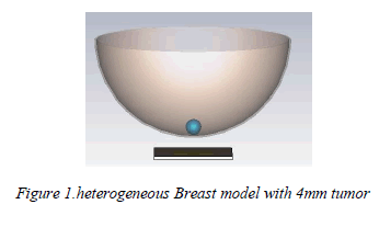
| In order to test the validation of any imaging method, a model that represents the main tissues of the breast is needed. Different models have been used by researchers to represent breast [5]-[12]. An isometric view of the threedimensional model that is utilized in this work is depicted in Figure 1. To emulate the realistic size of early tumors, three values (3 mm, 4 mm, and 5 mm) are assumed for the tumor radius. |
| Here the phantom model is created using CST microwave studio which is a full-featured software package for electromagnetic analysis and design in the high frequency range. The breast model designed to have heterogeneous breast tissue in hemi-spherical shape of radius 50mm. It includes a skin layer, fat layer and a tumor. A 4mm tumor is embedded into the breast model at four different frequencies. |
 |
B. Antenna Model
|
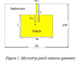
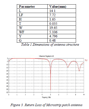
| The antenna consists of a rectangular patch, a substrate, feed line and a ground plane. The antenna is printed on a FR4 substrate with thickness of 0.1 mm and having dielectric constant of 4.3 with the ground on the other side. On the other side, a 50 Ohms micro strip feed line is placed symmetrically with respect to the center line of the patch. The antenna is excited with a Gaussian pulse. The dimensions of the antenna structure are shown in Table 1 |
 |
 |
C. Specific Absorption Rate (SAR)
|
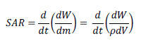
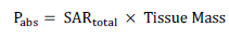
| The absorption loss is derived from Specific Absorption Rate. The Specific Absorption Rate (SAR) is defined as the time derivative of the incremental energy (dW) absorbed by an incremental mass (dm) contained in a volume element (dV) of a given mass density (ρ) [9]. It is expressed as |
 |
| As specified by IEEE C95.3 standard the typical local SAR value is averaged in 10 g tissue mass [11]. The total absorbed power in these models is calculated using the following equation |
 |
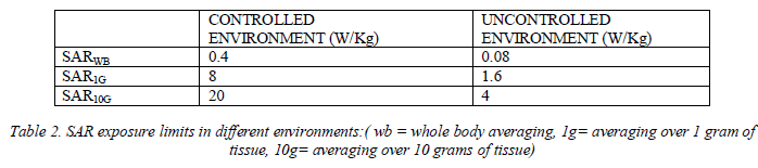 |
| Table 2 shows the limits for the peak local SAR values that is averaged over the whole-body (WB), 1 g of tissue (1G), or 10 g of tissue (10G) under different environments [10]. |
RESULTS
|
| The heterogeneous breast is illuminated with an electromagnetic signal from the microstrip patch antenna and the total SAR, 10-g averaged SAR and coordinates of maximum value of SAR are obtained for frequencies ranging from 4 to 7 GHz. Table 3 and 4 summarizes the values and coordinates of total SAR and maximum SAR obtained using equation in a normal and abnormal breast respectively. It is observed that both the total and maximum SAR values are higher in the tumor affected breast compared to the normal breast. |
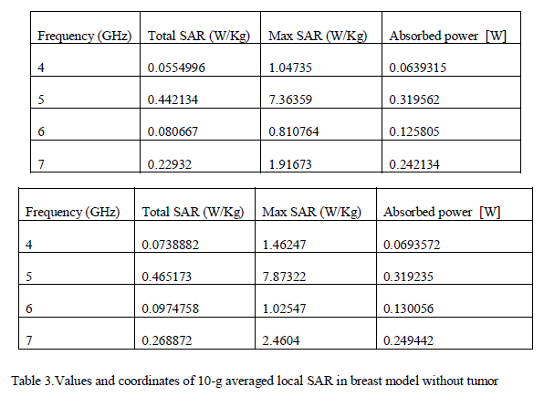 |
| It is observed that the coordinates of maximum value of SAR point to the position of the tumor within the tumor affected breast. This indicates that the maximum local SAR distributions occur in the tumor whereas in the normal breast the coordinates of maximum value of SAR point to locations close to the breast surface indicating that the maximum local SAR distributions occur in the breast layer close to the antenna. At 5 GHz the total and maximum SAR value for a normal breast is 0.442134 W/Kg and 7.36359 W/Kg respectively. Whereas for a tumor affected breast it is 0.465173W/Kg and 7.87322 W/Kg respectively. |
 |
| Table 5 shows the absorbed power for different sizes of the abnormal breast ranging from 25 mm to 50 mm at 5GHz frequency. The tumor is placed at the centre of the breast for all the varying sizes. It is observed that when the mass of the breast increases the absorbed power also increases for different frequencies. |
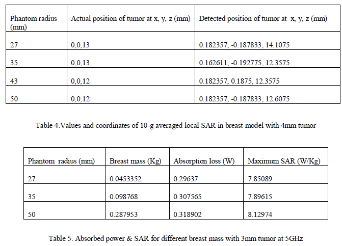
| Table 6 summarizes the coordinates of the maximum value of SAR when the tumor is placed inside the breast at 4 different locations. Out of the four, 2 locations of the tumor were detected successfully. On changing the size of the breast, it was observed that the coordinates of the maximum value of SAR was still able to detect the location of the tumor. For example, the absorption loss value for a 50 mm breast at 5 GHz is 0.318902 W. At the same frequencies however the absorbed power values for 35mm and 43mm breast are 0.307565 W and 0.312339 W. |
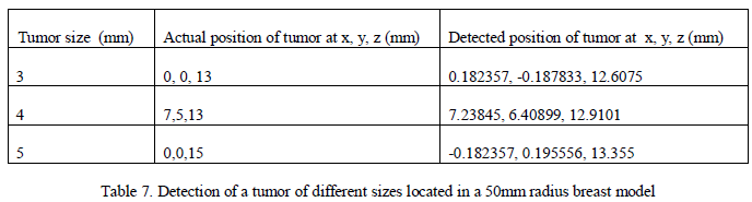 |
| Table 7 summarizes the coordinates of the maximum value of SAR when a tumor of 3mm, 4mm and 5mm is placed inside the 50mm radius breast at 3 different locations. On changing the size of the tumor, it was observed that the coordinates of the maximum value of SAR was still able to detect the location of the tumor. |
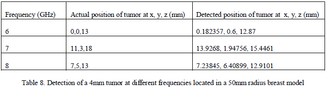 |
| Table8 summarizes the coordinates of the maximum value of SAR when a 4mm tumor is placed inside the 50mm radius breast at 3 different frequencies. The locations of the tumor were detected successfully. On changing the frequency, it was observed that the coordinates of the maximum value of SAR was still able to detect the location of the tumor. Thus, by first establishing that an abnormality is present we can then try to identify the location of the tumor. |
CONCLUSION
|
| The above simulated data clearly shows that the SAR and the absorption loss values are higher in a cancerous breast and these values increase as the mass of the breast increases. The coordinates for maximum value of SAR can be used as an indication for locating a tumor as it was able to detect the positions on changing the tumor location. The same was found on increasing the breast diameter. All these indicate a possibility of using EM absorption loss for detecting breast tumors. Future work is needed to develop realistic breast models and expand this method by determining an acceptable range of absorption values for normal breast tissues. |
References
|
- P. T. Huynh, A. M. Jarolimek, and S. Daye, “The false-negative mammogram,”Radiograph., vol. 18, no. 5, pp. 1137–1154, 1998.Poplack, and K. D. Paulsen, “A clinical prototype for active microwave imaging of the breast,” IEEE Trans. Microwave Theory Tech., vol. 48, pp.1841–1853, Nov. 2000.
- J. G. Elmore, M. B. Barton, V. M. Moceri, S. Polk, P. J. Arena, and S. W. Fletcher, “Ten-year risk of false positive screening mammograms andclinical breast examinations,” New Eng. J. Med., vol. 338, no. 16,pp. 1089–1096, 1998.
- AJ.Surowiec, S. S. Stuchly, J. R. Barr, and A. Swarup, “Dielectric properties of breast carcinoma and the surrounding tissues,” IEEE Trans.Biomed. Eng., vol. BME-35, pp. 257–263, Apr. 1988.
- P. M. Meaney, M. W. Fanning, D. Li, S. P.
- S. C. Hagness, A. Taflove, and J. E. Bridges, “Two-dimensional FDTD analysis of a pulsed microwave confocal system for breast cancerdetection: Fixed-focus and antenna-array sensors,” IEEE Trans. Biomed. Eng., vol. 45, no. 12, pp. 1470–1479, Dec. 1998.
- N.I.M. Yusoff, S. Khatun, S.A. AlShehri, Characterization of Absorption Loss for UWB Body Tissue Propagation Model,” in InternationalConference on Communications (MICC), pp. 254-258, 2009.
- Mohammed M. Elsewe, “Evaluation of EM Absorption Loss over Breast Mass for Breast Cancer Diagnosis,” in International Conference onEngineering in Medicine and Biological Society (EMBS), pp. 3897-3900, 2011.
- “International Commission on Non-Ionizing Protection (ICNIRP) 1998 “Guidelines for limited exposure to time varying electric, magnetic andelectromagnetic fields (up to 300 GHz)”, Health Physics, vol. 74, no. 4, pp. 494-522, Apr 1998.
- Recommended Practice for Measurements andComputations of Radio Frequency Electromagnetic Fields With Respect to Human Exposure to Such Fields, 100 kHz-300 GHz, IEEE StandardC95.3, 2002.
- International Commission on Non-Ionizing Protection (ICNIRP) 1998" Guidelines for limited exposure to time varying electric, magneticand electromagnetic fields (up to 300 GHz)”, Health Physics, vol. 74, no. 4, pp. 494-522, Apr 1998. 11)
- Begaud Xavier, âÃâ¬Ãâ¢Ultra wideband wide slot antenna with band-rejection characteristics,âÃâ¬Ãâ in European Conference on Antennas andPropagation (EUCAP), pp. 1-6, 2006.
- A. Abbosh âÃâ¬Ãâ¢Early breast cancer detection using Doppler frequency shift," Asia-Pacific Microwave Conference Proceedings (APMC), pp.275-278, Dec. 2010
|