ISSN ONLINE(2278-8875) PRINT (2320-3765)
ISSN ONLINE(2278-8875) PRINT (2320-3765)
Manikandan V1, Mohammed Farook I2, Dhanalakshmi S3
|
| Related article at Pubmed, Scholar Google |
Visit for more related articles at International Journal of Advanced Research in Electrical, Electronics and Instrumentation Engineering
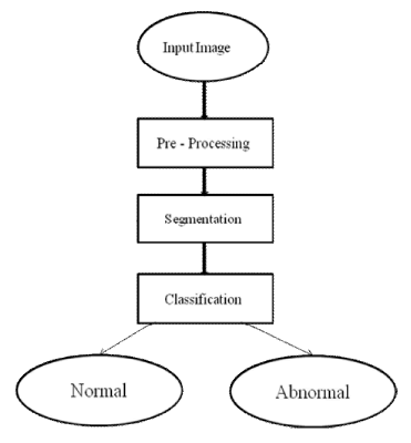
This work presents a novel approach on completely automated technique for recognition and segmentation of carotid-arteries (CA) in view of computing various dynamic properties such as intimamedia thickness (IMT) measurement and distal (far) wall segmentation. The goal of this work is to provide a clinical tool for cardiovascular diseases. Atherosclerosis is a generalized disease that causes lesions in large- and medium-sized elastic and muscular arteries and this is the major cause for cardio-vascular disease. The fully automated segmentation algorithm is based on active contours and active contours without edges and incorporates anatomical information to achieve accurate segmentation. The segmented regions are used to automatically achieve image normalization, which is followed by speckle removal. The resulting smoothed lumen-intima boundary combined with anatomical information provide an excellent initialization for parametric active contours that provide the final IMT segmentation. After segmentation the carotid artery ultrasound image is classified as normal or abnormal depends on the parametric values extracted.
Index Terms |
| IMT, Atherosclerosis, Segmentation |
INTRODUCTION |
| Images play the most important role in human perception. Medical imaging systems are based on the physical interaction between some energy source and the human body. A typical application field of medical image processing is in the diagnosis of atherosclerosis. Atherosclerosis is a cardio-vascular disease characterized by thickening of arterial walls which affects blood flow. Carotid artery disease is a disease in which a waxy substance called plaque builds up inside the carotid arteries. You have two common carotid arteries, one on each side of your neck. They each divide into internal and external carotid arteries. The internal carotid arteries supply oxygen-rich blood to your brain. The external carotid arteries supply oxygen-rich blood to your face, scalp, and neck. If plaque builds up in the body's arteries, the condition is called atherosclerosis. Over time, plaque hardens and narrows the arteries. This may limit the flow of oxygen-rich blood to your organs and other parts of your body .Atherosclerosis can affect any artery in the body. For example, if plaque builds up in the coronary (heart) arteries, a heart attack can occur. If plaque builds up in the carotid arteries, a stroke can occur. Carotid arteries are main blood suppliers to the brain. Narrowing of carotid artery generally blocks the blood flow into the brain and thus may cause a serious brain stroke. Consequently, early detection of the plaque in the arteries may help in preventing brain strokes .Further, manual intima–media thickness (IMT) measurement produces results that may not be reproducible. Thus, a computer-aided diagnostic (CAD) technique for segmentation, plaque detection, IMT measurement, and classification of segmented images is highly desirable for carotid artery ultrasound images. This may help the medical experts to extract significant information about the plaque in determining the stage of disease. |
OVERVIEW OF THE PROPOSED METHOD |
| The proposed method consists of removing the noise from the input image by anisotropic filter and enhancement is done using histogram equalization. The features are extracted from the input carotid artery ultrasound image. The image is segmented using active contour algorithm. The IMT values are measured for the image and classification is done by using SVM neural network. |
PREPROCESSING |
| The major negative aspects of ultrasound images are their poor quality, presence of speckle noise, and wave interferences. To obtain better segmented images, noise must be removed. For this purpose, we have used kaun-filter for noise removal as image preprocessing step because in case of carotid artery ultrasound images, it has been observed that it smoothes out noise and preserves image details in better way as compared to average and bilateral filters. |
 |
SEGMENTATION OF CAROTID ARTERY ULTRASOUND IMAGES |
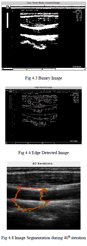
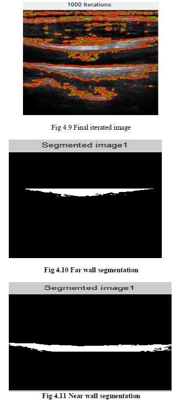
| In our study, we have employed active contour method to segment the carotid artery ultrasound images in an automatic way. The advantage of active contour model for segmentation is that the snakes are autonomous and self-adapting in search of a minimal energy state and can easily be manipulated. The active contour model can be used to track objects in both spatial and temporal domains. |
ACTIVE CONTOUR |
| Active contours, also called snakes, are used extensively in computer vision and image processing applications, particularly to locate object boundaries .Active contours, or snakes, are curves defined within an image domain that can move under the influence of internal forces coming from within the curve itself and external forces computed from the image data. The essential idea is to evolve a curve or a surface under constraints from image forces so that it is attracted to features of interest in an intensity image. Snakes are widely used in many applications, including edge detection, shape modelling, segmentation, and motion tracking. |
REGION BASED METHODS |
| Boundary based level set methods presented thus far provide efficient and stable algorithms to detect contours in a given image. The presented methods handle changes in topology and provide robust stopping terms to detect the goal contours. However, structures such as interior of objects, e.g. interior of discs are not segmented. Because of level set formulation the final contours are always closed contours. In addition, in images where the objects object boundaries are noisy and blurry these methods face some difficulties. Some recent works in active contours consider these issues. |
ACTIVE CONTOURS WITHOUT EDGES |
| There are some objects whose boundaries are not well defined through the gradient. For example, smeared boundaries and boundaries of large objects defined by grouping smaller ones. The main idea is to consider the information inside the regions not only at their boundaries. Chan and Vese define the following energy, |
 |
| Where is the original image, , are some constants and H(ø) is given by: |
| H(ø) = |
| This model looks for the best approximation of image as a set of regions with only two different intensities ( ). One of the regions represents the objects to be detected (inside of c ), and the other region corresponds to the background (outside of c ). The snake c will be the boundary between these two regions. |
CLASSIFICATION |
| Carotid artery disease diagnosis greatly depends upon accurate artery image segmentation and classification of the segmented images. Segmented images are classified into normal or abnormal. In this study, we have used SVM classifier for classification of the carotid artery images. Four different features are extracted from the IMT values obtained from carotid artery-segmented images to train and test the SVM classifier. |
RESULTS |
 |
 |
 |
 |
CONCLUSION |
| By this method the intima-media thickness of the carotid artery ultrasound image is measured. This is an non-invasive and sensitive technique for identifying and quantifying the presence of plaque in the carotid artery. Automatic segmentation of carotid artery ultrasound image poses a great challenge due to there complexity and various factors like operator dependence and acquisition settings. This work is fully automatic and achieves lower absolute mean IMT error compared to inter-observer variability in the evaluated data set. |
| The presented technique may represent a generalized and standard methodology towards completely automated and accurate IMT measurement. It may be used for aiding the clinicians through providing support in their examination, reducing not only reading times, but also the variability between readers whilst improving reproducibility. It can be used for evaluating the IMT in large multicenter studies for developing and evaluating risk models and screening methods. More importantly, future work will involve the use of the presented method for the evaluation of different features including wall irregularity and area texture measures, in addition to the IMT, for the establishment of risk models for the development of cardiovascular disease to aid the clinicians in screening and evaluation of treatment. |
References |
|