International Journal of Advanced Research in Electrical, Electronics and Instrumentation Engineering
ISSN ONLINE(2278-8875) PRINT (2320-3765)
ISSN ONLINE(2278-8875) PRINT (2320-3765)
Amruta Pandit1, Shrikrishna Kolhar2, Pragati Patil3
|
| Related article at Pubmed, Scholar Google |
Visit for more related articles at International Journal of Advanced Research in Electrical, Electronics and Instrumentation Engineering
Blood cell segmentation and identification is very important as blood being health indicator. A person’s health is determined using complete blood count. The contents of the blood in particular the white blood cells, the red blood cells and platelets define the state of health. For detection and treatment of diseases like anemia, leukemia etc. RBC count is required. In laboratory, blood cell counting is done by using hemocytometer and microscope. This method gives inaccurate and unreliable results that depend on physician skill. This task is laborious and time consuming. The aim of this research is to produce a survey on computer vision system that can detect and estimate the number of red blood cells in the blood sample image using image processing algorithms. This paper considers image processing for counting of blood cells. Image processing algorithms involve six major steps: image acquisition, preprocessing, image enhancement, image segmentation, feature extraction and counting algorithm. The objective is to study the different methodologies of RBC counting and identification of research directions.
Keywords |
| Image Processing, Image segmentation, Image enhancement, Morphology, Hough Transform |
INTRODUCTION |
| For overall health evaluation and diagnosis of many disorders including anemia, infection and leukemia, complete blood count is required. The human blood consists of three types of blood cells such as red blood cell (RBC), white blood cell (WBC) and platelets (PLT) [1]. A person’s health is determined using complete blood count. Blood cell segmentation and identification is important as blood being health indicator. Abnormal increase or decrease in cell count indicates that person has underlying medical condition. |
| Red blood cells, also known as enthrocytes are the most important and numerous blood cells in human body. Main function of RBCs is to carrying oxygen and delivering it to the cells in the body [2]. They are minute disc shaped. They does not contain nucleus but a protein called hemoglobin. Both inner and outer layers of cell are made of protein that gives red color to blood. Hemoglobin actually does the work of grabbing and carries oxygen. Usually level of hemoglobin is tested in blood test. Decrease in level may cause severe diseases including anemia, blood loss, leukemia and malnutrition. |
| A life span of RBC is of around 120 days for normal individual [3]. A normal RBC count for an adult male is between 4.6x10¹² and 6.2x10¹² per liter of blood. Production of red blood cells takes place in the bone marrow from precursor stem cells. Typical red blood cell count (RBC) levels are: |
| 4.2 to 5.4 million cells per micro liter for women |
| 2.6 to 4.8 million cells per micro liter for children |
| 4.5 to 6.2 million cells per micro liter of blood for men. |
| In diagnosis of several diseases, major step is automated detection and counting of red blood cells. In the conventional procedure, haematologist manually counts and classifies the cells with the help of a microscope. The task is to measure the red blood cells and assess the size and shape of red blood cells. But this procedure is time consuming, complex and tedious [4-6]. Also, the accuracy of recognition is affected by subjective factors like experience and fatigue due to human tiredness. As a solution to this problem, to provide automated, cost-effective and efficient alternative to detection and counting of RBCs, image processing techniques are used. |
| In this paper, review is done on some of the methods of RBC detection and counting. The paper is organized in following sections as: present the methodology used (Section II), related works (Section III) and discuss the conclusions (Section IV). |
METHODOLOGY |
| There are six major steps involved in the process of estimating the red blood cells. These are input image acquisition, preprocessing, enhancement, segmentation, feature extraction, RBC counting. These steps are shown in Fig. 1 |
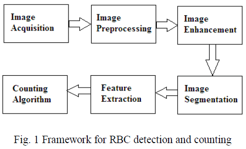 |
Image Acquisition |
| The first step in the process is image acquisition- that is, to acquire a digital image. Usually it is microscopic image that can be obtained from online medical library or hospital blood sample images. These images are in RGB color format. |
Image Preprocessing |
| Image pre-processing is a technique of adjusting images suitable for the next step of computational process. It is done in such a way that image quality improved for the success of the other processes. Pre-processing techniques usually include enhancing contrast, removing noise, isolating regions and use of different color models Grayscale image, hsv image. Original blood cells images are in color. To ease the process of ratio determination, the original images will be converted into grayscale color. Grayscale represents the intensity of the image. Acquired images have low contrast as all blood elements colors close to background color. Also noise is included due to clustered white blood cells. To overcome or reduce such effects contrast enhancement is done. |
Image Enhancement |
| After pre-processing, image enhancement is done. It is carried out for the improvement of image’s contrast and brightness characteristics all well as to reduce noise in the image or sharpen the details. These techniques include image negation, histogram plotting, image subtraction and filtering. |
Image Segmentation |
| The next stage deals with image segmentation. Segmentation partitions an input image into foreground and background region. There are various approaches for segmentation i.e. segmentation by using histogram and thresholding, Otsu adaptive thresholding and watershed transform, hough transform technique as well as segmentation by Means clustering. The objective of segmentation is extraction of desired objects from the background. Segmentation is more complex step and requires more processing time in comparison with other methods. However it is the most important and challenging step because the feature extraction and counting depends on the correct segmentation of RBC. |
Feature Extraction |
| Feature extraction includes morphological operations. It extracts features that contain quantitative information of objects of interest. Shape features are areas of cell and nucleus, cell perimeter, ratio of nucleus to overall cell area, boundary of the nucleus and circularity factor. Texture features include contrast, homogeneity and entropy derived from the gray-level co-occurrence matrix. Color histogram, mean and standard deviation of the color components in CIE-Lab domain, form the color features. Feature extraction techniques include classifiers like Artificial Neural network (ANN), support vector machine (SVM). |
Counting Algorithm |
| To measure number of RBCs counting algorithm is applied. Connected components labeling is the most popular method used for counting. Besides that circular Hough transform is also used to get the RBC count. |
LITERATURE SURVEY |
A.Hough Transform Based Methods |
| Vinutha H Reddy introduced an automatic RBC and WBC counting using computer vision [6]. The estimation of red blood cells involves several steps. These are input acquisition of input image, preprocessing of acquired image, segmentation, feature extraction, counting. The pre-processing step consists of conversion of the original blood smear image into saturation image. Histogram thresholding and morphological operations are used for segmentation. Feature extraction is done with the help of morphological operations to differentiate between RBCs, WBCs, Platelets and background. Last step is to measure number of Red Blood Cell by using Hough Transform from the blood smear image. |
| Venkatalakshmi. B et al. presented a method for automatic red blood cell counting using Hough transform [8]. The algorithm for estimating the red blood cells consists of five major steps: input image acquisition, pre-processing, segmentation, feature extraction and counting. In pre-processing step, original blood smear is converted into HSV image. As Saturation image clearly shows the bright components, it is further used for analysis. First step of segmentation is to find out lower and upper threshold from histogram information. Saturation image is then divided into two binary images based on this information. Morphological area closing is applied to lower pixel value image and morphological dilation and area closing is applied to higher pixel value image. Morphological XOR operation is applied to two binary images and circular Hough transform is applied to extract RBCs. |
| Siti Madihah Mazalan et al. also presented an approach for automatic RBC counting using circular Hough transform technique [9]. It contains two major steps viz. finding out minimum and maximum radius of RBC and hough transform. For measurement of minimum and maximum radius, sub steps are carried out that include: cropping the image, RBG to gray conversion, morphological processing, thresholding, noise removing and finally measuring mean, standard deviation and tolerance. With the help of known radius, circular Hough transform is applied to count RBCs in peripheral blood smear image. |
| Museum Maitra et al. introduced a method for automatic segmentation and counting of red blood cells in microscopic image using Hough Transform [10]. Preprocessing techniques include edge detection, spatial smoothing filtering and adaptive histogram equalization. Feature extraction has been done through the Hough Transform method which has been used to find out the red blood cells based on their sizes and their shapes. This isolates the red blood cells from the rest of the image of the blood sample so that further processes like counting can be applied exclusively on them. |
B.Thresholding Based Method |
| The WBCs and RBCs are counted by using the gray thresholding algorithm by Pooja R. Patil et al [5]. The first step of proposed method is RGB to gray conversion. After that, median filtering is done to remove noise in the background. Otsu’s method is used for binarization of image. For proper segmentation of blood cells, holes presented in the binary image are filled. The cells near border contain less information and are removed to reduce the complexity. Labeling algorithm is applied to count the connected objects. Form factor calculation is done and we get the total RBC count. |
C.Watershed Transform Based Method |
| Hemant Tulsani et al. presented a method for counting of blood cells [7]. The image processing techniques used for counting are spatial filtering, morphological operations and segmentation using watershed transformation. In the preprocessing step, smoothening of image is done using average filter. In next step, RGB image is converted to YCbCr image and Cb component is extracted to get nucleus and platelets from the image. Blood smear image is binarized and morphological opening is done to get the mask of WBCs and platelets. Individual images for WBCs and platelets can be obtained with another opening and image subtraction. Next, grayscale image of blood smear is applied to opening by reconstruction and closing by reconstruction. Finally, the binary base image containing all the cells is obtained and mask is subtracted from it to get RBC binary image. Binary images are segmented using Watershed transform. |
D. Cell Structure and Intensity Based Method |
| S. Kareem et al. introduced angular ring ratio method for counting of RBCs in thin blood films [11]. The method consists of conversion of RGB image to grayscale. Dilation of the image using a ring shaped structuring element is done. After that Erosion of the image using a disk shaped structuring is done. Next step is to convert the closed image to a ratio transformed image by calculating the ratio of average intensities of the annular concentric ring structuring element to disk shaped structuring element masked over the image. After that peak intensities of the ratio transformed image are calculated. Next, mapping the peaks on to the corresponding coordinates, which is actually the centre of each RBC is done. |
E.Comparative Method |
| Comparative analysis of Hough Transform and k-Means clustering algorithm for extraction and detection of RBCs was done by Monika Mogra et al [4]. K-means clustering algorithm contains six major steps: image acquisition, Clustering image, Histogram equalization, Image segmentation, Blood cell extraction and Counting of cells. While Hough transform algorithm contains following steps: Input image acquisition, Hough transforms edge linking, Image segmentation, Snake body detection, Output image and finally Counting of cells. |
CONCLUSION |
| Analysis of blood cell image is more accurate as well as efficient in terms of cost and time by use of image processing. Research work is increasing in the field of RBC counting and various image processing techniques are implemented in order to get more accurate result. Use of image processing techniques is useful and better than existing techniques of medical diagnosis and blood cell counting provided that standardization of blood smear is done properly to obtain blood cell image. |
References |
|