


 20 / 21

Pathology 2018
Research & Reviews: Journal of Medical and Health Sciences
ISSN: 2319-9865
Page 52
October 08-09, 2018
Edinburgh, Scotland
17
th
International Conference on
Pathology & Cancer
Epidemiology
H
ydatid disease is a parasitic infection caused by the larval
form of
Echinococcus granulosus.
Breast hydatid cyst is
extremely uncommon accounting for only 0.27% of all cases of
hydatiddiseaseandcanbeeasily confusedwithothermalignant
and benign breast lesions and therefore missed until an
operative diagnosis is made. We report a case of breast hydatid
cyst diagnosed pre-operatively in an 80 years old patient who
presented with a right breast nodule. Mammograms revealed
three well circumscribed lesions. The final diagnosis was
obtained by using breast needle core biopsy with pathological
examination showing typical hydatid laminated membranes.
Abdominal ultrasound and chest roentgenogram revealed no
similar or obviously cystic lesions. Hydatid disease can occur
in many organs, among them the liver (60% of hydatid cases),
the lungs (30%), the kidneys (2.5%), the heart (2.5%), the bone
(2%), the spleen (1.5%), the muscle (1%), and in the brain 0.5%.
Hydatic cyst occurs in the breast in only 0.27% of all cases.
Fine needle aspiration and core needle biopsy help establish
a preoperative diagnosis. Total surgical excision of the cystic
lesion is the standard treatment. Despite its rarity, it should
be included in the differential diagnosis of breast lumps for
patients living in endemic areas.
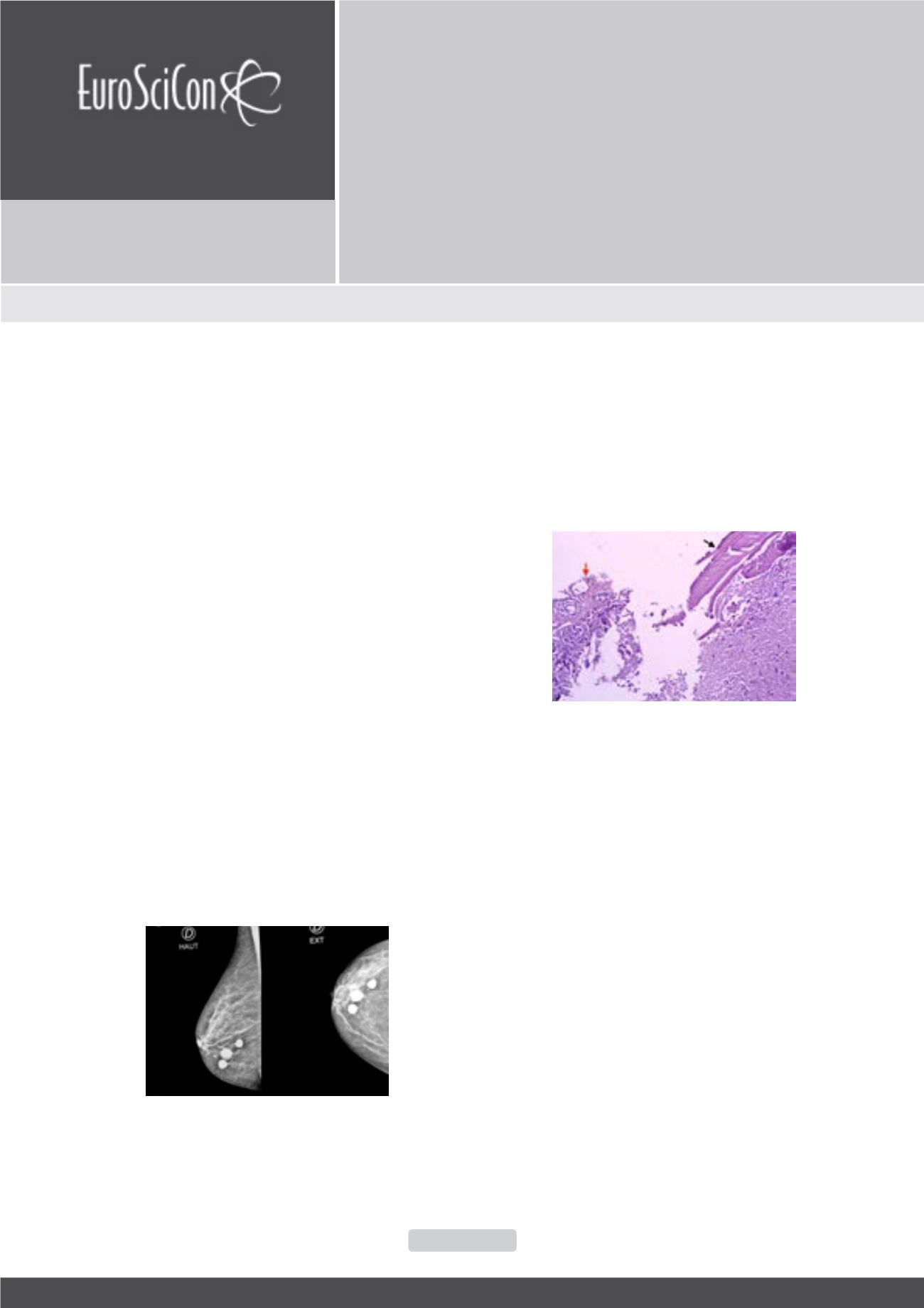
Figure 1:
Mammogram scan showing three well-circumscribed,
homogenous smooth nodules of the right breast.
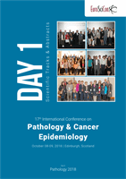
Figure 2:
Microphotography showing the presence of a
laminated membrane (black arrow) with occasional
residual terminal ductulo-lobular units (red arrow).
Biography
Miry Achraf is a second year Pathology resident, he has completed his
PhD from Oujda medical faculty.
achrafmiry@outlook.comPrimary breast hydatid cyst: case report and literature
review
Miry Achraf
and
Elfatemi Hinde
Hassan II University Hospital, Morocco
Miry Achraf et al., RRJMHS 2018
Volume: 7