Image compression is a method through which we can reduce the storage space of images, videos and is helpful to increase storage and transmission process’s performance. Wavelet based Image Compression scheme is used to compress the images which helps to reduce the size of images. The method uses different metrics such as peak signal to noise ratio, mean squared error, & percentage compression rate measured the performance to compare the results. The aim of compression is to achieve good quality compressed image making the storage and transmission more efficient. This paper studied the image compression comparison based on wavelets using different medical images and identified the best appropriate wavelet transform.
Keywords |
| Image Compression, Wavelet, Haar, Daubechies, Biorthogonal, Coiflet |
INTRODUCTION |
| With the increasing development and demand of multimedia products, the issue of insufficient bandwidth of network
and storage of memory device are found. So the data compression becomes more and more significant for reducing the
data redundancy to save more hardware space and transmission bandwidth. In computer science and information, data
compression or source coding is the process of encoding information using fewer bits or other information-bearing
units than an unencoded representation. Compression is used because it helps to reduce the consumption of expensive
resources such as hard disk space or transmission bandwidth. |
| The basic goal of image data compression is to reduce the bit rate for transmission and storage while either maintaining
the original quality or providing an acceptable fidelity. Image compression involves reducing the size of image files,
while retaining necessary information. The resultant image is called the compressed image and is used to reconstruct
the image which results as the decompressed image. Image Compression, the art and science of reducing the amount of
data required to represent an image is one of the most useful and commercially successful technologies in the field of
digital image processing. The number of images that are compressed and decompressed daily is staggering, and the
compressions and decompressions themselves are virtually invisible to the user. |
Image compression addresses the problem of reducing the amount of data required to represent a digital image. It is a
process that intend to compact representation of an image by reducing the image storage or transmission requirements.
The goal of image compression is to reduce the image file size without affecting the quality of an image. The term data
compression refers to the process of reducing the amount of data requirement to represent a given quantity of
information. In this definition, data and information is not the same thing; data means by which information is
conveyed. Because various amounts of data can be used to represent the same amount of information and
representations that contain repeated or irrelevant information are said to contain redundant data.Let b and b’ denote the
number of bits in two representations of the same information, the relative data redundancy R of the representation with
b bits is 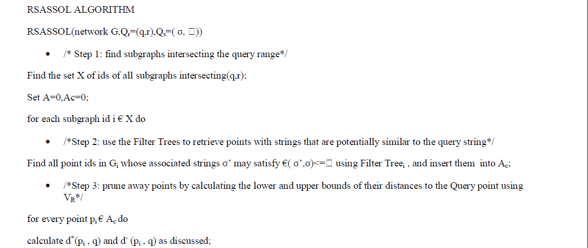 |
| Where C, commonly called the compression ratio, is defined as |
 |
| Compression is achieved by the removal of one or more of the three basic data redundancies: |
| Coding Redundancy |
| Interpixel Redundancy |
| Psychovisual Redundancy |
| Coding redundancy is present when less than optimal code words are used. Interpixel redundancy results from
correlations between the pixels of an image. Psycho visual redundancy is due to data that is ignored by the human
visual system (i.e. visually non essential information). The objective of compression is to reduce the number of bits as
much as possible, while keeping the resolution and the visual quality of the reconstructed image as close to the original
image as possible. |
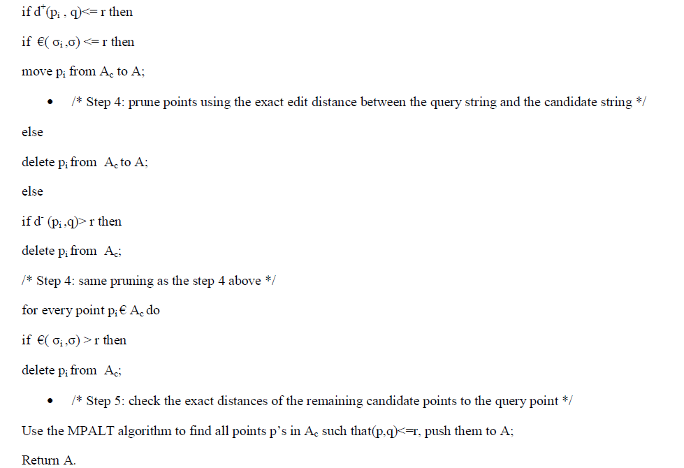
| Image compression systems are composed of two distinct structural blocks: an encoder and a decoder. As shown in the
fig 1, the encoder is responsible for reducing the coding, interpixel and psycho visual redundancies of input image. In
first stage, the mapper transforms the input image into a format designed to reduce interpixel redundancies. The second
stage, qunatizer block reduces the accuracy of mapper’s output in accordance with a predefined criterion. In third and
final stage, a symbol decoder creates a code for quantizer output and maps the output in accordance with the code.
These blocks perform, in reverse order, the inverse operations of the encoder’s symbol coder and mapper block. As
quantization is irreversible, an inverse quantization is not included. |
A. Need of compression |
| In a raw state, image can occupy a large amount of memory both in RAM and in storage. Image compression reduces
the storage space required by an Image and the bandwidth needed when streaming that image across a network. |
B. Benefits of compression |
| It provides a potential cost savings associated with sending less data over switched telephone network where
cost of call is really usually based upon its duration. |
| It not only reduces storage requirements but also overall execution time. |
| It also reduces the probability of transmission errors since fewer bits are transferred. |
| It also provides a level of security against illicit monitoring. |
C. Medical Imaging |
| Medical imaging is the technique and process used to create images of the human body (or parts and function thereof)
for clinical purposes (medical procedures seeking to reveal, diagnose, or examine disease) or medical science
(including the study of normal anatomy and physiology). Although imaging of removed organs and tissues can be
performed for medical reasons, such procedures are not usually referred to as medical imaging, but rather are a part
of pathology. As a discipline and in its widest sense, it is part of biological imaging and incorporates radiology (in the
wider sense), nuclear medicine, investigative radiological sciences, endoscopy, (medical) thermography, medical
photography, and microscopy (e.g. for human pathological investigations). A majority of diagnostic imaging centers
are located in California, followed by Texas and Florida. |
| Measurement and recording techniques which are not primarily designed to produce images, such
as electroencephalography (EEG), magneto encephalography (MEG), electrocardiography (EKG), and others, but
which produce data susceptible to be represented as maps (i.e., containing positional information), can be seen as forms
of medical imaging. Up until 2010, 5 billion medical imaging studies had been conducted worldwide. Radiation
exposure from medical imaging in 2006 made up about 50% of total ionizing radiation exposure in the United States. |
| In the clinical context, "invisible light" medical imaging is generally equated to radiology or "clinical imaging" and the
medical practitioner responsible for interpreting (and sometimes acquiring) the images are a radiologist. "Visible light"
medical imaging involves digital video or still pictures that can be seen without special equipment. Dermatology and
wound care are two modalities that utilize visible light imagery. Diagnostic radiography designates the technical
aspects of medical imaging and in particular the acquisition of medical images. The radiographer or radiologic
technologist is usually responsible for acquiring medical images of diagnostic quality, although some radiological
interventions are performed by radiologists. While radiology is an evaluation of anatomy, nuclear medicine provides
functional assessment. |
WAVELET BASED IMAGE COMPRESSION |
| A wave is an oscillating function of time or space and is periodic. In contrast, wavelets are localized waves [1].
Wavelet means a “small waves”. The smallness implies to a window function of finite length. Wavelets are functions
that satisfy certain mathematical requirements and are used in representing data or other functions. A wavelet is a
waveform of effectively limited duration that has an average value of zero. Wave in itself refers to the condition that
this function is oscillatory [4]. Wavelets are mathematical tools for hierarchically decomposing functions. Wavelet
Transform has been proved to be a very useful tool for image processing in recent years. It allows a function which
may be described in terms of a coarse overall shape, plus details that range from broad to narrow [3]. |
| Wavelets are mathematical functions which help in describing the original image into an image in frequency domain,
which can further divided into sub band images of different frequency components. Each component is studied with a
resolution matched to its scale [2]. Wavelets are mathematical functions that cut up data into different frequency
components, and then study each component with a resolution matched to its scale [10]. |
| The fundamental idea behind wavelets is to analyze according to scale. Wavelets are functions that satisfy certain
mathematical requirements and are used in representing data or other functions. In wavelet analysis, the scale that we
use to look at data plays a special role. Wavelet algorithms process data at different scales or resolutions. If we look at a
signal with a large "window," we would notice gross features. Similarly, if we look at a signal with a small "window"
we would notice small features. There are many members in the wavelet family, a few of them that are generally found
to be more useful, are as per the following Haar wavelet is one of the oldest and simplest wavelet. Therefore, any
discussion of wavelets starts with the Haar wavelet. Daubechies wavelets are the most popular wavelets. They represent
the foundations of wavelet signal processing and are used in numerous applications. |
| Haar- This wavelet is discontinuous, and resembles a step function. |
| Coiflets- The wavelet function has 2N moments equal to 0 and the scaling function has 2N-1 moments equal to 0. The
two functions have a support of length 6N-1. |
| Symlets- The symlets are nearly symmetrical wavelets. The properties of the two wavelet families are similar. |
| Meyer - The Meyer wavelet and scaling function are defined in the frequency domain. |
| Biorthogonal- This family of wavelets exhibits the property of linear phase, which is needed for signal and image
reconstruction. By using two wavelets, one for decomposition (on the left side) and the other for reconstruction (on the
right side) instead of the same single one, interesting properties are derived. |
| Daubechies- Daubechies are compactly supported orthonormal wavelets and found application in DWT. Its family has
got nine members in it [4]. |
COMPARATIVE STUDY |
| It has been already explained that the Image Compression is of two types, lossy and lossless. Different Scientists have
put their work in the field of compression. Our first problem is to select the most appropriate wavelet transform for a medical image compression. In this, we are going to analyze the behaviour of different type of wavelet transforms with
different type of medical images and identify the most appropriate wavelet transform that can perform optimum
compression for a given medical images. Thus, the problem taken for this research work is divided into some objectives
which are as follows. |
A. Objectives: |
| Study of Image Compression. |
| Study of different wavelet transformation. |
| Pointing out the pros and cons of the wavelet transformations. |
| Comparison of all different wavelet transformations. |
B. Comparison Model |
| The comparison model focuses on the above objectives which are helpful to find the most appropriate wavelet
transform that can perform optimum compression. In this paper, we have studied the performance of different wavelet
transforms on different medical images. Fig 2 shows the basic design of the comparison model. |
| There are number of phases while we are studying the image compression based on different wavelets. They are: |
C. Image Acquisition: |
| The first stage of any compression system is the image acquisition stage. After the image has been obtained, various
methods of processing can be applied to the image to perform the many different tasks required today. However, if the
image has not been acquired satisfactorily then the intended tasks may not be achievable. In this phase a number of
samples are collected which are different type of medical images that is CT Scan, ECG, Fundus, Infrared Image,
Mammography, MRI, US Image and X-Ray images. |
D. Image Compression based on Wavelets: |
| There are different members in the wavelet family, Haar wavelet is one of the oldest and simplest wavelet. Daubechies
wavelets are the most popular wavelets. They represent the foundations of wavelet signal processing and are used in
numerous applications. The Haar, Daubechies and Coiflets are compactly supported orthogonal wavelets. These wavelets along with Meyer wavelets are capable of perfect reconstruction. The Meyer, Morlet and Mexican Hat
wavelets are symmetric in shape. The wavelets are chosen based on their shape and their ability to analyze the signal in
a particular application. Biorthogonal wavelet exhibits the property of linear phase, which is needed for signal and
image reconstruction. By using two wavelets, one for decomposition (on the left side) and the other for reconstruction
(on the right side) instead of the same single one, interesting properties are derived. |
| E. Comparison: In the comparison phase, all the results of compression using Haar, Daubechies, Biorthogonal &
Coiflet Wavelet are compared and identify the best appropriate wavelet transform for compression using different type
of medical images. Comparison is on the basis of different parameters to measure the quality of image & their
compression ratio. |
CONCLUSION |
| A result from a comparative study of Wavelet based Image Compression is presented in this paper. In this paper,
Comparative study is on the basis of some parameters calculated in previous works. From the results we studied that
the Daubechies transform gives a higher percentage of compression for MRI, Fundus and Infrared images, Haar
transform gives a higher percentage of compression for ECG images, Biorthogonal transform gives a higher percentage
of compression for X-ray images and Coiflets transform gives a higher percentage of compression for CT, US and
Mammography images, at constant PSNR. |
| In this work we present the Comparative study of wavelet based compression quite successfully. Still there is some
hope of improvement. If we can use optimization algorithm with more image samples for the compression, the results
could have been better. So, future work could go on the direction of optimized systems using artificial intelligent
techniques. |
| |
Figures at a glance |
 |
 |
| Figure 1 |
Figure 2 |
|
| |
References |
- AboodKuther, AboudHayder, and A.H. Falih, “X-ray image compression using neural network,” ISSN 2229-5518,Vol.3, Issue 10, 2012.
- Ajay K.S, ShamikTiwari and V.P. Shukla, “Wavelet based Multi Class image classification using Neural Network,” International Journal of Computer Applications, Vol.37, No.4, 2012.C.
- Anuj Bhardwaj and Rashid Ali, “Image Compression Using Modified Fast Haar Wavelet Transform,” ISSN 1818-4952, Vol.7, No.5, 2009.
- BaluramNagaria, Mhd. FarukhHashmi and PradeepDhakad, “Comparative Analysis of Fast Transform for Image Compression for optimal Image Quality and Higher Compression Ratio,” International Journal of Engineering Science and Technology (IJEST), Vol.3, No.5, 2011.
- Ben Amar, and O. Jemai, “Wavelet Networks Approach for Image Compression,” ICGST International Journal on Graphics, Vision and Image Processing, pp. 37-45, 2007.
- K. H. Talukder, and K. Harada, “Haar Wavelet Based Approach for Image Compression and Quality Assessment of Compressed Image” International Journal of Applied Mathematics, Vol.36, pp.1-8, 2007.
- Khashman A. and Dimililer K, “Image Compression using Neural Networks and Haar Wavelet,” WSEAS Trans Signal Processing Vol.4, pp.330-339, 2008.
- N.Senthilkumaran, and J.Suguna, “Neural Network Techniques for Lossless Image Compression using X-ray Images,” International Journal of Computer and Electrical Engineering, Vol.3, No.1, 2011.
- R.Gonzalez and R.woods, “Digital image processing” 3rd edition, Pearson education, 2011.
- S N Sivanandam, S Sumathi and S N Deepa, “Introduction to Neural Networks using Matlab 6.0” McGraw Hill, 2012.
- Sindhu M and Rajkamal R, “Images and Its Compression Techniques,” International Journal of Recent Trends in Engineering, Vol.2, No. 4, 2009.
|