
Research & Reviews: Journal of Veterinary Sciences
 Menu
Menue-ISSN No.:2581-3897
e-ISSN No.:2581-3897
Emily Johnson*
1 Department of Veterinary Surgery, Oakridge University of Veterinary Medicine, Austin, Texas, USA
Received: 26-Dec-2024, Manuscript No. JVS-26-156564; Editor assigned: 31-Dec-2024, Pre QC No. JVS-26-156564 (PQ); Reviewed: 14-Jan-2025, QC No. JVS-26-156564; Revised: 17-Feb-2026, Manuscript No. JVS-26-156564 (R); Published: 24-Feb-2026, DOI: 10.4172/2581-3897.10.1.001
Citation: Johnson E. Diagnostic Imaging and Surgical Management of Pulmonary Choristoma in a 4-Day-Old Holstein Calf. RRJ Vet Sci. 2026;10:001.
Copyright:© 2026 Johnson E. This is an open-access article distributed under the terms of the Creative Commons Attribution License, which permits unrestricted use, distribution and reproduction in any medium, provided the original author and source are credited.
Visit for more related articles at Research & Reviews: Journal of Veterinary Sciences
A 4-day-old Holstein calf presented with a protruding mass on its back, diagnosed as spina bifida through radiography. Ultrasonography revealed bronchi-like structures and large vessels within the mass. Doppler ultrasonography showed pulsatile vessels entering the lumbar vertebrae, aiding surgical planning. Although complete resection was unfeasible due to deep invasion, histopathology confirmed a pulmonary choristoma with lunglike structures. Doppler ultrasonography helped differentiate between bronchi-like structures and arterial vessels, assisting diagnosis.
Holstein calf; Doppler ultrasonography; Pulmonary choristoma; Lumbar vertebrae
A 4-day-old Holstein calf, weighing approximately 44 kg, presented with a large swelling between the thirteenth thoracic and fourth lumbar vertebrae. The swelling, measuring 19 cm in length, 31 cm in width, and 23 cm in height, was diagnosed as a subcutaneous mass, but the calf showed no neurological weakness. Radiographs revealed deformities in the spinous processes, suggesting spina bifida. Ultrasonography demonstrated a large, 7-10 mm diameter vessel within the mass, accompanied by smaller vessels and anechoic tubular structures with bronchi-like lumens, indicative of lung-like tissue. Doppler ultrasonography confirmed pulsatile blood flow, supporting a diagnosis of Pulmonary Choristoma (PC) (Figure 1) [1].
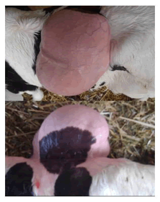
Figure 1. Holstein calf presented with a protruding mass on its back.
The calf had surgery on day 14. The tumor, which had winding blood arteries, was exposed by a 20 cm skin incision. Both ends of the major arterial vessel, which had a diameter of around 1 cm, were ligated. Even with meticulous dissection, part of the mass could not be removed because of its deep extension into soft tissue between the lumbar vertebrae. The calf experienced hindlimb paralysis four months after the surgical wound healed without incident, but the lump started to grow again [2-4].
The calf was put to death on day 196 after a second operation was carried out on day 187 to remove the regrown tumor, but the paralysis in its hindlimbs remained. A fibromatous, capsular mass with purulent material that extended far into the lumbar vertebrae was discovered during necropsy, along with a defect in the spinous process of the second lumbar vertebra. The hindlimb paralysis was found to be caused by the mass's pressing on the spinal cord.
The excised mass's histopathology revealed alveolar-like structures, cartilage, arteries, and branching bronchi-like structures with columnar ciliated epithelial cells. These characteristics supported the diagnosis of pulmonary choristoma, as did bleeding and macrophage infiltration. Peripheral nerve bundles, adipose tissue, and fibrous tissue were also present in the bulk [5-7].
The difficulties in diagnosing and treating congenital lumps in newborn calves are highlighted by this instance. Surgical planning was guided by the identification of the mass's structure, vascularity, and features using imaging techniques such radiography, ultrasonography, and Doppler ultrasonography. Despite surgical intervention, the mass's profound penetration of surrounding tissues and subsequent growth resulted in consequences, including neurological impairments and ultimately With an emphasis on their roles in preoperative planning and surgical outcomes, this study evaluates the diagnostic efficacy of radiography and ultrasonography in the diagnosis of Pulmonary Choristomas (PCs) in cows. The degree of the mass's invasion into soft tissues and the thoracic cavity can be determined via radiography, which was frequently employed in earlier cases involving cows. Skull radiographs can show bone flaws, like fissures, in head lesions, and they are especially useful for identifying gas and fluid-filled cystic cavities in intrathoracic lesions. However, because soft tissue masses frequently present as soft tissue opacities, radiographic findings can be limited for detecting soft tissue masses. In some cases, contrast radiography can enhance mass visibility, but it was not significantly helpful in this case. Additionally, lateral radiographs failed to detect abnormalities in the spinous processes due to the mass's overlap, and ventrodorsal or dorsoventral views are preferred for clearer assessment of vertebral malformations [8,9].
In contrast, ultrasonography offers superior soft tissue visualization and is invaluable for observing superficial masses such as abscesses, hematomas, and tumors. Pulmonary Choristomas (PCs) typically appear as well-defined or irregular echogenic solid masses, which can be distinguished from other conditions like abscesses or hematomas based on their capsular structure and variable echogenic content. Spina bifida cystica, which can macroscopically resemble PCs, is characterized by a cystic mass filled with anechoic or hypoechoic fluid, sometimes showing the brain or spinal cord if associated with meningoencephalocele or myelomeningocele. Similarly, bronchogenic cysts can appear as mixed echogenic cystic masses, but ultrasonography can help differentiate them from PCs.
A critical feature in diagnosing PCs with ultrasonography is the detection of large, tortuous, pulsatile vessels within the mass. Doppler ultrasonography shows these arteries, which usually originate from systemic systems like the descending thoracic or abdominal aorta. In this instance, Doppler ultrasonography verified the existence of aberrant vascularization, which helped differentiate PCs from other avascular subcutaneous masses such hamartomas or infiltrative lipomas, even if the precise source of the blood flow was not conclusively determined.
Although CT imaging was not utilized in this instance, it might have yielded important information, especially about the anatomical link between the mass and underlying skeletal systems. Abnormal vasculature, such as massive branches from the descending thoracic or abdominal aorta, can be seen on CT angiography.
In order to reduce arterial bleeding during surgery, preoperative imaging is essential for identifying big feeding arteries that need to be ligated prior to resection. Despite these improvements, ultrasonography's capacity to estimate the extent of resection was limited since it was unable to thoroughly evaluate the interface between the mass and underlying structures. Large subcutaneous PC tumors are still difficult to surgically remove, particularly when skeletal abnormalities are present. Previous examples have shown poor outcomes due to postoperative complications like spinal cord compression and mass regrowth. Despite undergoing surgery, the calf in this instance suffered progressive hindlimb paresis and was put to death five months later. This emphasizes how crucial imaging is for diagnosis and surgical planning, but it also shows that it cannot ensure successful surgical outcomes.
This example demonstrated the value of sophisticated imaging methods, including as radiography, ultrasonography, and Doppler ultrasonography, in the diagnosis and surgical planning of complicated congenital tumors, such as pulmonary choristomas in newborn calves. Although careful surgical intervention was performed, challenges such as deep tissue invasion and mass regrowth underscore the limitations of surgical treatment in these cases. Early diagnosis and thorough preoperative planning are essential for managing such conditions, though complete resolution may not always be achievable. The calf's eventual euthanasia emphasizes the need for ongoing monitoring and intervention in managing congenital disorders with progressive neurological involvement.
None.
[Crossref] [Google Scholar] [PubMed]
[Crossref] [Google Scholar] [PubMed]
[Crossref] [Google Scholar] [PubMed]
[Crossref] [Google Scholar] [PubMed]
[Crossref] [Google Scholar] [PubMed]
[Crossref] [Google Scholar] [PubMed]
[Crossref] [Google Scholar] [PubMed]
[Crossref] [Google Scholar] [PubMed]